Primary hepatic schwannoma: imaging and histological findings

Hong Kong Academy of Medicine. CC BY-NC-ND 4.0
 
PICTORIAL MEDICINE
Primary hepatic schwannoma: imaging and histological findings
HL Tsui, MB, ChB, FRCR1; SM Yu, MB, ChB, FHKAM (Radiology)1; CH Lau, MB, ChB2; Sherman SM Lam, MB, BS, FHKAM (Surgery)3; PY Chu, MB, ChB, FHKAM (Radiology)1; YH Hui, MB, BS, FHKAM (Radiology)1; KL Lo, MB, ChB, FHKAM (Radiology)1
1 Department of Radiology and Organ Imaging, United Christian Hospital, Hong Kong
2 Department of Pathology, United Christian Hospital, Hong Kong
3 Department of Surgery, United Christian Hospital, Hong Kong
 
Corresponding author: Dr HL Tsui (karen.tsuihl@gmail.com)
 
 Full paper in PDF
 
Schwannoma is a rare tumour in the liver. It is likely to arise from the hepatobiliary nerves among the hepatic plexus in the liver hilum as well as interlobular connective tissues and hepatic arteries. To the best of our knowledge, no prior publications have reported cases in Hong Kong.
 
We report the case of a 64-year-old man with a history of nasopharyngeal carcinoma and colon carcinoma and a new liver lesion detected on follow-up imaging for surveillance in 2018 following detection of a slightly elevated serum carcinoembryonic antigen level (4.7 μg/L). Alpha fetoprotein level was within the normal range (3.9 μg/L). Liver function tests were normal and he was asymptomatic with no history of neurofibromatosis.
 
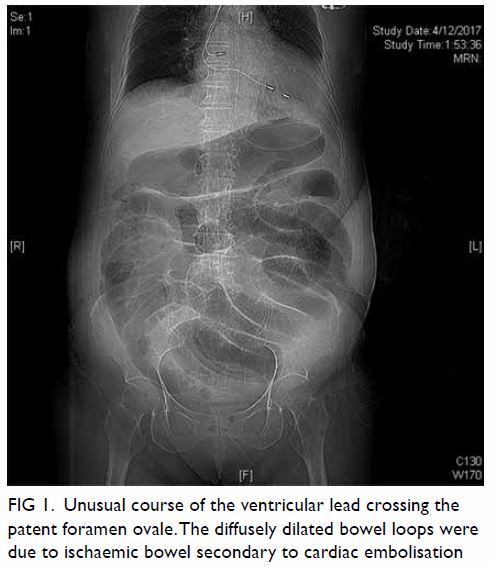
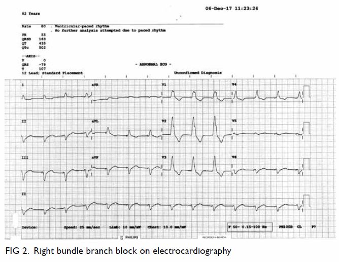
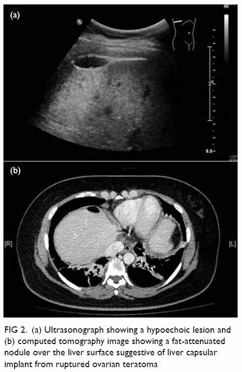
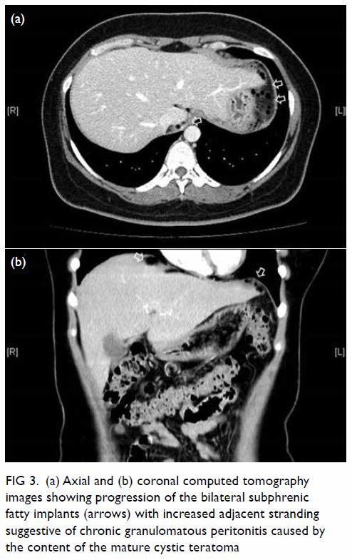
Triphasic contrast computed tomography (CT) of the liver revealed a 5.2-cm ovoid hypodense lesion with heterogeneous enhancement in the caudate lobe of the liver (Fig 1). No washout of contrast was evident in the portal venous or delayed phases. Fluorodeoxyglucose-18 positron emission tomography–CT showed a moderately hypermetabolic lesion at the caudate lobe with a maximum standardised update value of 5.8 (Fig 2).
 

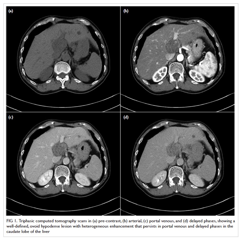
Figure 1. Triphasic computed tomography scans in (a) pre-contrast, (b) arterial, (c) portal venous, and (d) delayed phases, showing a well-defined, ovoid hypodense lesion with heterogeneous enhancement that persists in portal venous and delayed phases in the caudate lobe of the liver
 

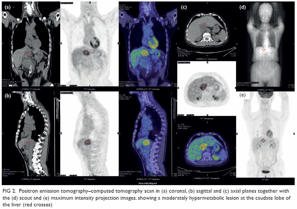
Figure 2. Positron emission tomography–computed tomography scan in (a) coronal, (b) sagittal and (c) axial planes together with the (d) scout and (e) maximum intensity projection images, showing a moderately hypermetabolic lesion at the caudate lobe of the liver (red crosses)
 
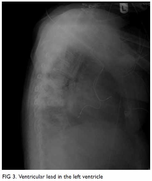
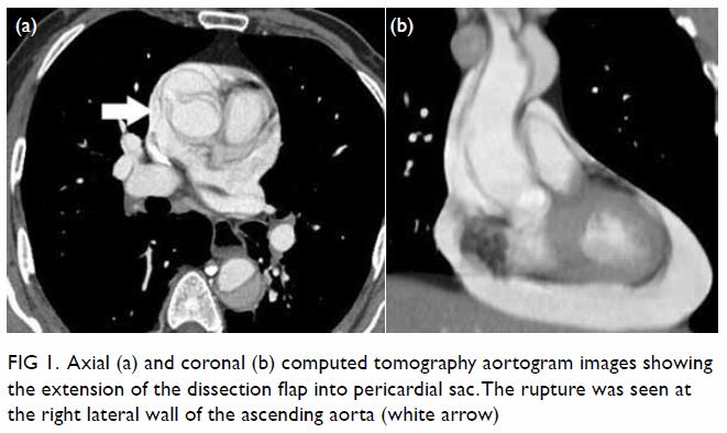
Surgical resection of the lesion was performed (Fig 3). Pathology showed a schwannoma and 
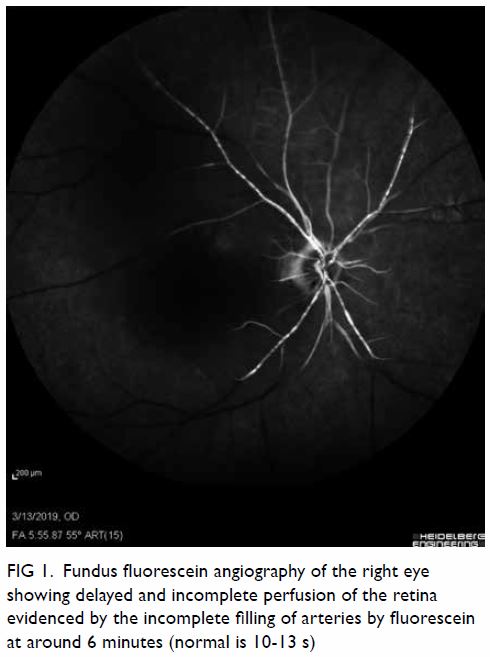
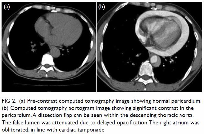
degenerative changes. Histological examination revealed an encapsulated tumour consisting of highly ordered Antoni type A and B areas (Fig 4). Immunohistochemical analysis showed the tumour cells to be diffusely positive for S100, consistent with neural differentiation (Fig 4). HerPar1, a mitochondrial antigen of hepatocytes, was negative. The CD34, a cell surface glycoprotein that is positive in gastrointestinal stromal tumour, was also negative.
 

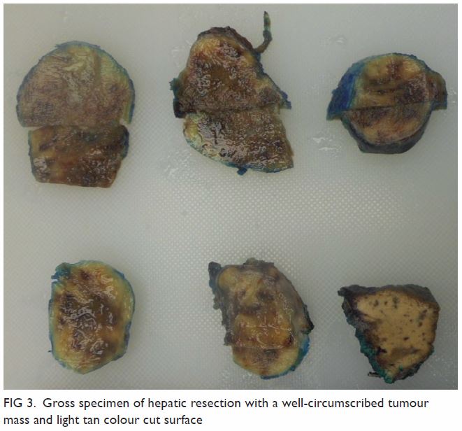
Figure 3. Gross specimen of hepatic resection with a well-circumscribed tumour mass and light tan colour cut surface
 

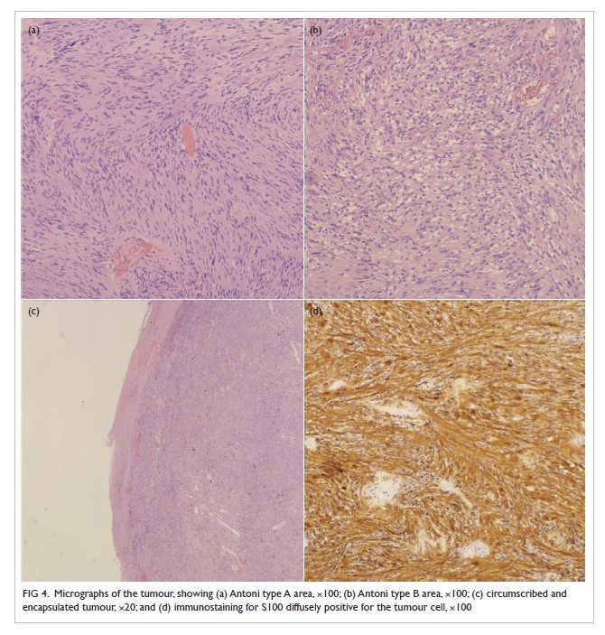
Figure 4. Micrographs of the tumour, showing (a) Antoni type A area, ×100; (b) Antoni type B area, ×100; (c) circumscribed and encapsulated tumour, ×20; and (d) immunostaining for S100 diffusely positive for the tumour cell, ×100
 
Schwannoma is most commonly found in the limbs and the head and neck region. A fifth of cases shows association with neurofibromatosis type 1. The mediastinum and retroperitoneum are other possible sites. It is uncommon in the gastrointestinal tract and extremely rare in the liver.1 It was first reported in 1978 by Pereira et al.2 A literature search through PubMed and MEDLINE revealed 32 reported cases. No cases have been published in Hong Kong.
 
The origin of hepatic schwannoma is the hepatobiliary nerves among the hepatic plexus in the liver hilum as well as interlobular connective tissues and the hepatic arteries.1 3 They are usually well-encapsulated and grow very slowly, usually smaller than 5 cm at the time of diagnosis. Larger schwannomas may undergo secondary degeneration with consequent pseudocystic regression, haemorrhage, and calcification. Malignant transformation is very rare.3
 
Pathologically, a schwannoma is an encapsulated tumour that arises within the nerve sheaths. It consists of a highly ordered cellular component (Antoni type A area) characterised by spindle cells with twisted nuclei arranged in short bundles, and a hypocellular area in a loose myxoid stroma (Antoni type B area) that comprises a loose meshwork of gelatinous and microcystic tissue.4
 
On imaging, hepatic schwannoma is usually well circumscribed with various signal characteristics, depending on the distribution of Antoni A and Antoni B areas.1 It is commonly of low density with heterogeneous enhancement on CT, hypointense on T1-weighted, and hyperintense on T2-weighted magnetic resonance imaging.5 There have also been reports of malignant tumours but there are no distinct radiological features that differentiate them from benign tumours.3 A hepatic schwannoma may be fluorodeoxyglucose-avid depending on inflammatory activity and cellularity. Fluorodeoxyglucose-18 positron emission tomography–CT alone may enable differentiation of a schwannoma from malignant lesions of the liver.1
 
Hepatic schwannoma is an extremely rare tumour and preoperative diagnosis with imaging is challenging. Biopsy or surgical resection is usually required for definitive diagnosis.
 
Author contributions
Concept or design: All authors.
Acquisition of data: HL Tsui and CH Lau.
Analysis or interpretation of data: HL Tsui, SM Yu, and CH Lau.
Drafting of the manuscript: HL Tsui, SM Yu, and CH Lau.
Critical revision of the manuscript for important intellectual content: All authors.
 
All authors had full access to the data, contributed to the study, approved the final version for publication, and take responsibility for its accuracy and integrity.
 
Conflicts of interest
All authors have disclosed no conflicts of interest.
 
Funding/support
This research received no specific grant from any funding agency in the public, commercial, or not-for-profit sectors.
 
Ethics approval
This study is approved by the cluster Research Ethics Committee (Ref KC/KE-19-0247/ER-3). Written patient consent was also obtained.
 
References
1. Hayashi M, Takeshita A, Yamamoto K, Tanigawa N. Primary hepatic benign schwannoma. World J Gastrointest Surg 2012;4:73-8. Crossref
2. Pereira Filho RA, Souza SA, Oliveira Filho JA. Primary neurilemmal tumour of the liver: case report. Arq Gastroenterol 1978;15:136-8.
3. Ozkan EE, Guldur M, Uzunkoy A. A case report of benign schwannoma of the liver. Intern Med 2010;49:1533-6. Crossref
4. Wan DL, Zhai ZL, Ren KW, Yang YC, Lin SZ, Zheng SS. Hepatic schwannoma: a case report and an updated 40-year review of the literature yielding 30 cases. Mol Clin Oncol 2016;4:959-64. Crossref
5. Yamamoto M, Hasegawa K, Arita J, et al. Primary hepatic schwannoma: a case report. Int J Surg Case Rep 2016;29:146-50. Crossref

A common but often neglected source of emboli

© Hong Kong Academy of Medicine. CC BY-NC-ND 4.0
 
PICTORIAL MEDICINE
A common but often neglected source of emboli
SY Au, MB, BS, FHKAM (Medicine); KM Fong, MB, ChB, MRCP (UK); Jack KC Shek, MB, BS, FHKAM (Anaesthesiology); SK Yung, MB, BS, FHKAM (Medicine); George WY Ng, MB, BS, FHKAM (Medicine)
Intensive Care Unit, Queen Elizabeth Hospital, Hong Kong
 
Corresponding author: Dr SY Au (h0145237@gmail.com)
 
 Full paper in PDF
 
Case
An 82-year-old woman was admitted to the intensive care unit with acute abdomen. She was initially admitted for chest infection but complained of sudden-onset abdominal pain and abdominal distension. She was confused with decreased consciousness. She had a medical history of sick sinus syndrome with dual-chamber pacing performed 4 years previously. She was being treated for hypertension but was prescribed neither aspirin nor anticoagulation. Initial investigations revealed an increased white cell count, coagulopathy, and deranged liver and renal function, with severe mixed respiratory and lactic acidosis. She was commenced on a high-dose dopamine infusion and was intubated prior to transfer to the intensive care unit. Further imaging investigations were performed. Abdominal plain radiograph showed dilated bowel and subsequent computed tomography of the abdomen confirmed diffuse bowel ischaemia (Fig 1). There was no abdominal aortic aneurysm. Computed tomography was also performed in view of her confusion and showed left middle cerebral artery infarction. Electrocardiography performed to identify cardiac causes of systemic embolisation revealed a paced rhythm of 80 beats per minute and right bundle branch block pattern (Fig 2). A lateral chest radiograph subsequently confirmed that the ventricular lead was in the left ventricle as it was pointing posteriorly (Fig 3). Transthoracic echocardiography showed no intracardiac clots but confirmed that the pacing lead was positioned in the left heart through the atrial septum. Transoesophageal echocardiography confirmed that the lead entered the left heart through a patent foramen ovale. The ischaemic bowel was too extensive and not amenable to surgery and the patient finally succumbed.
 

Figure 1. Unusual course of the ventricular lead crossing the patent foramen ovale.The diffusely dilated bowel loops were due to ischaemic bowel secondary to cardiac embolisation
 

Figure 2. Right bundle branch block on electrocardiography
 

Figure 3. Ventricular lead in the left ventricle
 
Confirmation of placement of a pacing lead by a posterior-anterior chest radiograph can be difficult. A left bundle branch paced rhythm on echocardiography and a lateral radiograph that shows an anteriorly pointing ventricular lead help confirm placement of a pacing lead in the right ventricle. A pacing lead in the left ventricle and the absence of anticoagulation places a patient at risk of clot formation that may lead to systemic embolisation. In our patient, an intracardiac clot was not well demonstrated on either computed tomography or echocardiography, and 4 years had passed between pacemaker insertion and this systemic embolic episode. Only a probable, not a definite causal relationship could be established. We postulate that her chest infection may have impacted her clotting profile and resulted in this systemic embolic event.
 
Author contributions
Concept or design: All authors.
Acquisition of data: All authors.
Analysis or interpretation of data: SY Au.
Drafting of the manuscript: SY Au.
Critical revision of the manuscript for important intellectual content: All authors.
 
All authors had full access to the data, contributed to the study, approved the final version for publication and take responsibility for its accuracy and integrity.
 
Conflicts of interest
All authors have no conflicts of interest to disclose.
 
Funding/support
This pictorial medicine received no specific grant from any funding agency in the public, commercial or not-for-profit sectors.
 
Ethics approval
The study was conducted in accordance with the Declaration of Helsinki. Consent for publication was obtained from the patient’sfamily.
 

Vision loss due to ophthalmic artery occlusion secondary to spontaneous internal carotid artery dissection

© Hong Kong Academy of Medicine. CC BY-NC-ND 4.0
 
PICTORIAL MEDICINE
Vision loss due to ophthalmic artery occlusion secondary to spontaneous internal carotid artery dissection
Sunny CL Au, MB, ChB, AFCOphthHK1; Simon TC Ko, MB, BS, FHKAM (Ophthalmology)2
1 Department of Ophthalmology, Hong Kong East Cluster Ophthalmic Service, Tung Wah Eastern Hospital, Hong Kong
2 Department of Ophthalmology, Tung Wah Eastern Hospital, Hong Kong
 
Corresponding author: Dr Sunny CL Au (kilihcua@gmail.com)
 
 Full paper in PDF
 
Case
A 47-year-old male smoker with hypertension, diabetes mellitus, and hyperlipidaemia complained of right eye vision loss on waking. His best-corrected visual acuity was reduced to light perception only. Physical examination revealed anisocoria without ptosis and no extraocular movement deficit. The right eye pupil was larger than the left, and the difference was more obvious under a light environment. Direct and consensual pupil reflexes were both present but there was a marked right relative afferent pupillary defect. Slit lamp examination revealed normal anterior segments of the eyes, but a right pale optic disc without rim thinning, and a typical cherry-red spot over the right fovea surrounded by generalised whitened retina. Vessels were not tortuous and no emboli were seen nor retinal haemorrhage over different layers (Figs 1 and 2). Left eye posterior segment was normal. The patient also reported right-sided headache and left-sided numbness. Thorough physical examination revealed no other cranial nerve deficit or systemic focal neurological deficit. Temporal pulse was easily palpable and non-tender, and no carotid bruit was heard. The patient worked on a construction site and denied any trauma. He commenced hyperbaric oxygen therapy but no improvement was observed. Blood tests and brain imaging were all normal, but carotid Doppler showed significant obstruction >90% of the internal carotid artery, accounting for the absence of bruit. Computed tomography angiography confirmed dissection of a long segment of the right internal carotid artery (Figs 3 and 4), not amendable to stenting or bypass surgery. A diagnosis of ophthalmic artery occlusion was made and explained the ineffectiveness of hyperbaric oxygen therapy due to choroidal ischaemia. Symptomatic internal carotid artery dissection is a rare but major cause of young-onset stroke, itself uncommon.1 Neck trauma is a major aetiology, and there is a slight male predominance with mean age of onset in the 40s.2 Apart from the neurological signs and symptoms of stroke, the ophthalmological presentation of internal carotid artery dissection is more similar to that of painful Horner’s syndrome due to compression of the adjacent third-order sympathetic chain fibres; followed by cranial nerve palsy, caused by direct local compression or compromise of feeder vessels.3 Ophthalmic artery occlusion is rare. Computed tomography angiography has 80% sensitivity for diagnosis and patients need to be closely monitored for massive stroke that may occur weeks to months after first presentation.4 Hyperbaric oxygen therapy is indicated for central retinal artery occlusion, but not ophthalmic artery occlusion. It aims to reperfuse the ischaemic retina with oxygen by diffusion from the choroidal circulation, bypassing the obstructed retinal vasculature. The choroidal arteries are supplied by the posterior ciliary arteries that branch from the ophthalmic artery. If the ophthalmic artery is occluded, hyperbaric oxygen therapy has no means to tackle the compromised posterior ciliary vessels.5
 

Figure 1. Fundus fluorescein angiography of the right eye showing delayed and incomplete perfusion of the retina evidenced by the incomplete filling of arteries by fluorescein at around 6 minutes (normal is 10-13 s)
 

Figure 2. Optical coherence tomography of the right eye showing generalised retinal oedema secondary to retinal ischaemia that marks the typical clinical sign of cherry red spot on fundus examination
 

Figure 3. Three-dimensional computed tomography angiography showing the presence of left, but the absence of right internal carotid artery (indicated by white arrow) perfusion by contrast
 

Figure 4. Coronal computed tomography angiography showing the presence of left, but the absence of right internal carotid artery perfusion by contrast
 
In conclusion, vision loss due to internal carotid artery dissection is uncommon. Multidisciplinary care is essential.
 
Author contributions
Concept or design: SCL Au.
Acquisition of data: SCL Au.
Analysis or interpretation of data: SCL Au.
Drafting of the manuscript: SCL Au.
Critical revision of the manuscript for important intellectual content: All authors.
 
All authors had full access to the data, contributed to the study, approved the final version for publication, and take responsibility for its accuracy and integrity.
 
Conflicts of interest
All authors have disclosed no conflicts of interest.
 
Funding/support
This pictorial medicine received no specific grant from any funding agency in the public, commercial, or not-for-profit sectors.
 
Ethics approval
This study was conducted in accordance with the principles outlined in the Declaration of Helsinki. Relevant patient consent was obtained for the purpose of this case study.
 
References
1. Thanvi B, Munshi SK, Dawson SL, Robinson TG. Carotid and vertebral artery dissection syndromes. Postgrad Med J 2005;81:383-8. Crossref
2. Blum CA, Yaghi S. Cervical artery dissection: a review of the epidemiology, pathophysiology, treatment, and outcome. Arch Neurosci 2015;2:e26670. Crossref
3. Kasravi N, Leung A, Silver I, Burneo JG. Dissection of the internal carotid artery causing Horner syndrome and palsy of cranial nerve XII. CMAJ 2010;182:E373-7. Crossref
4. Borgman CJ. Horner syndrome secondary to internal carotid artery dissection after a short-distance endurance run: a case study and review. J Optom 2012;5:209-16. Crossref
5. Kim SH, Cha YS, Lee Y, Kim H, Yoon IN. Successful treatment of central retinal artery occlusion using hyperbaric oxygen therapy. Clin Exp Emerg Med 2018;5:278-81. Crossref

Temporal changes in computed tomography of COVID-19 pneumonia with perilobular fibrosis

Hong Kong Med J 2020 Jun;26(3):250–1.e1-2  |  Epub 29 Apr 2020
© Hong Kong Academy of Medicine. CC BY-NC-ND 4.0
 
PICTORIAL MEDICINE
Temporal changes in computed tomography of COVID-19 pneumonia with perilobular fibrosis
FH Ng, MB, ChB, FHKCR; SK Li, MB, ChB, FHKCR; YC Lee, MB, ChB, FHKCR; Johnny KF Ma, MB, BS, FHKCR
Department of Radiology, Princess Margaret Hospital, Hong Kong
 
Corresponding author: Dr FH Ng (ngphonehim@gmail.com)
 
 Full paper in PDF
 
As the designated tertiary referral centre for infectious diseases, Princess Margaret Hospital, Hong Kong, received the city’s first influx of patients diagnosed with coronavirus disease 2019 (COVID-19). In January 2020, a 57-year-old man with a history of travel to Wuhan, China, presented to Princess Margaret Hospital with a 4-day history of respiratory symptoms and fever. Laboratory studies showed lymphopoenia 0.7 × 10⁹/L (reference range, 1.1-2.9 109/L) and elevated inflammatory markers. The patient’s C-reactive protein level was 33.2 mg/L (reference range, <5 mg/L) on the day of admission and peaked (123 mg/L) on day 3 after admission. The diagnosis of COVID-19 was made by the detection of severe acute respiratory syndrome coronavirus 2 RNA in a nasopharyngeal aspirate using reverse transcription polymerase chain reaction. High-resolution computed tomography (HRCT) images were acquired on days 2, 14, and 22 after admission. On day 2 after admission, HRCT showed peripheral subpleural ground-glass opacities (GGOs) and consolidation without zonal predominance in the absence of centrilobular nodules, pleural effusions, or lymph node enlargement, compatible with organising pneumonia,1 which correlated with the known radiological pattern of COVID-19 pneumonia.2
 
Concurrent with improvement of clinical symptoms and normalising inflammatory markers, serial HRCTs showed reduced GGOs (Fig 1). By day 22 after admission, the patient’s C-reactive protein level had normalised (5.3 mg/L). Instead of complete resolution, consolidations became curved or arched consolidation bands, with shaded margins, distributed around the structures surrounding the secondary pulmonary lobules. It was likely the result of perilobular inflammation and took the form of an arcade (Fig 2). This arcade-like sign is one of the typical features of perilobular fibrosis found in secondary organising pneumonia.3
 

Figure 1. High-resolution computed tomography images of a 57-year-old man with a history of travel to Wuhan, China, with a 4-day history of respiratory symptoms and fever. On days 2, 14, and 22 after admission, peripheral subpleural ground-glass opacities and consolidation without zonal predominance are visible, compatible with an organising pneumonia pattern. Serial computed tomography scans show reduction of the ground-glass opacities with residual fibrosis
 

Figure 2. Computed tomography images of a 57-year-old man with a history of travel to Wuhan, China, with a 4-day history of respiratory symptoms and fever. Arcade-like signs (black arrows) are shown on coronal, axial and sagittal and reformatted computed tomography images. Consolidations became curved or arched consolidation bands, with shaded margins, distributed around the structures surrounding the secondary pulmonary lobules, suggestive of perilobular fibrosis
 
Residual GGOs with irregular lines and interfaces are the second most common pattern in COVID-19 pneumonia, suggesting the presence of secondary organising pneumonia, and hence pulmonary fibrosis.4 The gold standard to confirm pulmonary fibrosis requires lung biopsy or bronchoalveolar lavage, which are invasive. Lung biopsy was not performed in our case.
 
The present case highlights the computed tomography findings of pulmonary fibrosis, one of the sequelae of COVID-19 pneumonia.
 
Author contributions
All authors contributed to the concept or design of the study, acquisition of the data, drafting of the manuscript, and critical revision of the manuscript for important intellectual content. All authors had full access to the data, contributed to the study, approved the final version for publication, and take responsibility for its accuracy and integrity.
 
Conflicts of interest
All authors have disclosed no conflicts of interest.
 
Acknowledgement
We would like to express our gratitude to the Infectious Disease Team and “dirty team” physicians of Princess Margaret Hospital, Hong Kong, for their professional patient care and invaluable contribution to the understanding of a novel disease.
 
Funding/support
This pictorial medicine paper received no specific grant from any funding agency in the public, commercial, or not-for-profit sectors.
 
Ethics approval
This study was approved by the Kowloon West Cluster Research Ethics Committee (Ref 144-20). The patient provided written informed consent for all procedures and treatments.
 
References
1. Ujita M, Renzoni EA, Veeraraghavan S, Wells AU, Hansell DM. Organizing pneumonia: perilobular pattern at thin-section CT. Radiology 2004;232:757-61. Crossref
2. Chung M, Bernheim A, Mei X, et al. CT imaging features of 2019 novel coronavirus (2019-nCoV). Radiology 2020;295:202-7. Crossref
3. Chiarenza A, Ultimo LE, Falsaperla D, et al. Chest imaging using signs, symbols, and naturalistic images: a practical guide for radiologists and non-radiologists. Insights Imaging 2019;10:114. Crossref
4. Wang Y, Dong C, Hu Y, et al. Temporal changes of CT findings in 90 patients with COVID-19 pneumonia: A longitudinal study. Radiology 2020:200843. Crossref

High-resolution computed tomography in a patient with COVID-19 with non-diagnostic serial radiographs

Hong Kong Med J 2020 Jun;26(3):248–9.e1–3  |  Epub 29 Apr 2020
© Hong Kong Academy of Medicine. CC BY-NC-ND 4.0
 
PICTORIAL MEDICINE
High-resolution computed tomography in a patient with COVID-19 with non-diagnostic serial radiographs
HM Kwok, MB, BS# 1; SC Wong, MB, BS# 1TF Ng, MB, BS, FHKAM (Radiology)1; KS Yung, MB, BS1; WH Luk, MB, BS, FHKAM (Radiology)1KF Ma, MB, BS, FHKAM (Radiology)1; Thomas SH Chik, MRCP (UK), FHKAM (Medicine)2
1 Department of Radiology, Princess Margaret Hospital, Hong Kong
2 Infectious Disease Team, Princess Margaret Hospital, Hong Kong
# The first two authors contributed equally to the work
 
Corresponding author: Dr HM Kwok (hmkwok15@hotmail.com)
 
 Full paper in PDF
 
Case
A 63-year-old Chinese man from Wuhan, China, presented to the emergency department of Princess Margaret Hospital, Hong Kong, in January 2020 with coryzal symptoms.
 
He was afebrile with unremarkable respiratory examination results. He was admitted to an isolation ward for further investigation. He subsequently developed a fluctuating fever up to 38.5°C after admission and passed loose stool intermittently. His oxygen saturation levels were normal throughout hospitalisation. Supportive treatment and empirical antibiotics (amoxicillin-clavulanate) were administered.
 
Coronavirus disease 2019 (COVID-19) was suspected, and severe acute respiratory syndrome coronavirus 2 (SARS-CoV-2) infection was confirmed the next day by throat swab and nasopharyngeal aspirate tests. Stool culture was negative for SARS-CoV-2 and other pathogens.
 
Blood test results revealed lymphopoenia (0.6 × 10⁹/L). Total white blood cell count (4.4 × 10⁹/L), absolute neutrophil count (3.4 × 10⁹/L), monocyte count (0.3 × 10⁹/L), and eosinophil count (0.0 × 10⁹/L) were normal. Liver and renal function test results, C-reactive protein level, and serum calcium and phosphate levels were normal.
 
Daily portable chest radiographs over the next 5 days were unremarkable (Fig 1). Chest high-resolution computed tomography (HRCT) was performed to assess for pulmonary involvement. High-resolution computed tomography performed 6 days after admission (Fig 2) demonstrated a few patchy subpleural ground-glass opacities in both lungs. No other abnormalities were detected. Subsequent follow-up chest radiograph (Fig 3) remained clear.
 

Figure 1. A 63-year-old Chinese male with suspected coronavirus disease 2019 infection presented with coryzal symptoms without fever. A portable chest radiograph 6 days after admission was unremarkable despite changes on high-resolution computed tomography scans
 

Figure 2. High-resolution computer tomography 6 days after admission showing small patchy areas of subpleural ground-glass opacities in the posterior right lower lobe, the right middle lobe, and the left lower lobe (arrows). No other abnormalities are visible. Diagnosis of severe acute respiratory syndrome coronavirus 2 infection was confirmed with nasopharyngeal aspirate and throat swab samples
 

Figure 3. Follow-up chest radiograph 11 days after admission remained clear
 
The patient’s fever subsided 11 days after admission. No supplemental oxygen was required. Serial nasopharyngeal aspirate sample tests turned negative 22 days after admission. The patient was discharged from the hospital 27 days after admission.
 
Discussion
The SARS-CoV-2 infection is diagnosed with reverse transcription polymerase chain reaction results of respiratory specimens.
 
Prior to availability of confirmatory microbiological test results, challenges exist in decision making for patients with suspected COVID-19.
 
We advocate that early HRCT should be considered in suspected cases to aid in clinico-radiological diagnosis of SARS-CoV-2 infection by demonstrating compatible radiological features, before microbiological confirmation has been established.
 
This can be especially beneficial if patients have negative preliminary virology or radiographic findings, as in the previously reported HRCT features in severe acute respiratory syndrome (SARS) and Middle East respiratory syndrome (MERS),1 2 as well as recent reports on COVID-19 from China3 and Hong Kong.4
 
Findings on serial chest radiographs before and after HRCT remain occult without demonstrable airspace opacity, limiting its value in initial assessment, monitoring or exclusion of pulmonary involvement.
 
This is consistent with experience during the SARS epidemic that HRCT was useful for early radiological assessment for patients with negative chest radiographs.5
 
Our patient subsequently demonstrated a favourable outcome with recovery and did not require supplemental oxygen throughout hospitalisation.
 
Mirroring experiences in previous coronavirus outbreaks such as SARS and MERS, we believe that HRCT will likely play a key role in aiding diagnosis, assessing the extent of pulmonary involvement and risk in the current COVID-19 pandemic. Early HRCT can be beneficial in patients with obscure clinical presentation or negative preliminary virological or radiographic findings.
 
Author contributions
All authors contributed to the concept or design of the study, acquisition of the data, drafting of the manuscript, and critical revision of the manuscript for important intellectual content. All authors had full access to the data, contributed to the study, approved the final version for publication, and take responsibility for its accuracy and integrity.
 
Conflicts of interest
All authors have disclosed no conflicts of interest.
 
Acknowledgement
We would like to express our gratitude to the Infectious Disease Team and “dirty team” physicians of Princess Margaret Hospital, Hong Kong, for their professional patient care and invaluable contribution to the understanding of a novel disease.
 
Funding/support
This pictorial medicine paper received no specific grant from any funding agency in the public, commercial, or not-for-profit sectors.
 
Ethics approval
This study was approved by the Kowloon West Cluster Research Ethics Committee (Ref KW/EX-20-049(145-08)).
 
References
1. Koo HJ, Lim S, Choe J, Choi SH, Sung H, Do KH. Radiographic and CT features of viral pneumonia. Radiographics 2018;38:719-39. Crossref
2. Franquet T. Imaging of pulmonary viral pneumonia. Radiology 2011;260:18-39. Crossref
3. Lei J, Li J, Li X, Qi X. CT imaging of the 2019 novel coronavirus (2019-nCoV) pneumonia. Radiology 2020;295:18. Crossref
4. World Health Organization. Clinical management of severe acute respiratory infection when COVID-19 is suspected. Interim guidance. Available from: https:// www.who.int/publications-detail/clinical-management-of-severe-acute-respiratory-infection-when-novel-coronavirus-(ncov)-infection-is-suspected. Accessed 6 Feb 2020.
5. Hui JY, Hon TY, Yang MK, et al. High-resolution computed tomography is useful for early diagnosis of severe acute respiratory syndrome-associated coronavirus pneumonia in patients with normal chest radiographs. J Comput Assist Tomogr 2004;28:1-9. Crossref

Bow hunter’s syndrome: a sinister cause of vertigo and syncope not to be missed

© Hong Kong Academy of Medicine. CC BY-NC-ND 4.0
 
PICTORIAL MEDICINE
Bow hunter’s syndrome: a sinister cause of vertigo and syncope not to be missed
SC Wong, MB, BS; TS Chan, FHKCR, FHKAM (Radiology); CH Chan, FHKCR, FHKAM (Radiology); Johnny KF Ma, FRCR (UK), FHKAM (Radiology)
Department of Radiology, Princess Margaret Hospital, Laichikok, Hong Kong
 
Corresponding author: Dr SC Wong (bennychun1021@gmail.com)
 
 Full paper in PDF
 
Case
In May 2015, a 59-year-old woman presented to Princess Margaret Hospital, Hong Kong, with chronic intermittent vertigo and syncope, aggravated by head rotation to the right. Physical, otoscopy, and nasal endoscopy examination results were unremarkable. Cervical spine plain radiographs demonstrated cervical spondylosis with marginal osteophytosis (Fig 1). In May 2015, computed tomography angiography of the head and neck revealed focal moderate (50%) stenosis at bilateral vertebral arteries at C5/6 levels due to extrinsic compression from hypertrophied uncovertebral joints (Fig 2). In January 2016, magnetic resonance imaging demonstrated disco-osteophytic protrusion at C5/6 level without evidence of cord compression. In March 2016, digital subtraction angiography, performed with the patient’s head in neutral and bilateral rotated positions, demonstrated dynamic deterioration of focal stenosis of right vertebral artery at C5/6 level to up to 80% stenosis during head rotation to the right (Figs 3 and Fig 4).
 

Figure 1. Bilateral oblique radiographs of the cervical spine, showing degenerative changes with marginal osteophytes and narrowing of multiple bilateral lower cervical neural foramina (arrows)
 

Figure 2. Axial computed tomography angiogram of the bilateral vertebral arteries at C5/6 level with extrinsic compression (arrows) of (a) left and (b) right vertebral artery by hypertrophied uncovertebral joints
 

Figure 3. Digital subtraction angiography of the right vertebral artery in frontal (AP) and lateral (Lat) views taken with head in neutral position. Note the focal stenosis of vertebral artery at C5/6 level (arrows)
 

Figure 4. Digital subtraction angiography of the right vertebral artery in frontal (AP) and lateral (Lat) views taken with head rotated to the right. Note the deterioration in vertebral artery stenosis at C5/6 level on head rotation to the right (arrows)
 
A static focal moderate (50%) stenosis of the left vertebral artery at C5/6 was also present. Overall findings were compatible with bow hunter’s syndrome (BHS) with dynamic deterioration of right vertebral artery stenosis on head rotation, related to extrinsic compression by hypertrophied facet joint and disc protrusion. In April 2016, the patient underwent anterior C5/6 cervical discectomy and anterior spinal fusion with smooth recovery and symptomaticresolution.
 
Discussion
Bow hunter’s syndrome was first reported in 1978 when a patient developed lateral medullary syndrome during archery practice with lateral head rotation.1 It refers to symptomatic vertebrobasilar insufficiency secondary to mechanical occlusion or stenosis of vertebral arteries upon head and neck rotation.
 
The pathogenesis of BHS is related to the tortuous anatomical course of the vertebral artery along the cervical spine, which renders the artery susceptible to extrinsic compression, repetitive shear stress resulting in haemodynamic events in at-risk patients during head and neck rotation.2 Osteophytes, disc herniation, ligamentous or neck muscle hypertrophy are risk factors for BHS.3
 
Though rare, BHS is a not to be missed cause of vertigo, owing to its specific relationship with head and neck rotation and its potential risk of posterior circulation ischaemic stroke.1 2 Bow hunter’s syndrome is more common among males and those aged between 50 and 70 years old.3 Common clinical manifestations include vertigo and syncope. Other symptoms include nystagmus, emesis, Horner’s syndrome, and rarely motor and sensory deficits.2
 
Imaging is crucial in establishing the diagnosis of BHS, delineating the cause and site of extrinsic compression and evaluating complications such as infarction. Dynamic digital subtraction angiography remains the preferred modality for prompt and accurate localisation of stenotic segment and establishing causal relationship with head rotation.4 5 Non-invasive computed tomography or magnetic resonance angiography in both neutral and rotated head positions are also used. Computed tomography can delineate the relationship with surrounding compressive skeletal structures and magnetic resonance would be sensitive in documenting early ischaemic event.3
 
In this case, the patient’s complaint of vertigo exacerbation with specific direction of head rotation should raise the suspicion of BHS. Surgical treatment was offered in view of failed conservative approach, repeated fall related to syncope, and underlying uncovertebral joint hypertrophy and disc protrusion as the aetiological factors of vertebral artery compression.
 
Author contributions
All authors had full access to the data, contributed to the study, approved the final version for publication, and take responsibility for its accuracy and integrity.
 
Concept or design: All authors.
Acquisition of data: TS Chan, JKF Ma.
Analysis or interpretation of data: All authors.
Drafting of the manuscript: SC Wong. TS Chan.
Critical revision of the manuscript for important intellectual content: All authors.
 
Conflicts of interest
All authors have disclosed no conflicts of interest.
 
Funding/support
This pictorial medicine paper received no specific grant from any funding agency in the public, commercial, or not-for-profit sectors.
 
Ethics approval
All patients were treated in accordance with the Declaration of Helsinki and provided consent for all investigations and procedures.
 
References
1. Sorensen BF. Bow hunter’s stroke. Neurosurgery 1978;2:259-61. Crossref
2. Duan G, Xu J, Shi J, Cao Y. Advances in the pathogenesis, diagnosis and treatment of bow hunter’s syndrome: a comprehensive review of the literature. Interv Neurol 2016;5:29-38. Crossref
3. Rastogi V, Rawls A, Moore O, et al. Rare etiology of bow hunter’s syndrome and systematic review of literature. J Vasc Interv Neurol 2015;8:7-16.
4. Taylor WB 3rd, Vandergriff CL, Opatowsky MJ, Layton KF. Bowhunter’s syndrome diagnosed with provocative digital subtraction cerebral angiography. Proc (Bayl Univ Med Cent) 2012;25:26-7. Crossref
5. Go G, Hwang SH, Park IS, Park H. Rotational vertebral artery compression: bow hunter’s syndrome. J Korean Neurosurg Soc 2013;54:243-5. Crossref

Recognising eye implants on radiological imaging: pear or fishball shapes

© Hong Kong Academy of Medicine. CC BY-NC-ND 4.0
 
PICTORIAL MEDICINE
Recognising eye implants on radiological imaging: pear or fishball shapes
Sunny CL Au, MB, ChB, AFCOphthHK; Simon TC Ko, FHKAM (Ophthalmology), FCOphth HK
Department of Ophthalmology, Tung Wah Eastern Hospital (Hong Kong East Cluster Ophthalmic Service), Causeway Bay, Hong Kong
 
Corresponding author: Dr Sunny CL Au (kilihcua@gmail.com)
 
 Full paper in PDF
 
Radiological imaging is now readily available in Hong Kong, in both the public and private sector. Imaging helps diagnose or screen for diseases, but can create noise or false alarms.1 Ophthalmology consultations are common in both in-patient and out-patient settings. Floaters, visual field defects, and abnormal incidental findings on computed tomography or magnetic resonance imaging (MRI) are all common reasons for consultations.2
 
In 2018, a 60-year-old man underwent an MRI scan for acoustic neuroma screening, which revealed an abnormal eyeball shape (Fig 1). Diligent consultation with an ophthalmologist was reassuring for both the physician and the patient. In December 2017, the patient had been referred to our eye department for visual disturbance and was diagnosed with right eye macula-on retinal detachment. He had no history of laser or other eye surgery, nor trauma to the eyes. The patient reported right eye floaters and loss of the inferior visual field. Fundus examinations found a large retinal U-shaped tear at the superior retina, with bullous retinal detachment over the right eye. There were also multiple small flat holes with lattice degeneration over the retina in the temporal and nasal aspect. Retinal detachment repair surgery with encircling band as scleral buckling, and intravitreal gas injection was done for the presence of multiple retinal holes.3 Given the patient’s older age, the presence of cataract, and perceived cataract progression with postoperative intravitreal gas,4 cataract extraction with intraocular lens insertion was also done in the same operation. These are all evidenced on the MRI scan contrasting the presence of a natural thickness lens over the left eye.
 

Figure 1. T2-weighted magnetic resonance imaging scan transverse cut of the brain showing two T2 hyperintense structures highlighting the position of the eyeballs: the pear-shaped right eyeball, in contrast to the normal fishball- shaped left eyeball
 
Retinal detachment surgery can be of external or internal approach to relieve the vitreous traction and flatten the retina. External approach can be localised or 360-degree encircling depending on the retinal status.
 
The formation of the pear-shaped eyeball was due to the buckling effect of the encircling band (Fig 2). With basic knowledge of normal organ shapes, all practitioners would be concerned by such a deformed organ on plain radiographs. Typically, intraocular tumours or eyeball ruptures are not regular nor symmetrical, except large ring melanoma of the ciliary body. The key to differentiating a genuine pathology from a congenital/postoperative variant lies heavily on clinical history.1 Operative implants usually appear regular, or even symmetrical on imaging.
 

Figure 2. Schematic diagram of the eyeball cross-sectional plane showing the effect of the encircling band on the shape of the right eyeball. Solid arrows indicate the direction of force exerted by the encircling band on indenting the eyeball
 
Author contributions
All authors had full access to the data, contributed to the study, approved the final version for publication, and take responsibility for its accuracy and integrity.
 
Concept or design: SCL Au.
Acquisition of data: SCL Au.
Analysis or interpretation of data: SCL Au.
Drafting of the manuscript: SCL Au.
Critical revision of the manuscript for important intellectual content: All authors.
 
Conflicts of interest
All authors have disclosed no conflicts of interest.
 
Funding/support
This pictorial medicine paper received no specific grant from any funding agency in the public, commercial, or not-for-profit sectors.
 
Ethics approval
This study was conducted in accordance with the principles outlined in the Declaration of Helsinki. Relevant patient consent was obtained for the purpose of this case study.
 
References
1. Leslie A, Jones AJ, Goddard PR. The influence of clinical information on the reporting of CT by radiologists. Br J Radiol 2000;73:1052-5. Crossref
2. Carter K, Miller KM. Ophthalmology inpatient consultation. Ophthalmology 2001;108:1505-11. Crossref
3. Shanmugam PM, Ramanjulu R, Mishra KC, Sagar P. Novel techniques in scleral buckling. Indian J Ophthalmol 2018;66:909-15. Crossref
4. Thompson JT. The role of patient age and intraocular gases in cataract progression following vitrectomy for macular holes and epiretinal membranes. Trans Am Ophthalmol Soc 2003;101:485-98.

Giant perivascular spaces: an uncommon cause of obstructive hydrocephalus

© Hong Kong Academy of Medicine. CC BY-NC-ND 4.0
 
PICTORIAL MEDICINE
Giant perivascular spaces: an uncommon cause of obstructive hydrocephalus
MH So, MB, BS FRCR; WK Lo, FRCR, FHKAM (Radiology)
Department of Diagnostic and Interventional Radiology, Kwong Wah Hospital, Yaumatei, Hong Kong
 
Corresponding author: Dr MH So (manhon.so@gmail.com)
 
 Full paper in PDF
 
A 55-year-old man with good past health presented to the emergency department with unsteady gait for 6 months with recent mild left-sided weakness. Urgent computed tomography (CT) scan of the brain showed a multiseptated cystic lesion in the right mesencephalothalamic region with pressure effect on the third ventricle causing obstructive hydrocephalus (Fig 1). Urgent magnetic resonance imaging (MRI) scan (Fig 2) of the lesion showed no post-gadolinium enhancement, no restricted diffusion, complete suppression of the cystic areas on T2 fluid attenuation inversion recovery (FLAIR) sequence and no abnormal parenchymal signal intensities compared with normal brain parenchyma. These imaging findings are consistent with tumefactive perivascular space. Invasive biopsy and surgical excision were avoided, and the patient underwent surgery for ventricular drain insertion.
 

Figure 1. Plain computed tomography scan of the brain showing a multicystic lesion (arrow) in right mesencephalothalamic region with dilated frontal horns of the lateral ventricles and periventricular white matter hypodensities suggestive of transependymal oedema
 

Figure 2. Magnetic resonance images showing tumefactive perivascular space (arrows): (a) T2-weighted sequence; (b) T2 fluid attenuation inversion sequence with gadolinium showing complete suppression of fluid signal in the multicystic lesion; (c) T1-weighted sequence with gadolinium showing no enhancement; and (d) diffusionweighted imaging showing no restricted diffusion
 
Dilated perivascular spaces (PVSs) in the brain are interstitial fluid-filled structures lined by pia-mater that have accompanying patent penetrating arteries within, most commonly seen along the lenticulostriate arteries.1 They can be unilocular or multilocular and may have a radial pattern along the course of the penetrating arteries. The PVSs occur across all age-groups and are more frequent and larger with advancing age. The cause of dilated PVS remains unknown though numerous theories have been postulated including increased permeability of arterial wall and obstruction/disturbance of interstitial fluid drainage/flow. Dilated PVSs may be associated with microvascular diseases, trauma, non-vascular dementia, multiple sclerosis, and the mucopolysaccharidoses.
 
Rarely PVSs are markedly expanded and are termed tumefactive or giant PVSs. Some authors define tumefactive PVSs as those >1.5 cm. Tumefactive PVSs are most commonly located in mesencephalothalamic region.2 They are also seen in cerebral white matter and in the cerebellar dentate nuclei. They can exhibit mass effect and cause obstructive hydrocephalus when occurring in the mesencephalothalamic region as in our case. The MRI signal intensities of typical PVSs should follow cerebrospinal fluid in all sequences including FLAIR imaging with no post-gadolinium enhancement. There is no restricted diffusion as the compartments are communicating. Tumefactive PVSs in cerebral white matter may have perilesional abnormal T2 and FLAIR hyperintensities in up to 50% of cases. The mass effect of the tumefactive PVS may cause chronic ischaemic change in adjacent white matter.3 Histopathological results typically show a pial-lined cyst with no evidence of neoplasm or infection.
 
Differential diagnoses include cystic infarction, tumour, and infection.4 Cystic infarctions assume a slit-like or ovoid shape whereas PVSs are more rounded or linear. The cystic content of tumours is usually not isointense to cerebrospinal fluid on MRI. Solid components are often present, which may enhance after contrast and are surrounded by oedema. Parasitic infections have a range of appearances on CT or MRI scans with contrast enhancement and oedema during the active phase and calcifications in the quiescent phase.
 
Asymptomatic tumefactive PVSs can be managed by follow-up imaging for stability in size. Spontaneous regression of tumefactive PVSs without surgical intervention is rare. Tumefactive PVSs with mass effect and obstructive hydrocephalus can be treated surgically with ventriculostomy, cyst fenestration, ventriculoperitoneal shunting, or cystoperitoneal shunting. When the appearance is typical, surgical biopsy or excision should be avoided.
 
Author contributions
All authors contributed to the concept, acquisition and interpretation of data, drafting of the manuscript, and revision for important intellectual content. All authors had full access to the data, contributed to the study, approved the final version for publication, and take responsibility for its accuracy and integrity.
 
Conflicts of interest
All authors have disclosed no conflicts of interest.
 
Funding/support
This research received no specific grant from any funding agency in the public, commercial, or not-for-profit sectors.
 
Ethics approval
The patient was treated in accordance with the Declaration of Helsinki. The patient provided informed consent for all procedures.
 
References
1. AI Abdulsalam H, Alatar AA, Elwatidy S. Giant tumefactive perivascular spaces: a case report and literature review. World Neurosurg 2018;112:201-4. Crossref
2. Choh NA, Shaheen F, Robbani I, Singh M, Gojwari T. Tumefactive Virchow–Robin spaces: a rare cause of obstructive hydrocephalus. Ann Indian Acad Neurol 2014;17:345-6. Crossref
3. Salzman KL, Osborn AG, House P, et al. Giant tumefactive perivascular spaces. AJNR Am J Neuroradiol 2005;26:298- 305.
4. Kwee RM, Kwee TC. Virchow-Robin spaces at MR imaging. Radiographics 2007;27:1071-86. Crossref

Ruptured ovarian teratoma with granulomatous peritonitis

© Hong Kong Academy of Medicine. CC BY-NC-ND 4.0
 
PICTORIAL MEDICINE
Ruptured ovarian teratoma with granulomatous peritonitis
WL Wong, MB, BS, FRCR1; Anthony WT Chin, MB, ChB, FRCR1; WM Yu, MB, ChB1; FH Ng, FRCR, FHKAM (Radiology)2
1 Department of Radiology, United Christian Hospital, Kwun Tong, Hong Kong
2 Department of Radiology, Caritas Medical Centre, Shamshuipo, Hong Kong
 
Corresponding author: Dr WL Wong (jesswong723@gmail.com)
 
 Full paper in PDF
 
Case
In January 2017, a 35-year-old woman was admitted to our hospital with insidious onset of upper abdominal pain. A computed tomography (CT) scan showed bilateral ovarian cysts with fat fluid level, calcifications, and Rokitansky protuberance, compatible with ovarian teratoma (Fig 1). Anti-dependent fatty pockets with soft tissue rim were found at the bilateral subphrenic space, likely representing reactive changes to spilt cyst content, which also explained the patient’s upper abdominal pain. The patient had stable vital signs and was therefore treated conservatively. Follow-up ultrasonography scan showed globular fatty locules on the liver surface, compatible with escaped fatty cyst content (Fig 2). Subsequently, the patient underwent bilateral ovarian cystectomy. Histology confirmed bilateral ovarian mature cystic teratoma. Intra-operatively, widespread flimsy adhesions and multiple sebum-like implants were seen in the peritoneal cavity, consistent with changes related to teratoma rupture. The peritoneal cavity was irrigated and her symptoms gradually subsided; however, follow-up CT showed mild interval enlargement of the fat-attenuated lesions (Fig 3).
 

Figure 1. (a) Ultrasonograph showing a partially echogenic mass with posterior acoustic attenuation at left adnexal region and (b) computed tomography image showing a partly fat-attenuated mass with tooth like calcification, characteristic of ovarian teratoma
 

Figure 2. (a) Ultrasonograph showing a hypoechoic lesion and (b) computed tomography image showing a fat-attenuated nodule over the liver surface suggestive of liver capsular implant from ruptured ovarian teratoma
 

Figure 3. (a) Axial and (b) coronal computed tomography images showing progression of the bilateral subphrenic fatty implants (arrows) with increased adjacent stranding suggestive of chronic granulomatous peritonitis caused by the content of the mature cystic teratoma
 
Discussion
Mature cystic teratomas (also known as dermoid cysts) are common ovarian germ cell neoplasms accounting up to 10% to 25% of all ovarian neoplasms.1 They are cystic tumours composed of well-differentiated derivations from at least two of the three germ cell layers. Tumours are bilateral in about 10% of cases. On ultrasonography, cystic teratoma commonly manifests as a cystic lesion with a densely echogenic tubercle projecting into the cystic lumen; or a diffusely or partially echogenic mass with posterior attenuation by sebaceous material and hair. Multiple thin echogenic bands caused by hair in cyst cavity can also been seen. Pure sebum within the cyst can be hypoechoic or anechoic, fluid-fluid level can result from sebum floating on aqueous fluid which appears more echogenic than the sebum layer. On CT, the diagnosis of mature cystic teratoma is rather straightforward; fat attenuation within a cyst is diagnostic of mature cystic teratoma. Teeth or other calcifications can be seen in 56% of cases.2
 
Spontaneous rupture is an uncommon complication of dermoid cysts owing to the presence of a thick capsule, and is only seen in 1% to 4% of cases.1 Acute peritonitis can result from sudden rupture of tumour contents as seen in the present case. Chronic granulomatous peritonitis is caused by chronically leaking teratoma and is characterised by multiple small white peritoneal implants and dense adhesions with variable ascites. Visualisation of fatty implants within the peritoneal cavity is diagnostic.3
 
The reported CT findings of granulomatous peritonitis or intraperitoneal rupture of teratoma are inconsistent.1 3 In one case, capsular fatty implants were seen in the dome of the liver, similar to the CT appearance in the present case. In other patients there can be a significant amount of intraperitoneal free fluid and an omental cake appearance mimicking peritoneal carcinomatosis.
 
Chronic granulomatous peritonitis is a potentially serious complication of ruptured dermoid cyst and can lead to bowel obstruction resulting from adhesion. Removal of cystic content and copious peritoneal lavage should be performed to prevent new adhesion and peritoneal granuloma, and can be a successful method for treating chemical peritonitis caused by ruptured ovarian teratoma.4 In the present case, progression of the right subphrenic fatty implant, possibly related to incomplete removal of the cystic content, resulted in chronic granulomatous peritonitis. The patient may have benefitted from further peritoneal lavage.
 
Author contributions
WL Wong contributed to acquisition of data. All authors contributed to concept or design, analysis or interpretation of data, drafting of the article, and critical revision for important intellectual content. All authors had full access to the data, contributed to the study, approved the final version for publication, and take responsibility for its accuracy and integrity.
 
Conflicts of interest
All authors have disclosed no conflicts of interest.
 
Funding/support
This research received no specific grant from any funding agency in the public, commercial, or not-for-profit sectors.
 
Ethics approval
This study was approved by the Kowloon Central/Kowloon East Research Ethics Committee (Ref KC/KE-19-0158/ER-4). Informed consent was obtained from the patient.
 
References
1. Erbay G. Ruptured ovarian dermoid cyst mimicking peritoneal carcinomatosis: CT and MRI. J Clin Anal Med 2016;6:701-3.
2. Outwater EK, Siegelman ES, Hunt JL. Ovarian teratomas: tumor types and imaging characteristics. Radiographics 2001;21:475-90. Crossref
3. Fibus TF. Intraperitoneal rupture of a benign cystic ovarian teratoma: findings at CT and MR imaging. AJR Am J Roentgenol 2000;174:261-2. Crossref
4. Shamshirsaz AA, Shamshirsaz AA, Vibhaka JL, Broadwell C, Van Voorhis BJ. Laparoscopic management of chemical peritonitis caused by dermoid cyst spillage. JSLS 2011;15:403-5. Crossref

Sudden cardiac arrest with pericardial contrast during computed tomography aortogram in a type A aortic dissection patient

© Hong Kong Academy of Medicine. CC BY-NC-ND 4.0
 
PICTORIAL MEDICINE
Sudden cardiac arrest with pericardial contrast during computed tomography aortogram in a type A aortic dissection patient
WM Yu, MB, ChB, FRCR1; WL Wong, MB, ChB, FRCR1; FH Ng, MB ChB, FRCR2
1 Department of Radiology, United Christian Hospital, Kwun Tong, Hong Kong
2 Department of Radiology, Caritas Medical Centre, Shamshuipo, Hong Kong
 
Corresponding author: Dr WM Yu (wenmingyu@hotmail.com)
 
 Full paper in PDF
 
A 69-year-old man with long-standing hypertension and history of pulmonary embolism on warfarin presented to Accident and Emergency Department with sudden upper back pain and transient loss of consciousness. On admission, the patient was in haemodynamic shock with blood pressure 83/57 mm Hg and pulse 42 beats per minute. Cardiovascular examination revealed radial-radial and radial-femoral delay. Electrocardiogram showed complete heart block and chest radiograph demonstrated a widened mediastinum. An immediate bedside echocardiogram found an intimal flap in the aortic root with evidence of aortic regurgitation and a thin rim of pericardial effusion.
 
Fluid resuscitation and inotropic support was given and urgent computed tomography (CT) aortogram was arranged to further delineate the extent of involvement of aortic dissection. However, the patient developed sudden cardiac arrest immediately after being given 80 mL intravenous Omnipaq at a rate of 3 mL/s via injector during CT aortogram. A review of CT images showed Stanford type A aortic dissection with an intimal flap extending into the pericardial sac (Fig 1). No obvious pericardial effusion was visible on the pre-contrast CT images (Fig 2a). However, significant contrast was seen in the pericardial sac on post-contrast CT images (Fig 2b), indicating a sudden rupture with haemopericardium had occurred during the CT scan. Despite prompt bedside pericardiocentesis and cardiopulmonary resuscitation, the patient died.
 

Figure 1. Axial (a) and coronal (b) computed tomography aortogram images showing the extension of the dissection flap into pericardial sac. The rupture was seen at the right lateral wall of the ascending aorta (white arrow)
 

Figure 2. (a) Pre-contrast computed tomography image showing normal pericardium. (b) Computed tomography aortogram image showing significant contrast in the pericardium. A dissection flap can be seen within the descending thoracic aorta. The false lumen was attenuated due to delayed opacification. The right atrium was obliterated, in line with cardiac tamponade
 
Aortic dissection is the most common acute emergency condition of the aorta. The mortality rate is high, and rupture is the cause of death in approximately one-third of affected patients.1 The pathology is due to a tear in the intimal layer allowing blood to propagate into the media and create a false lumen. In the ascending thoracic aorta, the primary tear is most often within 3 cm of the aortic cusps.2 The false lumen of aorta may rupture due to loss of elastic recoil and increased wall stress with dilatation. Often, the rupture site is close to the initial intimal-medial tear over the right lateral wall where it receives the ejected blood from the left ventricle.2 This ends up into the pericardial sac causing haemopericardium and subsequent fatal cardiac tamponade.2
 
Computed tomography aortogram is the first-line modality in the diagnosis of aortic dissection, delineation of its extent of involvement and end-organ ischaemia. In our case, rupture of the aorta into the pericardial sac was evidenced by significant contrast-enhanced haemopericardium on CT aortogram images which was absent in pre-contrast CT images. This could be related to the rapid injection of a large volume bolus of intravenous contrast by power-injector, resulting in a sudden elevation of left ventricular pressure, supported by previous study in human subjects demonstrating a significant increase in blood pressure after bolus injection of low osmolarity, non-ionic contrast agent.3
 
The treatment of type A aortic dissection with rupture is immediate surgical repair.4 5 Although it was previously considered controversial to perform pericardiocentesis as there is a risk of worsening the leak, recent evidence suggests that controlled pericardiocentesis may reduce haemodynamic instability in critical cardiac tamponade to allow sufficient time for urgent operative repair.5
 
Author contributions
WM Yu and FH Ng are responsible for the concept of study, acquisition and analysis of data, and drafting of the article. All authors are responsible for critical revision for important intellectual content. All authors had full access to the data, contributed to the study, approved the final version for publication, and take responsibility for its accuracy and integrity.
 
Conflicts of interest
All authors have disclosed no conflicts of interest.
 
Funding/support
This research received no specific grant from any funding agency in the public, commercial, or not-for-profit sectors.
 
Ethics approval
The present study was reviewed and approved by the Kowloon Central Cluster/Kowloon East Cluster Research Ethics Committee (KCC/KEC-2019-0179). Because the concerned patient was deceased, the requirement for consent was waived by the ethics board.
 
References
1. Mehta RH, Suzuki T, Hagan PG, et al. Predicting death in patients with acute type a aortic dissection. Circulation 2002;105:200-6. Crossref
2. Patel YD. Rupture of an aortic dissection into the pericardium. Cardiovascular Intervent Radiol 1986;9:222-4. Crossref
3. John AM, Yadar S. Evaluation of blood pressure variations during the administration of intravascular contrast media in CECT Abdomen. Asian J Pharm Clin Res 2018;11:309-11. Crossref
4. Adler Y, Charron P, Imazio M, et al. 2015 ESC Guidelines for the diagnosis and management of pericardial diseases: The Task Force for the Diagnosis and Management of Pericardial Diseases of the European Society of Cardiology (ESC). Endorsed by: The European Association for Cardio-Thoracic Surgery (EACTS). Eur Heart J 2015;36:2921-64. Crossref
5. Hayashi T, Tsukube T, Yamashita T, et al. Impact of controlled pericardial drainage on critical cardiac tamponade with acute type A aortic dissection. Circulation. 2012;126(11 Suppl 1):S97-S101. Crossref

Pages