Hong
Kong Med J 2020 Apr;26(2):149.e1–2
© Hong Kong Academy of Medicine. CC BY-NC-ND 4.0
PICTORIAL MEDICINE
Recognising eye implants on radiological imaging: pear or fishball shapes
Sunny CL Au, MB, ChB, AFCOphthHK; Simon TC Ko, FHKAM (Ophthalmology), FCOphth HK
Department of Ophthalmology, Tung Wah Eastern Hospital (Hong Kong East Cluster Ophthalmic Service), Causeway Bay, Hong Kong
Corresponding author: Dr Sunny CL Au (kilihcua@gmail.com)
 Full
paper in PDF
Full
paper in PDF
Radiological imaging is now readily available in Hong Kong, in both the public and private sector. Imaging helps diagnose or screen for diseases, but can create noise or false alarms.1 Ophthalmology consultations are common in both in-patient and out-patient settings. Floaters, visual field defects, and abnormal incidental findings on computed tomography or magnetic resonance imaging (MRI) are all common reasons for consultations.2
In 2018, a 60-year-old man underwent an MRI scan for acoustic neuroma screening, which revealed an abnormal eyeball shape (Fig 1). Diligent consultation with an ophthalmologist was reassuring for both the physician and the patient. In December 2017, the patient had been referred to our eye department for visual disturbance and was diagnosed with right eye macula-on retinal detachment. He had no history of laser or other eye surgery, nor trauma to the eyes. The patient reported right eye floaters and loss of the inferior visual field. Fundus examinations found a large retinal U-shaped tear at the superior retina, with bullous retinal detachment over the right eye. There were also multiple small flat holes with lattice degeneration over the retina in the temporal and nasal aspect. Retinal detachment repair surgery with encircling band as scleral buckling, and intravitreal gas injection was done for the presence of multiple retinal holes.3 Given the patient’s older age, the presence of cataract, and perceived cataract progression with postoperative intravitreal gas,4 cataract extraction with intraocular lens insertion was also done in the same operation. These are all evidenced on the MRI scan contrasting the presence of a natural thickness lens over the left eye.
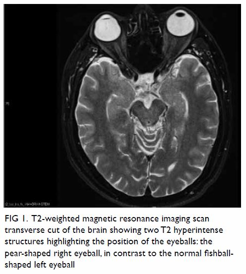
Figure 1. T2-weighted magnetic resonance imaging scan transverse cut of the brain showing two T2 hyperintense structures highlighting the position of the eyeballs: the pear-shaped right eyeball, in contrast to the normal fishball- shaped left eyeball
Retinal detachment surgery can be of external or internal approach to relieve the vitreous traction and flatten the retina. External approach can be localised or 360-degree encircling depending on the retinal status.
The formation of the pear-shaped eyeball was due to the buckling effect of the encircling band (Fig 2). With basic knowledge of normal organ shapes, all practitioners would be concerned by such a deformed organ on plain radiographs. Typically, intraocular tumours or eyeball ruptures are not regular nor symmetrical, except large ring melanoma of the ciliary body. The key to differentiating a genuine pathology from a congenital/postoperative variant lies heavily on clinical history.1 Operative implants usually appear regular, or even symmetrical on imaging.
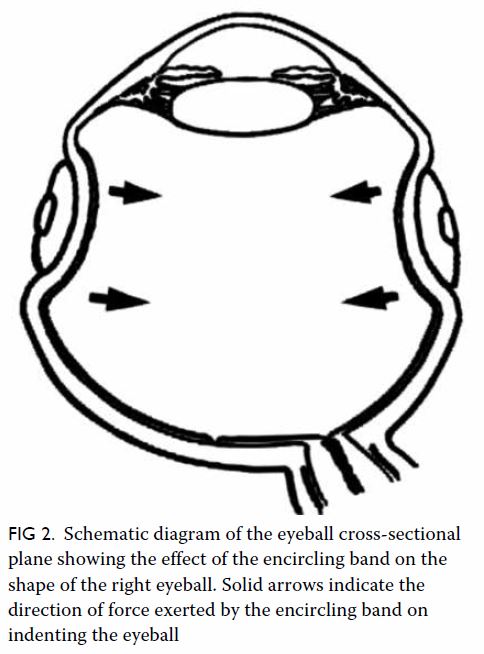
Figure 2. Schematic diagram of the eyeball cross-sectional plane showing the effect of the encircling band on the shape of the right eyeball. Solid arrows indicate the direction of force exerted by the encircling band on indenting the eyeball
Author contributions
All authors had full access to the data, contributed to the study, approved the final version for publication, and take responsibility for its accuracy and integrity.
Concept or design: SCL Au.
Acquisition of data: SCL Au.
Analysis or interpretation of data: SCL Au.
Drafting of the manuscript: SCL Au.
Critical revision of the manuscript for important intellectual content: All authors.
Acquisition of data: SCL Au.
Analysis or interpretation of data: SCL Au.
Drafting of the manuscript: SCL Au.
Critical revision of the manuscript for important intellectual content: All authors.
Conflicts of interest
All authors have disclosed no conflicts of interest.
Funding/support
This pictorial medicine paper received no specific grant from any funding agency in the public, commercial, or not-for-profit sectors.
Ethics approval
This study was conducted in accordance with the principles outlined in the Declaration of Helsinki. Relevant patient consent was obtained for the purpose of this case study.
References
1. Leslie A, Jones AJ, Goddard PR. The influence of clinical information on the reporting of CT by radiologists. Br J Radiol 2000;73:1052-5. Crossref
2. Carter K, Miller KM. Ophthalmology inpatient consultation. Ophthalmology 2001;108:1505-11. Crossref
3. Shanmugam PM, Ramanjulu R, Mishra KC, Sagar P. Novel techniques in scleral buckling. Indian J Ophthalmol 2018;66:909-15. Crossref
4. Thompson JT. The role of patient age and intraocular gases in cataract progression following vitrectomy for macular holes and epiretinal membranes. Trans Am Ophthalmol Soc 2003;101:485-98.