Breast lymphoma: a rare breast malignancy mimicking benign breast disease and carcinoma

© Hong Kong Academy of Medicine. CC BY-NC-ND 4.0
 
PICTORIAL MEDICINE
Breast lymphoma: a rare breast malignancy mimicking benign breast disease and carcinoma
Carol PY Chien, MB, BS, FRCR1; Kimmy KM Kwok, FRCR, FHKAM (Radiology)2; Eliza PY Fung, FRCR, FHKAM (Radiology)2
1 Department of Radiology, Queen Mary Hospital, Hong Kong
2 Department of Diagnostic and Interventional Radiology, Kwong Wah Hospital, Hong Kong
 
Corresponding author: Dr Carol PY Chien (chienpyc@gmail.com)
 
 Full paper in PDF
 
In July 2020, a 52-year-old woman presented with a 1-month history of palpable right breast lump. Clinical examination revealed a hard mass at the upper inner quadrant of the right breast. No skin changes or nipple retraction were evident. Ultrasonography confirmed multiple oval circumscribed hypoechoic masses at the upper right breast measuring up to 2 cm that were considered likely benign (Fig 1). The lesions were mammographically occult (Fig 2).
 

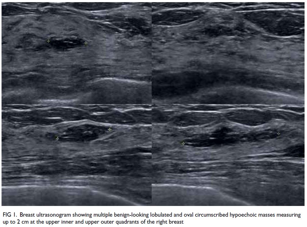
Figure 1. Breast ultrasonogram showing multiple benign-looking lobulated and oval circumscribed hypoechoic masses measuring up to 2 cm at the upper inner and upper outer quadrants of the right breast
 

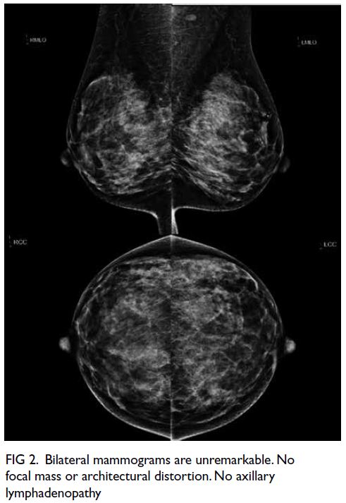
Figure 2. Bilateral mammograms are unremarkable. No focal mass or architectural distortion. No axillary lymphadenopathy
 
The patient also reported non-specific abdominal pain and distension for a few weeks. Positron emission tomography–computed tomography (PET-CT) showed multiple hypermetabolic masses at the right breast (maximum standardised uptake value [SUVmax] 19.0, 16 mm) and abdominal lymphadenopathy (Fig 3). Ultrasound-guided core biopsy of the right breast mass confirmed diffuse large B-cell lymphoma (Fig 4). The patient was diagnosed with stage IV lymphoma with diffuse extranodal involvement of the right breast. Reassessment PET-CT showed progressive disease despite R-CEOP chemotherapy with enlargement and increased metabolic activity of the right breast lymphoma (SUVmax 23.7, 41 mm) and abdominal lymphadenopathy (Fig 5). Second-line chemotherapy and radiotherapy of the breast was administered.
 

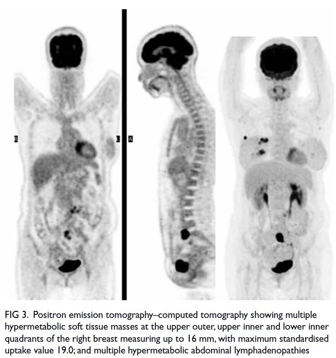
Figure 3. Positron emission tomography–computed tomography showing multiple hypermetabolic soft tissue masses at the upper outer, upper inner and lower inner quadrants of the right breast measuring up to 16 mm, with maximum standardised uptake value 19.0; and multiple hypermetabolic abdominal lymphadenopathies
 

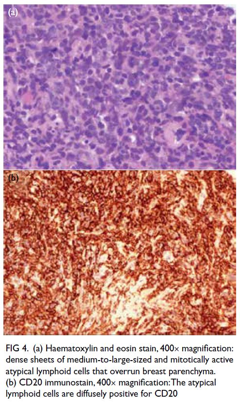
Figure 4. (a) Haematoxylin and eosin stain, 400× magnification: dense sheets of medium-to-large-sized and mitotically active atypical lymphoid cells that overrun breast parenchyma. (b) CD20 immunostain, 400× magnification: The atypical lymphoid cells are diffusely positive for CD20
 

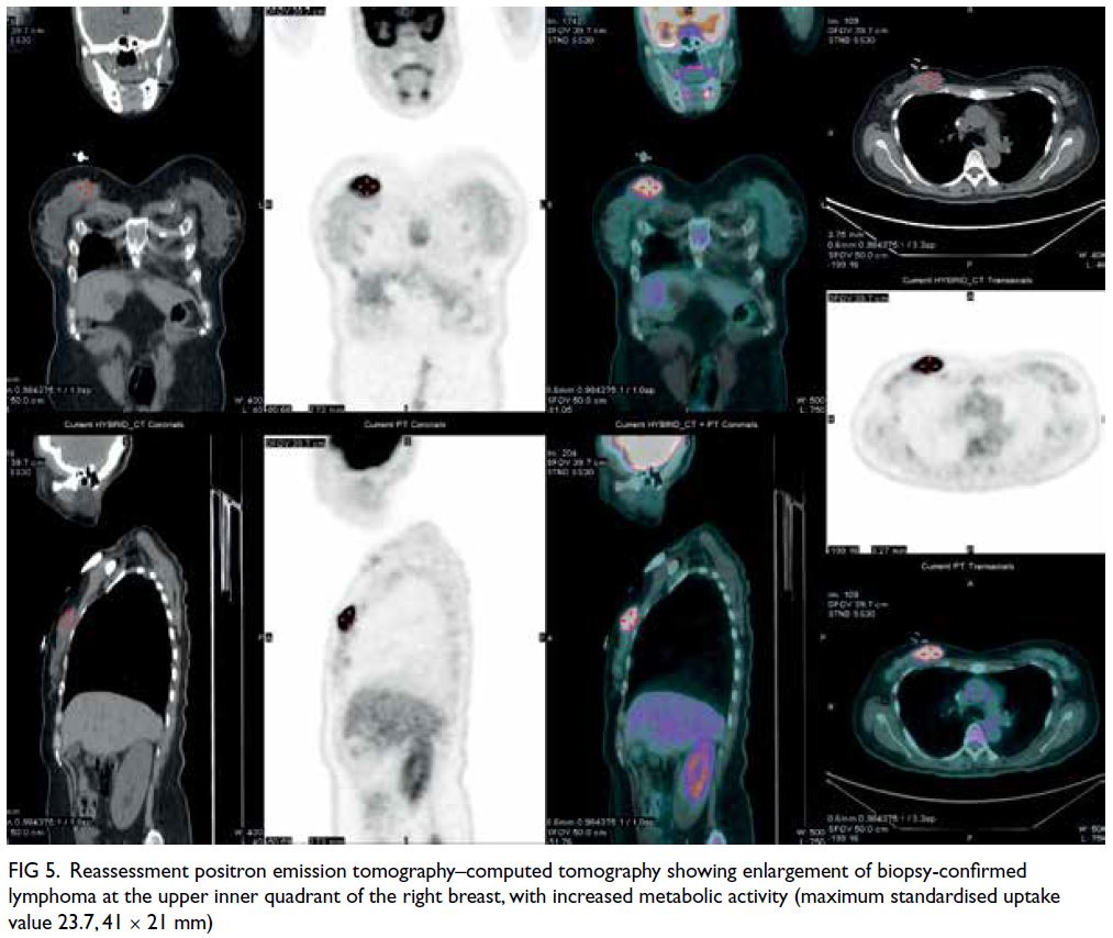
Figure 5. Reassessment positron emission tomography–computed tomography showing enlargement of biopsy-confirmed lymphoma at the upper inner quadrant of the right breast, with increased metabolic activity (maximum standardised uptake value 23.7, 41 × 21 mm)
 
The breast is an uncommon extranodal site of involvement by lymphoma because of the lack of lymphoid tissue. Breast lymphoma is a rare malignancy of the breast that accounts for <1% of all breast malignancies and <2% of all extranodal non-Hodgkin lymphoma.1 Diffuse large B-cell lymphoma is the most common histological type in both primary and secondary breast lymphoma with a median age at presentation of 60 to 70 years.1
 
The clinical presentation of breast lymphoma is highly variable, so diagnosis is challenging. It commonly presents as a mass that mimics carcinoma, or skin inflammatory changes that mimic benign inflammatory breast disease or inflammatory breast cancer.1 2 Axillary lymphadenopathy is uncommon.1 However, unlike with breast carcinoma, nipple retraction or discharge and other skin changes are rare with breast lymphoma.1 2
 
Mammographically, breast lymphoma commonly presents as a solitary mass (69%-76%) and asymmetry (20%). It may also present as skin thickening, architectural distortion or lymphedema.3 4 In contrast to breast carcinoma, spiculations, architectural distortion and microcalcifications are distinctively absent with breast lymphoma. Sonographically, breast lymphoma commonly presents as a parallel benign-looking hypoechoic mass without malignant features.3 4 Ultrasonography alone cannot distinguish breast lymphoma from breast carcinoma or benign breast lesions such as fibroadenoma. However, it can guide tissue diagnosis when mammogram is negative. This is particularly helpful in Asian patients with dense breast tissue that lowers mammogram sensitivity.
 
A recent study has demonstrated that 18F-fluorodeoxyglucose PET-CT can potentially differentiate between breast lymphoma and carcinoma.5 The authors found that the median SUVmax for breast lymphoma (10.96) was significantly higher than that for breast carcinoma (4.76; specificity up to 96.3%).5 Although the sonographic features of breast masses in our patient were absent, an SUVmax of 19.0 was unusually high for breast carcinoma. This raised the suspicion of secondary breast lymphoma rather than primary breast carcinoma with distant metastases. Histological diagnosis was required to guide appropriate treatment.
 
In conclusion, PET-CT can potentially differentiate between breast lymphoma and breast carcinoma. Increased awareness of breast lymphoma and correlation with radiological and pathological findings are essential for diagnosis.
 
Author contributions
Concept or design: All authors.
Acquisition of data: CPY Chien.
Analysis or interpretation of data: CPY Chien.
Drafting of the manuscript: CPY Chien.
Critical revision of the manuscript for important intellectual content: All authors.
 
All authors had full access to the data, contributed to the study, approved the final version for publication and take responsibility for its accuracy and integrity.
 
Conflicts of interest
All authors have disclosed no conflicts of interest.
 
Funding/support
This study received no specific grant from any funding agency in the public, commercial, or not-for-profit sectors.
 
Ethics approval
The patient was treated in accordance with the Declaration of Helsinki and provided informed consent for all investigations and procedures, and publication.
 
References
1. Raj SD, Shurafa M, Shah Z, Raj KM, Fishman MD, Dialani VM. Primary and secondary breast lymphoma: clinical, pathologic, and multimodality imaging review. Radiographics 2019;39:610-25. Crossref
2. Sabaté JM, Gómez A, Torrubia S, et al. Lymphoma of the breast: clinical and radiologic features with pathologic correlation in 28 patients. Breast J 2002;8:294-304. Crossref
3. Yang WT, Lane DL, Le-Petross HT, Abruzzo LV, Macapinlac HA. Breast lymphoma: imaging findings of 32 tumors in 27 patients. Radiology 2007;245:692-702. Crossref
4. Surov A, Holzhausen HJ, Wienke A, et al. Primary and secondary breast lymphoma: prevalence, clinical signs and radiological features. Br J Radiol 2012;85:e195-205. Crossref
5. Ou X, Wang J, Zhou R, et al. Ability of 18F-FDG PET/CT radiomic features to distinguish breast carcinoma from breast lymphoma. Contrast Media Mol Imaging 2019;2019:4507694. Crossref

Abdominal para-aortic ectopic thyroid tissue mimicking lymphadenopathy on computer tomography

© Hong Kong Academy of Medicine. CC BY-NC-ND 4.0
 
PICTORIAL MEDICINE
Abdominal para-aortic ectopic thyroid tissue mimicking lymphadenopathy on computer tomography
Charmaine HK Wong, MB, ChB, FRCR1; HL Tsui, MB, ChB, FHKAM (Radiology)1; CN Ling, MB, ChB2; Anthony WT Chin, MB, ChB, FHKAM (Radiology)1; PY Chu, MB, ChB, FHKAM (Radiology)1; CS Chan, MB, BS, FHKAM (Radiology)1
1 Department of Radiology and Organ Imaging, United Christian Hospital, Hong Kong
2 Department of Pathology, United Christian Hospital, Hong Kong
 
Corresponding author: Dr Charmaine HK Wong (charmainehwong@gmail.com)
 
 Full paper in PDF
 
A 70-year-old woman presented to our hospital for workup for chronic cough. A contrast computed tomography (CT) of the thorax and abdomen revealed an incidental finding of multilobulated soft tissue lesions in the abdomen along the left para-aortic region. The lesions measured up to 7.4 cm in diameter and demonstrated mild increased attenuation on pre-contrast images and vivid heterogenous enhancement on post-contrast study (Fig 1a and b). Appearance and location of these lesions raised concern about possible lymphadenopathy so fluorodeoxyglucose-18 positron emission tomography–CT was performed. The lesions demonstrated only mild metabolic activity (maximal standard uptake value=2.6) [Fig 1c]. They showed no interval change in appearance, and no other hypermetabolic lesion could be detected elsewhere. Tumour markers including alpha-fetoprotein, carcinoembryonic antigen, and cancer antigen 125 were not elevated.
 

Figure 1. (a) Pre-contrast and (b) post-contrast axial computed tomography images showing multilobulated lesions at left para-aortic region (white arrows). The lesions showing mild increased attenuation on pre-contrast images and vivid heterogenous enhancement on post-contrast images. (c) Axial fluorodeoxyglucose-18 positron emission tomography-computed tomography demonstrating mild metabolic activity over the lesions (maximum standard unit value=2.6)
 
The CT-guided biopsy of the para-aortic lesions subsequently showed cuboidal follicular cells arranged in architecture resembling thyroid parenchymal tissue (Fig 2). Immunostaining for thyroglobulin and thyroid transcription factor-1 were positive. There were no nuclear features of carcinoma.
 

Figure 2. (a, b) Histological images of the para-aortic lesions showing tissue consisting of colloid-containing follicles of different sizes, arranged in architecture resembling normal thyroid parenchymal tissue. Immunostaining for (c) thyroglobulin and (d) thyroid transcription factor-1 was positive
 
Thyroid scintigraphy using technetium-99m pertechnetate showed intense radiotracer uptake over the corresponding para-aortic lesions (Fig 3). Findings were consistent with ectopic thyroid tissue. The presence of an orthotopic thyroid at its normal pretracheal position was demonstrated on CT and on technetium-99m scan (Fig 4). The patient’s thyroid function was normal.
 

Figure 3. (a) Axial and (b) coronal thyroid scintigraphy images using technetium-99m pertechnetate showing intense uptake of radiotracer at the left side of the abdomen. Corresponding (c) axial and (d) coronal computed tomography fusion images showing the left para-aortic lesions
 

Figure 4. (a) Thyroid scintigraphy image and (b) corresponding computed tomography fusion image showing the presence of orthotopic thyroid gland, with a focal defect at the lower pole corresponding to a cold nodule
 
Ectopic thyroid gland is a rare developmental abnormality with a prevalence of approximately 1 per 100 000 to 300 000 population.1 It results from aberrant embryogenesis of the gland during its migration from the foramen cecum at the posterior aspect of the tongue to its final pretracheal position. Frequent locations include its path of embryological descent along the midline of the neck, with a lingual thyroid at the base of the tongue being most common, accounting for up to 90% of reported cases.2 Other locations include the lateral neck and superior mediastinum. More distant locations such as subdiaphragmatic locations have also been reported but are exceedingly rare. To the best of the authors’ knowledge, this is the first report of ectopic thyroid tissue occurring at the para-aortic region of the abdomen, mimicking the appearance of lymphadenopathy on CT.
 
Thyroid scintigraphy is the most important and sensitive imaging tool to detect ectopic thyroid tissue. It also has the advantage of being able to demonstrate the presence or absence of an orthotopic thyroid gland. In patients with ectopic thyroid tissue, it is important to evaluate for the presence of orthotopic thyroid and thyroid function. Hypothyroidism may be present, particularly in those without a normal thyroid gland, and more common in patients with lingual ectopic thyroid.2 Ultrasound is also useful to locate and evaluate the orthotopic thyroid gland.
 
Computed tomography and magnetic resonance imaging are important adjuncts in evaluation. Ectopic thyroid tissue may show similar imaging characteristics to normal thyroid gland with high attenuation on non-contrast CT due to its high iodine content and vivid post-contrast enhancement. When ectopic thyroid tissue is found in a location not consistent with embryologic development, the possibility of malignant metastasis needs to be considered, occurring in 7% to 23% of patients.3 Biopsy is invaluable and the orthotopic gland should be evaluated for possible malignant change. Pathological changes that affect a normal thyroid including malignant change have been reported in ectopic tissues,1 and should be considered in the management and follow-up of ectopic thyroid tissue.
 
Author contributions
Concept or design: All authors.
Acquisition of data: CHK Wong, HL Tsui, CN Ling.
Analysis or interpretation of data: CHK Wong, HL Tsui, CN Ling.
Drafting of the manuscript: CHK Wong, HL Tsui, CN Ling.
Critical revision of the manuscript for important intellectual content: All authors.
 
Conflicts of interest
All authors have disclosed no conflicts of interest.
 
Funding/support
This study received no specific grant from any funding agency in the public, commercial, or not-for-profit sectors.
 
Ethics approval
The patient was treated in accordance with the Declaration of Helsinki. The patient provided written informed consent for all treatments and procedures and consent for publication.
 
References
1. Noussios G, Anagnostis P, Goulis DG, Lappas D, Natsis K. Ectopic thyroid tissue: anatomical, clinical, and surgical implications of a rare entity. Eur J Endocrinol 2011:165:375-82. Crossref
2. Guerra G, Cinelli M, Mesolella M, et al. Morphological, diagnostic and surgical features of ectopic thyroid gland: a review of literature. Int J Surg 2014;12 Suppl 1:S3-11. Crossref
3. Ballard D, Patel P, Schild SD, Ferzli G, Gordin E. Ectopic thyroid presenting as supraclavicular mass: a case report and literature review. J Clin Transl Endocrinol Case Rep 2018;10:17-20. Crossref

Liver and kidney toxicity caused by wild mushroom poisoning

© Hong Kong Academy of Medicine. CC BY-NC-ND 4.0
 
PICTORIAL MEDICINE
Liver and kidney toxicity caused by wild mushroom poisoning
LF Yang, MD1; LP Zhu, MD1; YT Li, MD, PhD1; XB Zhong, MD1; ZG Chen, MD, PhD1; Karen KY Leung, MB, BS, MRCPCH2; KL Hon, MB, BS, MD2
1 Department of Pediatrics, The Third Affiliated Hospital of Sun Yat-sen University, Guangzhou, PR China
2 Department of Paediatrics and Adolescent Medicine, The Hong Kong Children’s Hospital, Hong Kong
 
Corresponding author: Dr KL Hon (ehon@hotmail.com)
 
 Full paper in PDF
 
In May 2020, a 2-year-old girl was admitted to our hospital with a 2-day history of persistent vomiting, diarrhoea, abdominal pain, and headache. Her family reported that she had eaten half of a large mushroom, about the size of a palm (Fig a) 17 hours before symptom onset.
 

Figure. Photographs of wild mushroom taken by the patient’s family and identified by mycologists as (a) possibly Amanita spp, which are hepatotoxic; and (b, c) Macrolepiota spp, which are edible
 
Physical examination was unremarkable. Laboratory investigations revealed liver disfunction, with elevated aspartate transaminase (AST) 228 U/L and alanine transaminase (ALT) 211 U/L rising to 2591 U/L and 2815 U/L, respectively. The patient was admitted to the paediatric intensive care unit for further treatment. After hospitalisation, AST and ALT further increased to 4005 U/L and 4335 U/L, respectively. Plasma exchange was performed along with treatments including 20% albumin, high-dose penicillin, N-acetylcysteine injection and Ganoderma lucidum. Liver function gradually normalised over the following week and the patient was discharged home.
 
The patient’s grandmother and aunt consumed the other half of the same mushroom (Fig a) and other mushrooms (Fig b and c), and both were admitted to hospital with serious vomiting, diarrhoea and mild abdominal pain. Her aunt had normal liver function and kidney function. The grandmother had mild liver and renal impairment with AST 169 U/L, ALT 147 U/L, blood urea nitrogen 12.1 mmol/L and creatinine 168 μmol/L, made a rapid and recovery following rehydration, a bicarbonate infusion, furosemide, and hepatoprotective drugs for 3 days.
 
Cyclopeptides are the major lethal toxins worldwide, often found in Amanita spp, Cyclospora and galea.1 2 In Guangzhou from 2007 to 2011, the most common species causing mushroom poisoning was Amanita spp. In mainland China 95% of fatal cases of mushroom poisoning are caused by amanitin. In Hong Kong, 90% of wild mushrooms are poisonous.
 
Ultra-performance liquid chromatography-mass spectrometry has high accuracy, and good sensitivity and specificity, and was used to identify the toxin in our patient. However, it was already 2 days after the girl ate the wild mushrooms, and the toxin might have been excreted so that the concentration was too low for detection.
 
Prognosis is often poor for patients with long incubation period of gastrointestinal symptoms (>6 hr), early manifestations of liver and kidney dysfunction, and multiple organ dysfunction.3 4 Plasma exchange effectively removes hepatotoxins and improves survival, drugs such as silybin, N-acetylcysteine, putatively ceftazidime/penicillin and Ganoderma lucidum can also have a significant benefit.5 Ganoderma lucidum contains triterpenoids that can protect the liver and reduce oxidative stress and apoptosis. Ganoderma lucidum decoction is effective in the treatment of amanita poisoning at a recommended dosage of 200 g/day decocted with 600 mL water, taking one-third of this decoction (200 mL per dose) 3 times per day, for 7 to 14 days. Liver transplantation is the final option for liver failure caused by mushroom poisoning.
 
Mushroom poisoning is usually the result of ingestion of wild mushrooms after misidentification of a toxic mushroom as an edible species. Public health education on identification of wild mushrooms and the dangers of wild mushroom consumption is particularly important to prevent mushroom poisoning.
 
Author contributions
Concept or design: KL Hon, LF Yang.
Acquisition of data: LF Yang, LP Zhu.
Analysis or interpretation of data: YT Li, XB Zhong.
Drafting of the manuscript: LF Yang.
Critical revision of the manuscript for important intellectual content: ZG Chen, KKY Leung, KL Hon.
 
All authors had full access to the data, contributed to the study, approved the final version for publication, and take responsibility for its accuracy and integrity.
 
Conflicts of interest
As an editor of the Journal, KL Hon was not involved in the peer review process. Other authors have disclosed no conflicts of interest.
 
Funding/support
This study received no specific grant from any funding agency in the public, commercial, or not-for-profit sectors.
 
Ethics approval
This study was approved by the Ethics Committee of The Third Affiliated Hospital of Sun Yat-sen University (Ref [2022]-02-064-01). Written informed consent was obtained from the patient’s guardian for the publication of this case report and all images.
 
References
1. Schenk-Jaeger KM, Rauber-Lüthy C, Bodmer M, Kupferschmidt H, Kullak-Ublick GA, Ceschi A. Mushroom poisoning: a study on circumstances of exposure and patterns of toxicity. Eur J Intern Med 2012;23:e85-91. Crossref
2. Brandenburg WE, Ward KJ. Mushroom poisoning epidemiology in the United States. Mycologia 2018;110:637-41. Crossref
3. Trabulus S, Altiparmak MR. Clinical features and outcome of patients with amatoxin-containing mushroom poisoning. Clin Toxicol (Phila) 2011;49:303-10. Crossref
4. Bonacini M, Shetler K, Yu I, Osorio RC, Osorio RW. Features of patients with severe hepatitis due to mushroom poisoning and factors associated with outcome. Clin Gastroenterol Hepatol 2017;15:776-9.Crossref
5. Lu Z, Hong G, Sun C, Chen X, Li H, Yu X. Chinese clinical guideline for the diagnosis and treatment of mushroom poisoning [in Chinese]. J Clin Emerg (China) 2019;20:583-98.

Unusual gallbladder disease: spontaneous gallbladder haematoma

© Hong Kong Academy of Medicine. CC BY-NC-ND 4.0
 
PICTORIAL MEDICINE
Unusual gallbladder disease: spontaneous gallbladder haematoma
Se-kook Kee, MD1; Dongwook Je, MD2; Woo-young Nho, MD1,2
1 Department of Surgery, CHA University, Republic of Korea
2 Department of Emergency Medicine, CHA University, Republic of Korea
 
Corresponding author: Prof WY Nho (wooyoung.nho@gmail.com)
 
 Full paper in PDF
 
In September 2020, a 29-year-old woman presented to the emergency department with a 2-day history of upper abdominal pain. The patient had no history of recent trauma around the abdominal area. The patient’s vital signs were stable upon arrival but physical examination revealed tenderness over the right upper quadrant area. The patient reported moderate to heavy alcohol consumption (10-15 standard drinks per week). Laboratory findings revealed a white blood cell count of 3030/mm3, haemoglobin level 12.8 g/dL, and low platelet count of 34 000/mm3. The prothrombin time was mildly prolonged to 14.2 s. Elevated levels of liver enzymes, aspartate transaminase (113 U/L), direct bilirubin (6.18 mg/dL), and gamma-glutamyltransferase (350 U/L), were reported. Computed tomography (CT) scans showed a distended gallbladder with several gallstones and bile duct stones. Moreover, the intraluminal space of the gallbladder was filled with heterogeneous material, and extravasation of the contrast agent was suspected in the arterial phase (Fig 1). Emergency laparoscopic exploration was planned with a preoperative diagnosis of gallbladder haematoma. At the first laparoscope view, cirrhotic change to the liver with ascites and an oedematous gallbladder were evident (Fig 2a). The surgeon tried gallbladder decompression by needle aspiration and made a small opening to remove the blood but it was unsuccessful (Fig 2b). Laparoscopic cholecystectomy was successful without complications such as perforation (Fig 2c and d). Endoscopic retrograde cholangiopancreatography with endoscopic sphincterotomy was performed 5 days after the initial surgery and remnant common bile duct stones were removed. Pathological evaluation revealed a gallbladder haematoma with 30 to 40 gallstones (Fig 3). Moreover, venous vessels were remarkably dilated in the vicinity of the haemorrhagic site on CD31 immunostaining. The patient was discharged on postoperative day 10 with no complications.
 

Figure 1. Computed tomography scans. (a) Non-enhanced image; heterogeneous shadow of intraluminal space (arrow). (b) Arterial phase image; extravasation of contrast was suspected (arrow). (c) Venous phase image. (d) Coronal section image (the arrow indicates contrast material)
 

Figure 2. Operative findings. (a) The first laparoscope view revealed a cirrhotic change to the liver with ascites. (b) A small opening to remove the blood. (c, d) Laparoscopic cholecystectomy was performed
 

Figure 3. Resected gallbladder with haematoma and gallstones
 
The gallbladder is normally physiologically empty or filled with bile juice. Gallbladder haematoma is defined as filling of the gallbladder with blood. The definition can vary from gallbladder bleeding that comprises active bleeding inside the gallbladder to gallbladder haematoma when already-formed blood clots are evident. Haemorrhagic cholecystitis may be an appropriate term in cases of a gallbladder showing inflammation with blood. Reported risk factors for spontaneous bleeding include atherosclerosis or aneurysm, biliary malignancy, renal failure, cirrhosis, and coagulopathy, or anticoagulant medication.1 2 3 Moreover, abdominal blunt trauma can lead to gallbladder injury and cause gallbladder bleeding.3 Clinical manifestations of gallbladder haematoma similarly represent acute gallbladder disease.2 Upper abdominal pain is frequent due to distension of the gallbladder, and melena or haematemesis may also be present. In cases of cholecystitis, fever may develop, and laboratory study will demonstrate inflammation. The conventional radiological methods to evaluate gallbladder disease like ultrasonography or CT scan have diagnostic value.3 Ultrasonography may reveal a blood clot in the lumen with wall thickening and fluid accumulation around the gallbladder.2 In the CT scan, hyperdense blood and bile shadow with or without fluid level may be suspicious of gallbladder haematoma. Extravasation of contrast material into the lumen of the gallbladder during the arterial phase of contrast-enhanced CT is related to active bleeding.3,4 Surgical treatment produces superior results and the minimally invasive method of a laparoscopic approach has been widely adopted.1 3 Other endoscopic or percutaneous approaches for decompression may be considered before surgery.1 Gallbladder haematoma is associated with high morbidity and mortality. Delayed management potentially increases the risk of perforation and concomitant unfavourable outcomes.4 5
 
Author contributions
Concept or design: WY Nho.
Acquisition of data: D Je.
Analysis or interpretation of data: SK Kee.
Drafting of the manuscript: SK Kee.
Critical revision of the manuscript for important intellectual content: WY Nho.
 
All authors had full access to the data, contributed to the study, approved the final version for publication, and take responsibility for its accuracy and integrity.
 
Conflicts of interest
All authors have disclosed no conflicts of interest.
 
Funding/support
This study received no specific grant from any funding agency in the public, commercial, or not-for-profit sectors.
 
Ethics approval
The study was reviewed and approved by the Institutional Review Board (IRB) of CHA Gumi Medical Center (GM20-11).
 
1. Leaning M. Surgical case report—acalculous hemorrhagic cholecystitis. J Surg Case Rep 2021;2021:rjab075. Crossref
2. Itagaki H, Katuhiko S. Gallbladder hemorrhage during orally administered edoxaban therapy: a case report. J Med Case Rep 2019;13:383. Crossref
3. Parekh J, Corvera CU. Hemorrhagic cholecystitis. Arch Surg 2010;145:202-4. Crossref
4. Kwon JN. Hemorrhagic cholecystitis: report of a case. Korean J Hepatobiliary Pancreat Surg 2012;16:120-2. Crossref
5. López V, Alconchel F. Hemorrhagic cholecystitis. Radiology 2018;289:316. Crossref

Transmural perforation by fish bone from stomach to liver

Hong Kong Academy of Medicine. CC BY-NC-ND 4.0
 
PICTORIAL MEDICINE
Transmural perforation by fish bone from stomach to liver
X Liu, MD1; J Shi, MD2
1 Department of Radiology, Hospital of Chengdu University of Traditional Chinese Medicine, Chengdu, China
2 Department of Nursing, Hospital of Chengdu University of Traditional Chinese Medicine, Chengdu, China
 
Corresponding author: Ms J Shi (267126966@qq.com)
 
 Full paper in PDF
 
A 46-year-old man was admitted to the emergency department with a 3-day history of epigastric abdominal pain associated with nausea. His temperature was 36.5°C, pulse 74 beats per minute and blood pressure 135/70 mm Hg. Physical examination revealed mild epigastric tenderness and rebound tenderness in the upper abdomen. Basic laboratory test results were normal. Abdominal computed tomography (CT) scan (Fig) revealed a radiodense linear foreign body measuring 3 cm, extending transmurally through the lesser curvature of the stomach and penetrating the liver capsule. Inflamed tissue surrounding the pylorus was also noted. Pneumoperitoneum was evident as free gas under the diaphragm. The patient was kept nil by mouth and transferred for emergency laparoscopy. An oesophagogastroduodenoscopy was performed and a fish bone penetrating the stomach wall was found. Clipping was performed after fish bone removal. Empiric intravenous antibiotic (tazobactam 4 g/8 h) was prescribed before and after the oesophagogastroduodenoscopy. The patient made an uneventful recovery and was discharged home 6 days after surgery.
 

Figure. (a, b) Axial and (c) coronal computed tomography images showing a linear hyperdense structure, extending transmurally through the lesser curvature of the stomach and penetrating the liver capsule, local fat stranding was identified (asterisk). (d) Axial computed tomography image showing pneumoperitoneum, evident as freeing gas (arrows) under the diaphragm, suggesting the possibility of perforation
 
After ingestion, most fish bones pass through the gastrointestinal tract within a week and cause no serious complications. Gastrointestinal perforation by fish bone is a rare medical emergency. Clinical symptoms are often atypical and may vary from mild to severe depending on the site of perforation and the degree of inflammation.1 In addition to a non-specific clinical presentation, a history of foreign body ingestion is rarely available. The patient in this report was unaware of fish bone ingestion. Laboratory findings are also non-specific and usually demonstrate elevated inflammatory markers. Thus, a diagnosis of fish bone perforation can be challenging and is often delayed. The radiologist plays an essential role in the detection of fish bone perforation and associated complications.
 
Computed tomography is considered the most effective means by which to identify foreign bodies and their associated complications due to its high resolution and high-quality multiplanar capabilities.2 The main imaging features of fish bone perforation on CT scan are a linear hyperdense structure in the gastrointestinal tract, inflammatory changes surrounding the perforation site, and pneumoperitoneum.2 3 Pneumoperitoneum is a rare yet important finding that can signal the possibility of perforation. In our case, multiplanar CT reconstructions revealed that the fish bone had partially perforated the stomach wall into the adjacent hepatic parenchyma, causing perigastric inflammatory changes. In addition, pneumoperitoneum was identified under the diaphragm. This report provides a reference for clinicians. Early endoscopic or surgical removal of a foreign body from the stomach causing complications is recommended.
 
Author contributions
Concept or design: X Liu.
Acquisition of data: All authors.
Analysis or interpretation of data: All authors.
Drafting of the manuscript: All authors.
Critical revision of the manuscript for important intellectual content: All authors.
 
All authors had full access to the data, contributed to the study, approved the final version for publication, and take responsibility for its accuracy and integrity.
 
Conflicts of interest
The authors declared no potential conflicts of interest.
 
Funding/support
This study received no specific grant from any funding agency in the public, commercial, or not-for-profit sectors.
 
Ethics approval
This study was approved by the Hospital of Chengdu University of Traditional Chinese Medicine Research Ethics Committee (Ref: 2021524). Informed consent was obtained from the patient.
 
References
1. Yi L, Cheng Z, Zhou Y, et al. Fishbone foreign body ingestion in duodenal papilla: a cause of abdominal pain resembling gastric ulcer. BMC Gastroenterol 2020;20:323. Crossref
2. Paixão TS, Leão RV, de Souza Maciel Rocha Horvat N, et al. Abdominal manifestations of fishbone perforation: a pictorial essay. Abdom Radiol (NY) 2017;42:1087-95. Crossref
3. Davarpanah AH, Eberhardt LW. Case 282: fishbone pylephlebitis. Radiology 2020;297:239-43. Crossref

Indocyanine green angiography and lymphography in microsurgical subinguinal varicocelectomy with evolving video microsurgery and fluorescence imaging platforms

Hong Kong Academy of Medicine. CC BY-NC-ND 4.0
 
PICTORIAL MEDICINE
Indocyanine green angiography and lymphography in microsurgical subinguinal varicocelectomy with evolving video microsurgery and fluorescence imaging platforms
CL Cho, FRCSEd (Urol), FHKAM (Surgery)1; Ringo WH Chu, FRCSEd (Urol), FHKAM (Surgery)2
1 SH Ho Urology Centre, Department of Surgery, The Chinese University of Hong Kong, Hong Kong
2 Private Practice
 
Corresponding author: Dr CL Cho (chochaklam@yahoo.com.hk)
 
 Full paper in PDF
 
Intra-operative use of indocyanine green (ICG) angiography and lymphography has been reported as a valuable adjunct during microsurgical subinguinal varicocelectomy (MSV).1 The development of a video microsurgery platform and fluorescence imaging technology further facilitates identification of testicular arteries and lymphatics. We report the intra-operative imaging of two patients who underwent varicocele repair for grade 3 left varicoceles in February and March 2021 using the new platform.
 
The operations were performed under three-dimensional (3D) optical magnification images on the television monitors using the video microsurgery platform with VITOM® 3D system (KARL STORZ SE, Tuttlingen, Germany) as previously described.2 A pack of 25 mg ICG (Diagnogreen; Daiichi Sankyo Co, Tokyo, Japan) was dissolved in 10 mL water. Identification of testicular arteries was assisted by ICG angiography with intravenous injection of 2 mL (5 mg) ICG solution. After preservation of the testicular arteries, the differentiation between lymphatics and small veins was facilitated by ICG lymphography performed following intra-parenchymal testicular injection of 0.5 mL (1.25 mg) ICG solution followed by gentle testicular massage.3 The setting of ICG fluorescence imaging consisted of an IMAGE1 S™ 4U RUBINA™ with 4K 3D monitor system (KARL STORZ SE, Tuttlingen, Germany) and a HOPKINS™ Straight Forward Telescope 0° (10-mm diameter/20-cm length) [KARL STORZ SE, Tuttlingen, Germany]. We demonstrate intra-operative ICG angiography of patient 1, a 35-year-old man with primary infertility and oligoasthenoteratozoospermia. The testicular artery appeared green on the overlay image mode with clear simultaneous visualisation of white light microscopy images in the background (Fig 1a and b). In addition to the testicular artery, the small (<1 mm) cremasteric artery could also be identified in the monochromatic mode that further enhanced the contrast (Fig 1c). After successful preservation of the testicular arteries in patient 2, a 28-year-old man with left scrotal pain, two probable lymphatics were identified under white light microscopy (Fig 2a). The strong green colouration seen in the overlay mode after ICG injection unambiguously confirmed the successful preservation of lymphatics in MSV (Fig 2b).
 

Figure 1. Intra-operative indocyanine green angiography by IMAGE1 S™ 4U RUBINA™ fluorescence imaging system. (a) The testicular artery is exposed under the white light microscopic view after retraction of the dilated internal spermatic veins. (b) The testicular artery is confirmed and appears green in the overlay mode with background white light images. (c) The testicular and cremasteric arteries are clearly demonstrated in monochromatic mode. The testicular and cremasteric arteries are denoted by arrows and stars, respectively
 

Figure 2. Intra-operative indocyanine green lymphography by IMAGE1 S™ 4U RUBINA™ fluorescence imaging system (a) before and (b) after injection of indocyanine green
 
Microsurgical subinguinal varicocelectomy is the standard for varicocele repair with excellent surgical outcomes reported.4 Nonetheless identification of testicular arteries and lymphatics under white light microscopy alone is operator-dependent and remains challenging for novice surgeons. Several adjuncts have been introduced to facilitate artery- and lymphatic-sparing procedures during MSV. One such adjunct is ICG fluorescence imaging.1 In our opinion, the new overlay mode provided by the latest platform is particularly useful for MSV. Without the need to switch between different modes, the combined regular white light image and near-infrared/ICG data allow accurate localisation of even the smallest vessel without ambiguity (Fig 3). Moreover, the display of ICG signal alone in white on a black background in the monochromatic mode maximises contrast and further improves identification of target vessels (Fig 3). We believe the advances of this surgical platform and imaging technology play a role in enhancing patient safety by increasing the success of arterial and lymphatic preservation in MSV.
 

Figure 3. Intra-operative indocyanine green (ICG) angiography with the previous fluorescence imaging platform utilising VITOM II ICG system with SPIES camera (KARL STORZ SE, Tuttlingen, Germany). Without the overlay mode, the images of (a) white light microscopy view and (b) Chroma mode can only be analysed separately. Correlation between the testicular arteries identified on ICG angiography and white light microscopy can be difficult
 
Author contributions
Concept or design: CL Cho.
Acquisition of data: All authors.
Analysis or interpretation of data: All authors.
Drafting of the manuscript: All authors.
Critical revision of the manuscript for important intellectual content: All authors.
 
All authors had full access to the data, contributed to the study, approved the final version for publication, and take responsibility for its accuracy and integrity.
 
Conflicts of interest
The authors have disclosed no conflict of interest.
 
Funding/support
This study received no specific grant from any funding agency in the public, commercial, or not-for-profit sectors.
 
Ethics approval
Patients were treated in accordance with the Declaration of Helsinki. Patients provided written informed consent for the procedures, and verbal consent for publication.
 
References
1. Cho CL. Is there any role for indocyanine green angiography in testicular artery preservation during microsurgical subinguinal varicocelectomy? In: Esteves SC, Cho CL, Majzoub A, Agarwal A, editors. Varicocele and Male Infertility: a Complete Guide. Switzerland AG: Springer Nature; 2019: 603-14. Crossref
2. Cho CL, Chu RW. Use of video microsurgery platform in microsurgical subinguinal varicocelectomy with indocyanine green angiography. Surg Prac 2019;23:20-4. Crossref
3. Cho CL, Chiu PK, Chu RW. Preliminary experience with indocyanine green lymphography during microsurgical subinguinal varicocelectomy. Surg Prac 2021;25:207-10. Crossref
4. Zini A. Varicocelectomy: microsurgical subinguinal technique is the treatment of choice. Can Urol Assoc J 2007;1:273-6.

Cullen sign in childhood malignancies

© Hong Kong Academy of Medicine. CC BY-NC-ND 4.0
 
PICTORIAL MEDICINE
Cullen sign in childhood malignancies
CC Au, MB, BS, MRCPCH1; Karen KY Leung, MB, BS, MRCPCH1; KL Hon, MB, BS, MD1; Junita KY Tung2; Carol LS Yan, MB, BS, MRCPCH1; WF Hui, MB, BS, MRCPCH1; WY Leung, MB, ChB, FRCSEd(Paed)3
1 Department of Paediatrics and Adolescent Medicine, The Hong Kong Children’s Hospital, Hong Kong
2 Faculty of Medicine, The Chinese University of Hong Kong, Hong Kong
3 Department of Surgery, The Hong Kong Children’s Hospital, Hong Kong
 
Corresponding author: Dr CC Au (aucc@ymail.com)
 
 Full paper in PDF
 
Introduction
We report four consecutive cases of young children with Cullen sign in a paediatric intensive care unit with abdominal malignancies and complications due to treatment. Two children had solid tumours with hepatic rupture and two had pancreatitis secondary to asparaginase use for leukaemia.
 
Case 1
A 23-month-old boy with ruptured liver metastasis and stage 4 neuroblastoma presented with abdominal distension due to a large retroperitoneal mass invading the liver and encasing the aorta and inferior vena cava. He required emergency laparotomy for haemorrhagic shock due to ruptured liver metastasis and endovascular embolisation of tumoural arteries. Subsequently there was an extensive subcutaneous haematoma around the surgical wound. Cullen sign was detected (Fig 1). Recurrent tumoural haemorrhage was excluded and surgical exploration performed to remove blood clots.
 

Figure 1. 23-month-old boy who required emergency laparotomy for haemorrhagic shock due to ruptured liver metastasis and endovascular embolisation of tumoural arteries. Subsequently there was an extensive subcutaneous haematoma around the surgical wound. Cullen sign was detected
 
Case 2
A 5-year-old girl had asparaginase-associated pancreatitis and common B-cell acute lymphoblastic leukaemia. She was 6 months into her treatment protocol and received re-induction phase chemotherapy including pegaspargase (Oncaspar). She presented with abdominal pain and vomiting due to severe acute necrotising pancreatitis. Cullen sign (peri-umbilicus), Grey Turner sign (flank), and Fox sign (inguinal ligament) were detected (Fig 2). She required vasopressor and non-invasive ventilation for systemic inflammatory response syndrome. She later developed pancreatic pseudocyst and underwent ultrasound-guided drainage.
 

Figure 2. 5-year-old girl presented with abdominal pain and vomiting due to severe acute necrotising pancreatitis. Cullen sign (peri-umbilicus), Grey Turner sign (flank), and Fox sign (inguinal ligament) were detected
 
Case 3
A 5-year-old boy with asparaginase-associated pancreatitis and B-cell acute lymphoblastic leukaemia had received re-induction phase chemotherapy with pegaspargase (Oncaspar) and presented with abdominal pain and vomiting. He had severe acute necrotising pancreatitis and systemic inflammatory response syndrome. Cullen sign (peri-umbilicus) and Bryant sign (scrotum) were detected (Fig 3). He required vasopressor support. Later he developed multiple intrapancreatic and peripancreatic fluid collections and required repeated computed tomography-guided aspiration and drainage.
 

Figure 3. 5-year-old boy presented with abdominal pain and vomiting due to severe acute necrotising pancreatitis and systemic inflammatory response syndrome. Cullen sign (peri-umbilicus) and Bryant sign (scrotum) were detected
 
Case 4
A 7-year-old boy with ruptured hepatoblastoma presented with abdominal distension. Hepatoblastoma involved all liver segments, extended into the inferior vena cava and had metastasised to the lungs. He required selective embolisation of left hepatic artery segments 2 and 3 for active haemorrhage. He had abdominal compartment syndrome and gross ascites that required drainage. Cullen sign was detected. (Fig 4) He was prescribed chemotherapy and consequently the tumour reduced in size.
 

Figure 4. 7-year-old boy with abdominal compartment syndrome and gross ascites that required drainage. Cullen sign was detected
 
Discussion
These four cases illustrate Cullen sign in paediatric malignancies. General practitioners and paediatricians should be aware of its diagnostic implications during clinical examination.
 
First described in 1918, Cullen sign referred to periumbilical ecchymosis due to retroperitoneal haemorrhage of ruptured ectopic pregnancy.1 It has become one of the classic abdominal signs in acute pancreatitis. Cullen sign or Grey Turner sign has been reported present in 3% of acute necrotising pancreatitis cases. In 1984, these signs were reported to have a significant mortality rate of 37%,2 but clinical outcomes of severe acute pancreatitis have since improved with intensive care. Nevertheless Cullen sign remains a pointer to serious intra-abdominal haemorrhage. Haemorrhage can originate anywhere along an anatomical pathway. Pathophysiologically, blood tracks from the retroperitoneum through the gastrohepatic ligament to the falciform ligament of the liver, then reaches the umbilicus through the round ligament of liver to form Cullen sign.3 Cullen sign has been reported in intra-abdominal malignancies, liver cirrhosis, and rectus sheath haematoma.4 5
 
Author contributions
All authors contributed to the concept or design of the study, interpretation of the data, drafting of the manuscript, and critical revision of the manuscript for important intellectual content. All authors had full access to the data, contributed to the study, approved the final version for publication, and take responsibility for its accuracy and integrity.
 
Conflicts of interest
As an editor of the journal, KL Hon was not involved in the peer review process for this article. Other authors have no conflicts of interest to disclose.
 
Funding/support
This study received no specific grant from any funding agency in the public, commercial, or not-for-profit sectors.
 
Ethics approval
The patients were treated in accordance with the Declaration of Helsinki. This study was approved by the Hong Kong Children’s Hospital Research Ethics Committee (Ref HKCH-REC-2019-009). Patients’ parents consented for publication of clinical photographs.
 
References
1. Cullen TS. A new sign in ruptured extrauterine pregnancy. Am J Obstet Gynecol 1918;78:457.
2. Dickson AP, Imrie CW. The incidence and prognosis of body wall ecchymosis in acute pancreatitis. Surg Gynecol Obstet 1984;159:343-7.
3. Bem J, Bradley EL 3rd. Subcutaneous manifestations of severe acute pancreatitis. Pancreas 1998;16:551-5. Crossref
4. Mabin TA, Gelfand M. Cullen’s sign, a feature in liver disease. Br Med J 1974;1:493-4. Crossref
5. Harris S, Naina HV. Cullen’s sign revisited. Am J Med 2008;121:682-3. Crossref

Emphysematous cystitis complicated by liver abscess

© Hong Kong Academy of Medicine. CC BY-NC-ND 4.0
 
PICTORIAL MEDICINE
Emphysematous cystitis complicated by liver abscess
L Zeng, MD1; Q Wang, MD2
1 Department of Radiology, Hospital of Chengdu University of Traditional Chinese Medicine, Sichuan, China
2 Department of Ultrasound, Hospital of Chengdu University of Traditional Chinese Medicine, Sichuan, China
 
Corresponding author: Dr Q Wang (444028177@qq.com)
 
 Full paper in PDF
 
In July 2019, a 54-year-old woman with type 2 diabetes mellitus and suboptimal drug compliance with metformin and gliclazide presented to the emergency department complaining of abdominal pain, progressive weakness, and confusion. Her body temperature was 39.1°C and blood tests revealed serum glucose 29.1 mmol/L; haemoglobin A1c 10.4%; serum glucose 29.1 mmol/L; haemoglobin A1c 10.4%; C-reactive protein 224.3 mg/L; and white cell count 11.9 × 10⁹/L with 92.8% segmented neutrophils; and serum creatinine 437.4 mmol/L. Computed tomography scan of the abdomen and pelvis without contrast revealed a cystic gas-producing liver lesion and diffuse intramural gas dissecting the urinary bladder wall with extension to the left kidney (Fig). A diagnosis was made of acute emphysematous cystitis complicated by liver abscess.
 

Figure. Computed tomography of the abdomen and pelvis. The patient had a gas-producing liver lesion (a) and diffuse intramural gas dissecting the urinary bladder wall (b) with extension to the left kidney
 
Percutaneous drainage of the liver abscess was performed under ultrasound guidance. Klebsiella pneumoniae, sensitive to meropenem, was cultured from the urine and drained pus. The patient was transferred to the intensive care unit and treated with resuscitation, glycaemic control, and broad-spectrum antibiotics with intravenous meropenem. Meropenem was continued for the initial 7 days and then changed to ceftazidime plus amikacin according to susceptibility testing results. Her symptoms subsided and blood parameters improved gradually. She made an uneventful recovery and was discharged from the hospital on day 20.
 
Emphysematous cystitis is an uncommon type of infection characterised by gas collections within the bladder wall and lumen. It is usually caused by gas-producing pathogens such as Escherichia coli and Klebsiella pneumoniae.1 Predisposing factors are diabetes, female sex, obstructive uropathy and possibly immunosuppression. Gas is believed to be produced by fermentation of albumin or glucose by the infecting organisms. Emphysematous cystitis combined with liver abscess is rare. The mechanisms of emphysematous cystitis are not well understood. Some reports emphasise that pathogenesis may be related to hematogenous transmission from the liver abscess.2 3 Immunosuppression related to poorly controlled diabetes is another contributing factor.
 
Clinical presentation of emphysematous cystitis varies. Our patient presented with systemic manifestations of septic shock and severe upper abdominal pain secondary to the liver abscess. She reported no lower urinary tract symptoms. Computed tomography is the best diagnostic modality with its high sensitivity when assessing the extent of gas patterns. Appropriate antibiotic therapy, correction of the underlying disorder, and adequate drainage is the recommended treatment.
 
Author contributions
Concept or design: Both authors.
Acquisition of data: L Zeng.
Analysis or interpretation of data: L Zeng.
Drafting of the manuscript: L Zeng.
Critical revision of the manuscript for important intellectual content: Q Wang.
 
Both authors had full access to the data, contributed to the study, approved the final version for publication, and take responsibility for its accuracy and integrity.
 
Conflicts of interest
The authors declared no potential conflicts of interest.
 
Funding/support
This study received no specific grant from any funding agency in the public, commercial, or not-for-profit sectors.
 
Ethics approval
This study was approved by the Hospital of Chengdu University of Traditional Chinese Medicine Research Ethics Committee. Informed consent was obtained from the patient.
 
References
1. Amano M, Shimizu T. Emphysematous cystitis: a review of the literature. Intern Med 2014;53:79-82. Crossref
2. Lai CC. Concomitant emphysematous cystitis and liver abscess. Korean J Intern Med 2018;33:839-40. Crossref
3. Su YC, Chen CC. Emphysematous cystitis complicating Klebsiella pneumoniae liver abscess. Am J Emerg Med 2006,24:256-7.Crossref

Gastric emphysema

© Hong Kong Academy of Medicine. CC BY-NC-ND 4.0
 
PICTORIAL MEDICINE
Gastric emphysema
G Liang, MMed; LC Zeng, MMed; MG Xie, BMed; MX Zhang, MMed (TCM); ZH Hou, MMed (TCM)
Department of Radiology, Hospital of Chengdu University of Traditional Chinese Medicine, Chengdu, Sichuan, China
 
Corresponding author: Prof MG Xie (xmg6806@163.com)
 
 Full paper in PDF
 
Case
A 47-year-old woman was admitted to the intensive care unit following craniectomy for intracranial haematoma. She reported tenderness in the epigastric region with no evidence of peritonitis. Enhanced computed tomography (CT) of the abdomen revealed a distended stomach and diffuse circumferential air in the stomach wall, with prominent enhancement of the stomach wall mucosa. Pneumoperitoneum was detected on identification of extraluminal stomach air along the greater curvature (Fig 1). Laboratory results showed no abnormality. The patient had no obvious predisposing factors or infection and inflammatory markers in blood cultures were normal. Based on the clinical presentation and relevant laboratory examinations, assessment of predisposing factors and CT findings, gastric emphysema (GE) was diagnosed and the patient was managed conservatively. She underwent gastric decompression with nasogastric tube placement and fluid resuscitation, and was prescribed a proton pump inhibitor and broad-spectrum antibiotics. Non-enhanced CT scan 11 days later revealed complete resolution of gas within the submucosal layer (Fig 2).
 

Figure 1. A 47-year-old woman with gastric emphysema. (a) Axial enhanced computed tomography and (b) sagittal multiplanar reconstruction images revealing a distended stomach and diffuse circumferential air in the stomach wall extending to the lower oesophagus, with prominent enhancement of the stomach wall mucosa. Pneumoperitoneum was evidenced by the presence of extraluminal stomach air along the greater curvature (white arrow)
 

Figure 2. Same patient 11 days after treatment. Non-enhanced computed tomography scan revealed complete resolution of gas within the submucosal layer
 
Discussion
Gastric pneumatosis is a rare finding identified by accumulation of gas within the stomach wall. Both GE and emphysematous gastritis (EG) are important differential diagnoses of intramural gastric air. They differ in their aetiology, clinical course, radiographic findings, management, and prognosis. However, it is important to differentiate the much more benign GE from the highly lethal EG.
 
Gastric emphysema is caused by a disruption in gastric mucosal integrity without underlying infection. Most patients with GE have no or mild symptoms, and the prognosis is excellent.1 Gastric emphysema is a relatively benign condition and usually self-limiting. The causes of the mucosal defect in GE include increased intragastric pressure, instrumentation such as gastroscopy, severe vomiting, and dissection of air from the mediastinum and ischaemia. The management of GE is usually non-surgical and includes bowel rest with nasogastric tube placement, fluid resuscitation and nutritional support.1 We think our case of GE was related to stress-related mucosal erosions of the stomach mucosa, and possibly increased gastric distension.
 
In contrast, EG, resulting from gas-forming organisms and associated with systematic toxicity, is a devastating infectious process with a mortality rate of 60%.2 3 Patients with EG usually display severe clinical signs including severe abdominal pain, severe abdominal tenderness, haematemesis, and occult gastric bleeding. The patient may need to be transferred to the intensive care unit and treated with broad-spectrum antibiotics if there is evidence of bacterial infection. Patients should undergo oesophagogastroduodenoscopy and enhancement CT as early as possible when EG is suspected. Surgical intervention is more commonly indicated for EG and is directed at removal of the septic organ, whereas the primary indication for surgical intervention in GE is uncertainty of diagnosis.3 4
 
In summary, despite similar radiographic findings, GE is typically secondary to mechanical injury of the stomach mucosa, whereas EG is an acute infection of the stomach wall. The differentiation of these two entities depends on the patient’s clinical presentation, assessment of predisposing factors, and CT findings.
 
Author contributions
Concept or design: All authors.
Acquisition of data: G Liang, LC Zeng.
Analysis or interpretation of data: All authors.
Drafting of the manuscript: G Liang.
Critical revision of the manuscript for important intellectual content: All authors.
 
All authors had full access to the data, contributed to the study, approved the final version for publication, and take responsibility for its accuracy and integrity.
 
Conflicts of interest
All authors have disclosed no conflicts of interest.
 
Acknowledgement
We would like to thank two anonymous reviewers and the journal editor, who have provided excellent comments and significantly contributed to the improvement of the article.
 
Funding/support
This study received no specific grant from any funding agency in the public, commercial, or not-for-profit sectors.
 
Ethics approval
This study was approved by the Hospital of Chengdu University of Traditional Chinese Medicine Research Ethics Committee. Informed consent was obtained from the patient.
 
References
1. Matsushima K, Won EJ, Tangel MR, Enomoto LM, Avaella DM, Soybel DI. Emphysematous gastritis and gastric emphysema: similar radiographic findings, distinct clinical entities. World J Surg 2015;39:1008-17. Crossref
2. Misro A, Sheth H. Diagnostic dilemma of gastric intramural air. Ann R Coll Surg Engl 2014;96:e11-3. Crossref
3. Guillén-Morales C, Jiménez-Miramón FJ, Carrascosa-Mirón T, Jover-Navalón JM. Emphysematous gastritis associated with portal venous gas: medical management to an infrequent acute abdominal pain. Rev Esp Enferm Dig 2015;107:455-6. Crossref
4. Inayat F, Zafar F, Zaman MA, Hussain Q. Gastric emphysema secondary to severe vomiting: a comparative review of 14 cases. BMJ Case Rep 2018;2018: bcr2018226594. Crossref

Picture-in-picture video demonstration of systematic transperineal prostate biopsy

© Hong Kong Academy of Medicine. CC BY-NC-ND 4.0
 
PICTORIAL MEDICINE
Picture-in-picture video demonstration of systematic transperineal prostate biopsy
KL Lo, FHKAM (Surgery); David Leung, MRCS; Zoe Lai, RN; Crystal Li, RN; SF Ma, MB, ChB; Julius Wong, MRCS; KK Yuen, FHKAM (Surgery); Joseph Li, FHKAM (Surgery); Peter Chiu, FHKAM (Surgery); SK Mak, FHKAM (Surgery); Joseph Wong, FHKAM (Surgery); CF Ng, FHKAM (Surgery)
Department of Surgery, The Chinese University of Hong Kong, Hong Kong
 
Corresponding author: Dr KL Lo (lokalun@surgery.cuhk.edu.hk)
 
  A video clip demonstrating systematic transperineal prostate biopsy is avaialble at www.hkmj.org
 
 Full paper in PDF
 
We first performed 10-core transperineal prostate biopsy at our institution in 2018. After having acquired the technique and experience, we developed a biopsy protocol that could be modified according to prostate size (Fig): 10 cores for prostate size <30 cc, 16 cores for prostate size 30 to 50 cc, and 20 cores for prostate size >50 cc.
 

Figure. Templates for prostate biopsy locations, longitudinal view: (a) 10-core template for prostate size <30 cc (5 cores in each lobe); (b) 16-core template for prostate size 30-50 cc (8 cores in each lobe); (c) 20-core template for prostate size >50 cc (10 cores in each lobe); (d) 24-core template (12 cores in each lobe)
 
We currently take a 24-core prostate biopsy (Fig d), irrespective of prostate size, to further improve the detection of prostate cancer. There is no gold standard protocol for total number of cores required in any patient; it is dictated by hospital policy, availability of resources, and the experience of the urologist.
 
This video demonstrates systematic transperineal prostate biopsy.
 
The instruments used during the procedure comprised an ultrasound machine with long side-fire sensor probe, a bed with two leg supports to facilitate lithotomy position, one disposable biopsy gun, two needles for local anaesthesia, two metal trocars, and eight specimen bottles.
 
Before the procedure and in the absence of any contraindication, patients are prescribed a single dose per oral 1 g Augmentin and 500 mg ciprofloxacin. One tube of per rectal 4.5 oz Fleet Enema is administered as bowel preparation. Local anaesthetic (EMLA) cream is applied to the patient’s perineum 1 hour before the procedure.
 
Step 1: the perineum is disinfected with chlorhexidine.
 
Step 2: 10 mL of 1% lidocaine is injected through each side of the perineum into the periprostatic plane as local anaesthesia, at an angle of 45° and 15 mm away from the anus as shown.
 
It is vital to maintain the needle parallel to the ultrasound probe to ensure continued visualisation of the needle.
 
Step 3: a 14-gauge metal trocar is inserted through the right side of the perineum under ultrasound guidance.
 
Step 4: anterior sector prostate biopsies are taken by tilting an 18-gauge disposable biopsy gun downwards.
 
It is important not to hit the urethra during the procedure.
 
Step 5: basal sector prostate biopsies are taken.
 
Step 6: central sector prostate biopsies are taken.
 
Step 7: posterior sector prostate biopsies are taken.
 
Step 8: Left lobe prostate biopsies are also taken using a technique similar to that for the right lobe.
 
Final step: After removing the metal trocars, haemostasis is achieved by compression. A transparent adhesive film dressing is sprayed over the two puncture sites.
 
No post-procedure antibiotic is required.
 
Author contributions
All authors contributed to the concept or design of the study, acquisition of the data, analysis or interpretation of the data, drafting of the manuscript, and critical revision of the manuscript for important intellectual content. All authors had full access to the data, contributed to the study, approved the final version for publication, and take responsibility for its accuracy and integrity.
 
Conflicts of interest
The authors have no conflicts of interest to disclose.
 
Funding/support
This study received no specific grant from any funding agency in the public, commercial, or not-for-profit sectors.
 
Ethics approval
This study was approved by the Joint Chinese University of Hong Kong–New Territories East Cluster Clinical Research Ethics Committee (Ref CREC 2018.323). The patient provided written informed consent.
 

Pages