Unilateral vocal cord palsy in a patient with jugular foramen schwannoma

© Hong Kong Academy of Medicine. CC BY-NC-ND 4.0
 
PICTORIAL MEDICINE
Unilateral vocal cord palsy in a patient with jugular foramen schwannoma
WK Wong, BDS, MD, CY Cheng, MD, WC Cheng, MD
Department of Neurosurgery, Chang Gung Memorial Hospital, Chiayi, Taiwan
 
Corresponding author: Dr WC Cheng (wancheng7511@yahoo.com.tw)
 
 Full paper in PDF
 
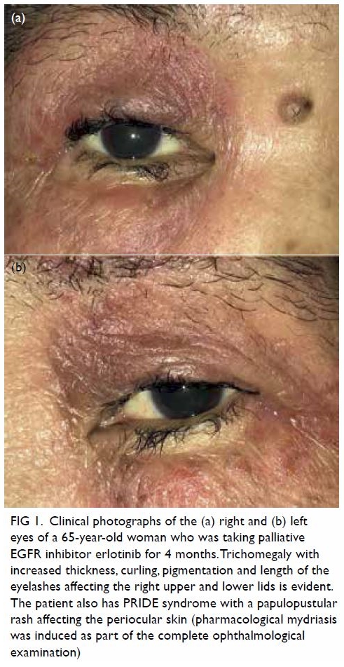
A 74-year-old man presented with a history of hoarseness of voice and choking for a few weeks. Laryngoscopy revealed left vocal cord palsy (Fig 1). 
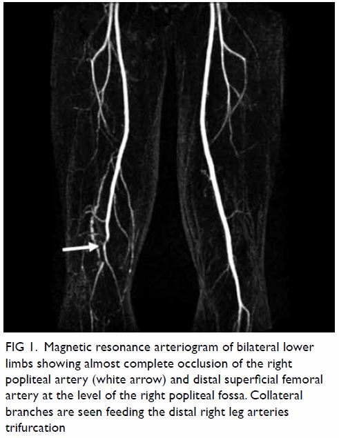
Non-contrast computed tomography (CT) showed no pulmonary or neck lesion, but one lobulated, centrally low attenuating mass. T2-weighted magnetic resonance images confirmed a 3.8-cm soft tissue mass with heterogeneous enhancement in the left cerebellopontine angle. In both images there was extension of the mass through an expansion of the left jugular foramen, widened left internal acoustic meatus and skull base destruction (Fig 2), compatible with jugular foramen tumour. Subsequently the patient developed progressive globus sensation, tinnitus, left sternocleidomastoid muscle wasting, and left neurosensory hearing loss. The tumour was resected via a retrosigmoid approach. Surgical assessment of the tumour revealed its origin from cranial nerves IX and X and compression of cranial nerves VIII and XI. The histopathological diagnosis with immunohistochemistry staining was that of schwannoma. Postoperative imaging demonstrated adequate resection of the tumour. Improvement in swallowing and hearing function test from severe to moderate impairment were noted postoperatively.
 

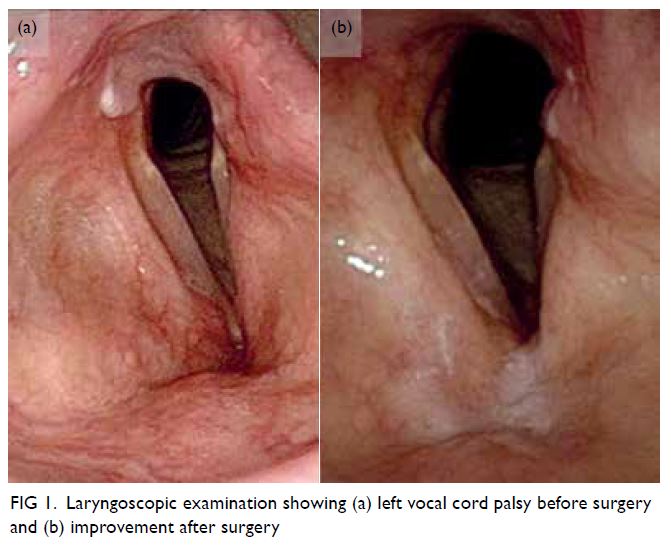
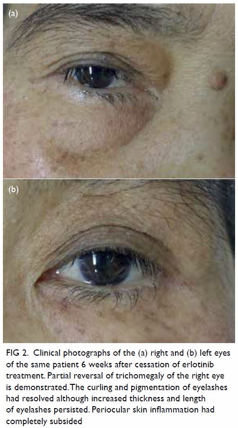
Figure 1. Laryngoscopic examination showing (a) left vocal cord palsy before surgery and (b) improvement after surgery
 

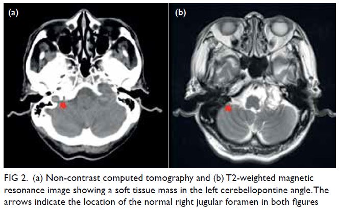
Figure 2. (a) Non-contrast computed tomography and (b) T2-weighted magnetic resonance image showing a soft tissue mass in the left cerebellopontine angle. The arrows indicate the location of the normal right jugular foramen in both figures
 
Discussion
Schwannoma is a benign tumour of the nerve sheath. The most common intracranial schwannoma is vestibular schwannoma (acoustic neuroma) that arises from cranial nerve VIII.1 Jugular foramen schwannomas, which mainly arise from cranial nerves IX and X, account for around 3% to 4% of all intracranial schwannomas. They are more prevalent in women and occur between the third and sixth decades of life. Clinical presentation is variable because of their slow-growing nature and proximity to other cranial nerves. Symptoms appear when the tumour is sufficiently large and most commonly consist of hearing loss, tinnitus, dysphagia, ataxia, and hoarseness. Other symptoms include dysarthria, dysphonia, aspiration, vertigo, dizziness, shoulder weakness, and headache.2 Differential diagnoses of unilateral vocal cord palsy are usually divided into malignancies of the lung and the neck and non-malignant lesions of traumatic, neurological, inflammatory or infectious origin. A CT scan is indicated to evaluate the more common pulmonary malignancies and to ensure presence of a rarer lesion along the course of the recurrent laryngeal and vagus nerve up to the skull base is not missed, especially if signs and symptoms of multiple cranial nerve involvement are present.3
 
Imaging studies can help differentiate vestibular and lower cranial nerve schwannomas, meningioma, glomus jugulare paraganglioma, ependymomas, and metastatic tumour. Imaging findings of jugular foramen schwannoma include scalloped and sclerotic expansion of the temporal bone instead of a lytic pattern. On magnetic resonance imaging, the lesion is T1 iso- or hypo-intense or T2 iso- or hyper-intense with gadolinium enhancement, whereas on CT, it is isodense to brain parenchyma and enhanced with contrast.4 The site of origin is classified as cisternal, foraminal (intraosseous) or extracranial. However, it is difficult to ascertain the exact site radiologically and clinically because of the variable location and nerve involvement. Assessment of the nerve root during surgery is required to confirm the origin of the tumour. The selection of surgical approach for treatment is determined by the location, pattern and extension of the tumour, the degree of bone destruction/erosion and the neurological and preoperative hearing status.5 Risks of operation include damage to other cranial nerves, especially facial nerve palsy, and incomplete removal of the tumour leading to recurrence.
 
Author contributions
Concept or design: All authors.
Acquisition of data: WK Wong, CY Cheng.
Analysis or interpretation of data: All authors.
Drafting of the manuscript: WK Wong.
Critical revision of the manuscript for important intellectual content: All authors.
 
All authors had full access to the data, contributed to the study, approved the final version for publication, and take responsibility for its accuracy and integrity.
 
Conflicts of interest
All authors have disclosed no conflicts of interest.
 
Funding/support
This study received no specific grant from any funding agency in the public, commercial, or not-for-profit sectors.
 
Ethics approval
The patient was treated in accordance with the Declaration of Helsinki and provided consent for all investigations and procedures.
 
References
1. Suri A, Bansal S, Singh M, Mahapatra AK, Sharma BS. Jugular foramen schwannomas: a single institution patient series. J Clin Neurosci 2014;21:73-7. Crossref
2. Bakar B. The jugular foramen schwannomas: review of the large surgical series. J Korean Neurosurg Soc 2008;44:285-94. Crossref
3. Stimpson P, Patel R, Vaz F, et al. Imaging strategies for investigating unilateral vocal cord palsy: how we do it. Clin Otolaryngol 2011;36:266-71. Crossref
4. Lee M, Tong K. Jugular foramen schwannoma mimicking paraganglioma: case report and review of imaging findings. Radiol Case Rep 2016;11:25-8. Crossref
5. Samii M, Alimohamadi M, Gerganov V. Surgical treatment of jugular foramen schwannoma: surgical treatment based on a new classification. Neurosurgery 2015;77:424-32. Crossref

Primary orbital melanoma

© Hong Kong Academy of Medicine. CC BY-NC-ND 4.0
 
PICTORIAL MEDICINE
Primary orbital melanoma
Vivian WK Hui, MB, ChB1; TC Lau, PhD2; Lawrence PW Ng, MSc2; Hunter KL Yuen, FRCOphth1; W Cheuk, FHKCPath2
1 Department of Ophthalmology, Hong Kong Eye Hospital, Hong Kong
2 Department of Pathology, Queen Elizabeth Hospital, Hong Kong
 
Corresponding author: Dr W Cheuk (cwzz01@ha.org.hk)
 
 Full paper in PDF
 
A 68-year-old Chinese man presented with proptosis, epiphora and discomfort in the right eye associated with impaired visual acuity. Contrast computed tomography scan revealed a 3.3 cm × 2.1 cm heterogeneously enhancing mass at the superotemporal aspect of the right orbit with displacement of the lateral rectus muscle (Fig 1). A pigmented tumour with dense adhesions to the surrounding structures was found in orbitotomy with leakage of the pigmented content upon surgical exploration. The lesion was excised as much as possible. Detailed clinical examination showed no evidence of intraocular melanoma, conjunctival or eyelid melanoma, or melanoma anywhere else. Systemic examination and whole-body positron emission tomography-computed tomography scan were unremarkable. The patient was alive with no evidence of disease at 9-month follow-up.
 

Figure 1. Contrast computed tomography scan showing a contrast-enhancing tumour behind the right globe displacing the optic nerve
 
The tumour was a solid, dark brownish multinodular mass covered by fat and skeletal muscle, comprising lobulated sheets of epithelioid and spindly melanocytes with vesicular chromatin, prominent nucleoli, and variable amounts of melanin pigment (Figs 2, 3, and 4). The mitotic count was 1 per 10 HPFs. The neoplastic melanocytes were positive for S100 and SOX10 but negative for BRAF. BRCA1-associated protein 1 immunostaining was intact. Sanger sequencing revealed a GNA11 Q209L mutation. No mutation was found in GNAQ, NRAS, KRAS, BRAF or KIT.
 

Figure 2. Micrograph showing multinodular mass with variable amount of melanin pigment in different areas (×40)
 

Figure 3. Large sheets of epithelioid (left upper field) and spindly (right lower field) melanoma cells with pale chromatin and prominent nucleoli were found. There were scattered histiocytes with abundant cytoplasmic melanin pigment (×200)
 

Figure 4. Melanoma cells were positive on SOX10 (red chromogen) immunohistochemical staining (×200)
 
Ocular melanoma most frequently involves the uveal tract (choroid, ciliary body, and iris) and the conjunctiva, where melanocytes are normally present. According to The Cancer Genome Atlas uveal melanoma project, mutually exclusive mutations in GNAQ, GNA11, CYSLTR2 and PLCB4 are the tumour-initiating mutations whereas BAP1, EIF1AX and SF3B1/SRSF2 mutations are associated with prognosis in uveal melanoma.1 In contrast, conjunctival melanoma typically exhibits BRAF, NRAS and NF1 mutations of the MAPK pathway with UV-induced C→T mutation signature.2 Orbital soft tissue melanoma is uncommon with >90% representing secondary tumours as a result of contiguous spread or metastasis from uveal, conjunctival, cutaneous or sinonasal melanomas.3 Primary orbital melanoma is exceedingly rare and has been postulated to arise in embryological remnants of melanocytes arrested in the region since a substantial proportion of cases are associated with blue nevus, orbital melanocytosis or oculodermal melanocytosis (nevus of Ota). Alternatively, it may arise from isolated melanocytes found in the optic nerve sheath, orbital fat, extraocular muscles or orbital periosteum. The majority of patients are white Europeans in their fourth to fifth decade. Histologically, it comprises epithelioid, spindle or mixed populations of neoplastic melanocytes that are frequently pigmented but with a lesser degree of nuclear pleomorphism than their cutaneous and mucosal counterparts. The prognosis is generally poor but a small proportion of patients survive long term. The most frequent site of metastasis is the liver.3 4 The clinicopathological features of primary orbital melanoma are very similar to those of uveal melanoma. The findings of GNAQ, SF3B1, EIF1AX mutations5 and GNA11 mutation in this case provide further evidence that primary orbital melanoma has a close pathogenic relationship with uveal melanoma.
 
Author contributions
All authors contributed to the concept or design of the study, acquisition of the data, analysis or interpretation of the data, drafting of the manuscript, and critical revision of the manuscript for important intellectual content. All authors had full access to the data, contributed to the study, approved the final version for publication, and take responsibility for its accuracy and integrity.
 
Conflicts of interest
As an adviser of the journal, HKL Yuen was not involved in the peer review process. Other authors have disclosed no conflicts of interest.
 
Declaration
This case was presented as a poster at the Annual Scientific Meeting of The College of Ophthalmologists of Hong Kong and Hong Kong Ophthalmological Society on 14 to 15 December 2019.
 
Funding/support
This pictorial medicine paper received no specific grant from any funding agency in the public, commercial, or not-forprofit sectors.
 
Ethics approval
The patient was treated in accordance with the tenets of the Declaration of Helsinki. Informed consent was obtained from the patient.
 
References
1. Bakhoum MF, Esmaeli B. Molecular characteristics of uveal melanoma: insights from the Cancer Genome Atlas (TCGA) Project. Cancers (Basel) 2019;11:1061.Crossref
2. Swaminathan SS, Field MG, Sant D, et al. molecular characteristics of conjunctival melanoma using whole-exome sequencing. JAMA Ophthalmol 2017;135:1434-7. Crossref
3. Rose AM, Luthert PJ, Jayasena CN, Verity DH, Rose GE. Primary orbital melanoma: presentation, treatment, and long-term outcomes for 13 patients. Front Oncol 2017;7:316. Crossref
4. Shields JA, Shields CL. Orbital malignant melanoma: the 2002 Sean B Murphy lecture. Ophthalmic Plast Reconstr Surg 2003;19:262-9. Crossref
5. Rose AM, Luo R, Radia UK, et al. Detection of mutations in SF3B1, EIF1AX and GNAQ in primary orbital melanoma by candidate gene analysis. BMC Cancer 2018;18:1262. Crossref

Tattoo-associated uveitis

© Hong Kong Academy of Medicine. CC BY-NC-ND 4.0
 
PICTORIAL MEDICINE
Tattoo-associated uveitis
CY Mak, MB, BS, MRCSEd (Ophth)1; Mary Ho, FCOphth HK, FHKAM (Ophthalmology)1; Angela Z Chan, MB, BS2; Lawrence PL Iu, FRCSEd (Ophth), FHKAM (Ophthalmology)1; Christina MT Cheung, FHKCP, FHKAM (Medicine)3; Marten E Brelen, BMBCh (Oxon), FRCOphth1; Paul CL Choi, FRCPA, FHKAM (Pathology)2; Alvin L Young, FRCOphth, FHKAM (Ophthalmology)1
1 Department of Ophthalmology and Visual Sciences, Prince of Wales Hospital, Hong Kong
2 Department of Anatomical and Cellular Pathology, Prince of Wales Hospital, Hong Kong
3 Division of Dermatology, Department of Medicine and Therapeutics, Prince of Wales Hospital, Hong Kong
 
Corresponding author: Prof Alvin L Young (youngla@ha.org.hk)
 
 Full paper in PDF
 
A 19-year-old man with extensive body tattooing presented with recurrent episodes of reduced vision, bilateral photophobia and concomitant swelling of body tattoos. He had multiple tattoos over his entire body with mainly black pigment and occasional red and yellow pigment, performed over a period of 3 years prior to the presentation of ocular symptoms. He enjoyed good past health with no history of autoimmune diseases. There was no joint pain, skin rash or chest symptoms.
 
Ophthalmological examination revealed bilateral injection, anterior chamber cells, posterior synechiae (Fig 1) and marked vitritis, consistent with anterior and intermediate uveitis. There were no mutton-fat keratic precipitates or iris nodules. Presenting visual acuity was 20/200 in both eyes. Dermatological examination showed prominent induration of skin with mild tenderness in areas of body tattoo containing black pigment (Fig 2). Non-tattooed skin was unremarkable with no signs of inflammation. Incisional skin biopsy was taken from an area of prominently indurated tattoo. Histopathology showed marked non-caseating granulomatous reaction within the dermis and abundant black pigment deposition (Fig 3). Periodic acid-Schiff staining showed no fungal elements. Chest radiograph was clear with no hilar lymphadenopathy and interferon-gamma releasing assay was negative. Syphilis and human immunodeficiency virus serology was negative. Immune markers including antinuclear antibodies, antineutrophil cytoplasmic antibodies and anti-extractable nuclear antigens antibody were negative. The patient declined a blood test for angiotensin-converting enzyme level due to the associated cost. Serum calcium was not elevated.
 

Figure 1. Slit lamp photo of the right eye showing ciliary injection, posterior synechiae formation. There were no iris nodules
 

Figure 2. Prominent induration of part of a tattoo containing black pigment. Surrounding non-tattooed skin was unremarkable
 

Figure 3. Skin biopsy of indurated tattoo under haematoxylin and eosin staining showing dermal deposition of black pigment and marked non-caseating granulomatous reaction
 
He was treated with topical prednisolone and oral prednisolone 60 mg daily after exclusion of infectious uveitides. Body tattoo swelling subsided rapidly after systemic steroid and the uveitis was brought under control gradually with significant improvement in bilateral vision. He was maintained on mycophenolate mofetil 1g twice a day as a steroid-sparing agent for uveitis control. His oral prednisolone was tapered to below 15 mg daily. His visual acuity improved and maintained at 20/30 bilaterally. There were no features of systemic sarcoidosis. Overall clinicopathological features were compatible with tattoo-associated uveitis, a rare dermato-ophthalmological complication of body tattooing.
 
Systemic sarcoidosis, a rare disease in Asians, can occasionally cause tattoo granuloma and uveitis.1 Tattoo-associated uveitis without systemic sarcoidosis is a rare entity first described in a case series half a century ago.2 The disease is characterised by recurrent episodes of uveitis in conjunction with raised and indurated tattoo, while histology of affected skin demonstrates florid non-caseating granulomatous reaction indistinguishable from tattoo granuloma in systemic sarcoidosis.3 The exact pathogenesis is unknown, but it was believed to be a type of delayed hypersensitivity reaction to tattoo pigments.3 Treatment is mainly to control ocular inflammation by topical and systemic steroid, with or without steroid-sparing agent. Tattoo excision has been reported to be useful in limiting recurrences.1 3 However, the extensive tattoo involvement in our patient rendered excision impractical.
 
In summary, the clinical photos illustrate a rare case of tattoo-associated uveitis, highlighting the importance of inquiry into tattoo history and skin examination of tattoos in a patient with recurrent uveitis.
 
Author contributions
Concept or design: CY Mak, ME Brelen, AL Young.
Acquisition of data: CY Mak, AZ Chan, CMT Cheung.
Analysis or interpretation of data: CY Mak, M Ho, LPL Iu, PCL Choi.
Drafting of the manuscript: CY Mak, M Ho, AZ Chan.
Critical revision of the manuscript for important intellectual content: All authors.
 
All authors had full access to the data, contributed to the study, approved the final version for publication, and take responsibility for its accuracy and integrity.
 
Conflicts of interest
All authors have disclosed no conflicts of interest.
 
Funding/support
This pictorial medicine paper received no specific grant from any funding agency in the public, commercial, or not-forprofit sectors.
 
Ethics approval
This patient was treated in accordance with the Declaration of Helsinki. The patient provided written informed consent for all treatments and procedures and for publication of clinical photos.
 
References
1. Kluger N. Tattoo-associated uveitis with or without systemic sarcoidosis: a comparative review of the literature. J Eur Acad Dermatol Venereol 2018;32:1852-61. Crossref
2. Rorsman H, Brehmer-Andersson E, Dahlquist I, et al. Tattoo granuloma and uveitis. Lancet 1969;2:27-8. Crossref
3. Ostheimer TA, Burkholder BM, Leung TG, Butler NJ, Dunn JP, Thorne JE. Tattoo-associated uveitis. Am J Ophthalmol 2014;158:637-43.e1. Crossref

Eyelash trichomegaly induced by erlotinib for metastatic lung cancer

© Hong Kong Academy of Medicine. CC BY-NC-ND 4.0
 
PICTORIAL MEDICINE
Eyelash trichomegaly induced by erlotinib for metastatic lung cancer
Julia YY Chan, AFCOphth HK, MRCSEd (Ophth)1,2; Tracy YT Kwok, FCOphth HK, FHKAM (Ophthalmology)1,2; Hunter KL Yuen, FRCOphth, FRCSEd1,2
1 Department of Ophthalmology and Visual Sciences, The Chinese University of Hong Kong, Hong Kong
2 Hong Kong Eye Hospital, Hong Kong
 
Corresponding author: Dr Julia YY Chan (cyyj717@gmail.com)
 
 Full paper in PDF
 
A 65-year-old Chinese woman was referred to the ophthalmology clinic complaining of grittiness in both eyes for 1 month. She had been diagnosed with stage IV metastatic pulmonary adenocarcinoma with positive mutation in epidermal growth factor receptor (EGFR) gene (L858R) and had been prescribed erlotinib 4 months previously as palliative treatment (Table).
 

Table. Timeline of the patient
 
Physical examination revealed bilateral long thick curly eyelashes (Figs 1 and 2) affecting all four eyelids. A papulopustular rash over the periocular region was also evident. The patient denied use of topical prostaglandin analogue for either medical or cosmetic use. A diagnosis was made of drug-induced trichomegaly. Corneal punctate epithelial erosion as a result of the misdirected lashes was treated symptomatically with regular epilation and topical lubricants.
 

Figure 1. Clinical photographs of the (a) right and (b) left eyes of a 65-year-old woman who was taking palliative EGFR inhibitor erlotinib for 4 months. Trichomegaly with increased thickness, curling, pigmentation and length of the eyelashes affecting the right upper and lower lids is evident. The patient also has PRIDE syndrome with a papulopustular rash affecting the periocular skin (pharmacological mydriasis was induced as part of the complete ophthalmological examination)
 

Figure 2. Clinical photographs of the (a) right and (b) left eyes of the same patient 6 weeks after cessation of erlotinib treatment. Partial reversal of trichomegaly of the right eye is demonstrated. The curling and pigmentation of eyelashes had resolved although increased thickness and length of eyelashes persisted. Periocular skin inflammation had completely subsided
 
Upon follow-up, gradual partial reversal of trichomegaly was evident at 6 weeks after erlotinib cessation. There was also improvement of the periorbital papulopustular rash. The reversal of trichomegaly after EGFR inhibitor cessation is not well documented in the literature. This case illustrates the presentation and partial resolution of drug-induced trichomegaly.
 
Eyelash trichomegaly is a rare condition characterised by increase in length, thickness, pigmentation, and curling of the eyelashes. The eyelash follicle, just as the scalp hair follicle, undergoes the cycle of anagen, catagen, and telogen phase. The anagen phase of eyelashes (ie, the growth phase) typically spans 8 weeks in Asian patients and occurs in 18% of eyelashes at any given time.1
 
Drug-induced trichomegaly is the most common form of trichomegaly to present in a general ophthalmology setting. Prostaglandin analogues such as latanoprost and bimatoprost, although commonly used as antiglaucomatous drugs, are well known for their side-effect of eyelash trichomegaly. The effect is due to up-regulation of prostaglandin E2, D2 receptor expressed in hair follicles.2
 
The occurrence of EGFR inhibitor–induced trichomegaly is seen less often but is not uncommon in the ophthalmology setting. It has been postulated that EGFR inhibitors inactivate the nuclear factor of activated T-cells. This leads to activation of stem cell bulge and suprabulbar region of the eyelash hair follicles.3 In a typical course, trichomegaly develops between the second and fifth month of EGFR inhibitor treatment.4 An Asian study reported that 2% of patients prescribed EGFR inhibitor treatment had trichomegaly.5 The EGFR inhibitor is also well known to cause a series of cutaneous adverse effects known as the PRIDE syndrome (papulopustules and/or paronychia, regulatory abnormalities of hair growth, itching, and dryness).6 In this case, the patient exhibited PRIDE syndrome affecting the periocular skin as demonstrated in Figures 1 and 2.
 
Trichomegaly is a benign condition, but long misdirected lashes may lead to corneal punctate epithelial erosion and corneal abrasion. Frequent trimming, epilation, and topical lubricants serve as first-line treatment. Electro-epilation or cryoablation directed at the affected hair follicles may be considered in instances of recurrent misdirected lashes with corneal complications such as infective corneal ulcer.
 
This case highlights the clinical course of EGFR inhibitor–induced trichomegaly upon cessation of drug therapy, which is not well documented in the literature. Partial reversal of trichomegaly was achieved after stopping erlotinib for 6 weeks. It is evident that the time required for trichomegaly resolution follows the typical eyelash follicle cycle.
 
Author contributions
All authors contributed to the design, acquisition of data, analysis of data, drafting of the manuscript, and critical revision of the manuscript for important intellectual content. All authors had full access to the data, contributed to the study, approved the final version for publication, and take responsibility for its accuracy and integrity.
 
Conflicts of interest
As an adviser of the journal, HKL Yuen was not involved in the peer review process. Other authors have disclosed no conflicts of interest.
 
Funding/support
This pictorial medicine received no specific grant from any funding agency in the public, commercial, or not-for-profit sectors.
 
Ethics approval
The patient was treated in accordance with the Declaration of Helsinki. The patient provided written informed consent for the treatment/procedures, and consent for publication.
 
References
1. Na JI, Kwon OS, Kim BJ, et al. Ethnic characteristics of eyelashes: a comparative analysis in Asian and Caucasian females. Br J Dermatol 2006;155:1170-6. Crossref
2. Colombe L, Michelet JF, Bernard BA. Prostanoid receptors in anagen human hair follicles. Exp Dermatol 2008;17:63- 72. Crossref
3. Dalal A, Sharma S, Kumar A, Sharma N. Eyelash trichomegaly: a rare presenting feature of systemic lupus erythematosus. Int J Trichology 2017;9:79-81. Crossref
4. Jeon SH, Ryu JS, Choi GS, et al. Erlotinib induced trichomegaly of the eyelashes. Tuberc Respir Dis (Seoul) 2013;74:37-40. Crossref
5. Chanprapaph K, Pongcharoen P, Vachiramon V. Cutaneous adverse events of epidermal growth factor receptor inhibitors: A retrospective review of 99 cases. Indian J Dermatol Venereol Leprol 2015;81:547. Crossref
6. Lacouture ME, Lai SE. The PRIDE (Papulopustules and/or paronychia, Regulatory abnormalities of hair growth, Itching, and Dryness due to Epidermal growth factor receptor inhibitors) syndrome. Br J Dermatol 2006;155:852-4. Crossref

Popliteal artery entrapment syndrome: a rare diagnosis for calf pain

© Hong Kong Academy of Medicine. CC BY-NC-ND 4.0
 
PICTORIAL MEDICINE
Popliteal artery entrapment syndrome: a rare diagnosis for calf pain
Stephanie C Woo, MB, BS, FRCR; TS Chan, FHKCR, FHKAM (Radiology); NY Pan, FHKCR, FHKAM (Radiology); Johnny KF Ma, FRCR (UK), FHKAM (Radiology)
Department of Radiology, Princess Margaret Hospital, Hong Kong
 
Corresponding author: Dr SC Woo (stephaniecheriwoo@gmail.com)
 
 Full paper in PDF
 
An 18-year-old man presented with a long history of occasional right calf pain and fullness. He also complained that over the last year his right foot
 
An 18-year-old man presented with a long history of occasional right calf pain and fullness. He also complained that over the last year his right foot became pale and numb after exercising for few minutes. There was no history of trauma and the patient had no constitutional symptoms. On physical examination, the right calf was non-tender with no mass although right posterior tibial and dorsalis pedis pulses were weaker than the left. Radiograph of the right knee showed static right proximal tibial exostosis, which had been monitored since the patient was aged 10 years. Magnetic resonance arteriogram showed almost complete occlusion of the right popliteal artery and distal superficial femoral artery at the level of the right popliteal fossa (Fig 1). Collateral branches were seen feeding the distal right leg arteries trifurcation. Magnetic resonance imaging scan of the knee revealed the medial head of the gastrocnemius inserting at a more lateral position than usual (Fig 2). The popliteal artery was separated from the popliteal vein, passing medial to and underneath the medial head of the gastrocnemius, and was severely compressed between the medial head of the gastrocnemius and distal femur. Loss of normal flow-related signal void was noted in the right popliteal artery distal to the compression.
 

Figure 1. Magnetic resonance arteriogram of bilateral lower limbs showing almost complete occlusion of the right popliteal artery (white arrow) and distal superficial femoral artery at the level of the right popliteal fossa. Collateral branches are seen feeding the distal right leg arteries trifurcation
 

Figure 2. Magnetic resonance image of the knee. (a) T2-weighted, (b) T2-weight fat saturated and (c) T1-weighted fat-saturated post-contrast images showing popliteal artery (white arrows) passing medial to and underneath the medial head of the gastrocnemius. It is severely compressed between the medial head of the gastrocnemius (asterisk) and distal femur into a slitlike configuration. Findings are consistent with popliteal artery entrapment syndrome
 
Popliteal artery entrapment syndrome (PAES) is a rare1 and frequently underdiagnosed disease entity. It typically affects young male athletes who commonly have hypertrophied musculature without significant cardiovascular risk factors. The classic presentation of PAES is of symptoms related to vascular compression, which is intermittent lower limb claudication. Other symptoms can include numbness, pain, discoloration, or even paralysis.2 Symptoms in the early stages typically occur during or following physical activity but can progress to symptoms at rest if complications develop.
 
In addition to a careful history, proper physical examination aids in diagnosis. Usual findings include calf muscle hypertrophy,2 and reduced posterior tibial and dorsalis pedis pulses on passive dorsiflexion or active plantar flexion of the foot.3 In addition, resting ankle-brachial index tests will usually be normal but will show a decrease with exercise.4 Differential diagnoses include other vascular diseases such as atherosclerosis, exertional syndrome, and cystic adventitial disease. Further diagnostic testing is usually needed to make a confident diagnosis of PAES.
 
Doppler ultrasonography is one of the first-line imaging modalities. It may demonstrate popliteal artery stenosis, increased velocity, or reduced peak systolic velocity during stress manoeuvres. However, it plays a limited role since imaging findings with this modality are non-specific and show only the consequences of the abnormal anatomy.4
 
Conventional angiography has been long used for the diagnosis of PAES.1 Typical findings include medial deviation of the proximal segment, occlusion in the middle segment, and post-stenotic dilatation at the distal segment.5 However, it is invasive and is unable to demonstrate surrounding soft tissue structures leading to the occlusion of the popliteal artery. It has recently been replaced by diagnostic modalities that are non-invasive such as computed tomography angiography and magnetic resonance imaging with magnetic resonance arteriogram.
 
Computed tomography angiography offers good soft tissue contrast and can provide diagnostic evaluation of surrounding muscular anomalies.4 It may also be used to evaluate the contralateral limb to exclude bilateral entrapment.
 
Magnetic resonance imaging and MR angiography are promising imaging modalities for the diagnosis of PAES6 due to their superior capability to demonstrate surrounding anatomy and soft tissue compared with computed tomography angiography, with no ionising radiation required.
 
In this case, timely diagnosis was made and treatment given. However, delay in diagnosis and management may lead to irreversible effects of lower limb ischaemia. This case illustrates the importance of considering this rare diagnosis when encountering young patients with lower limb claudication or calf pain symptoms. This will facilitate early surgical intervention to minimise the risk of complications.
 
Author contributions
All authors contributed to the design, acquisition of data, analysis of data, drafting of the manuscript, and critical revision of the manuscript for important intellectual content. All authors had full access to the data, contributed to the study, approved the final version for publication, and take responsibility for its accuracy and integrity.
 
Conflicts of interest
All authors have disclosed no conflicts of interest.
 
Funding/support
This pictorial medicine received no specific grant from any funding agency in the public, commercial, or not-for-profit sectors.
 
Ethics approval
The patient was treated in accordance with the Declaration of Helsinki. The patient provided written informed consent for the treatment/procedures, and consent for publication.
 
References
1. Thanila AM, Johnson CM, Hallett JW Jr, Breen JF. Popliteal artery entrapment syndrome: role of imaging in the diagnosis. AJR Am J Roentgenol 2003;181:1259-65. Crossref
2. Davis DD, Shaw PM. Popliteal Artery Entrapment Syndrome. In: StatPearls [Internet]. Treasure Island (FL): StatPearls Publishing; 10 May 2019. Available from: https://www.ncbi.nlm.nih.gov/books/NBK441965/. Accessed 1 Apr 2020.
3. Tercan F, Oğuzkurt L, Kizilkiliç O, Yeniocak A, Gülcan O. Popliteal artery entrapment syndrome. Diagn Interv Radiol 2005;11:222-4.
4. Eliahou R, Sosna J, Bloom AI. Between a rock and a hard place: clinical and imaging features of vascular compression syndromes. Radiographics 2012;32:33-49. Crossref
5. Zhong H, Liu C, Shao G. Computed tomographic angiography and digital subtraction angiography findings in popliteal artery entrapment syndrome. J Comput Assist Tomogr 2010;34:254-9. Crossref
6. Atilla S, Ilgit ET, Akpek S, Yücel C, Tali ET, Işik S. MR imaging and MR angiography in popliteal artery entrapment syndrome. Eur Radiol 1998;8:1025-9. Crossref

Megacalycosis: a rare radiological finding

Hong Kong Academy of Medicine. CC BY-NC-ND 4.0
 
PICTORIAL MEDICINE
Megacalycosis: a rare radiological finding
CL Cho, FRCS Ed (Urol), FHKAM (Surgery)1,2; CK Shiu, FRCR, FHKAM (Radiology)3
1 Department of Surgery, Union Hospital, Hong Kong
2 SH Ho Urology Centre, Department of Surgery, The Chinese University of Hong Kong, Hong Kong
3 Medical Imaging Department, Union Hospital, Hong Kong
 
Corresponding author: Dr CL Cho (chochaklam@yahoo.com.hk)
 
 Full paper in PDF
 
In April 2019, a 22-year-old woman was admitted to Union Hospital, Hong Kong, with right loin pain and fever. She had developed acute cystitis symptoms 1 week prior to the admission and was prescribed a course of oral antibiotic. She had good past health with no history of urinary tract infection. On admission she had a temperature of 39°C. The abdomen was soft and non-tender. Laboratory tests revealed a normal white cell count of 6.09 × 10⁹/L and normal serum creatinine level of 54 μmol/L. The C-reactive protein level was elevated at 45.5 mg/L. Urine tests revealed a slightly turbid urine with elevated white blood cell count of 260 cells/µL. There was no significant growth on urine culture. An urgent contrast computed tomography scan revealed features suggestive of pyelonephritis at the lower pole of the right kidney (Fig 1). There was no obstructive urinary stone. In addition, the imaging demonstrated an atypical right pelvicalyceal system (Fig 2). A course of antibiotic was prescribed with good clinical response. Repeat urine microscopy was unremarkable 8 weeks after the initial presentation. Follow-up computed tomography scan confirmed resolution of right pyelonephritis but the unusual finding of the right pelvicalyceal system remained unchanged (Fig 3).
 

Figure 1. Arterial phase contrast-enhanced computed tomography in (a) coronal and (b) axial planes demonstrating focal renal parenchymal swelling and hypoenhancement (asterisk) with associated perinephric fat stranding (arrow) consistent with acute pyelonephritis. Note the presence of a small renal stone (arrowhead) in a dilated calyx
 

Figure 2. Maximum intensity projection computed tomography urogram showing features of congenital megacalycosis with increased number and dilated, semilunar-shaped calyces in the right kidney. The right renal calyces are well opacified. The right renal pelvis (asterisk) and right ureter (arrow) are normal. Contralateral normal left upper tract urogram is shown for comparison
 

Figure 3. Follow-up computed tomography urogram with three-dimensional volume rendering showing persistent features of congenital megacalycosis with increased number and dilated right renal calyces
 
The radiological features were diagnostic of congenital megacalycosis. The anomaly is characterised by caliectasis with malformation. The classic triangular or conical shape of the renal calyces is replaced by a semilunar configuration. The pyramids of Malpighi are hypoplastic and the tip of each papilla is flat. The calyces have a rounded appearance with neither fornix nor papillae impressions. Polycalycosis is another feature of the condition and typically 20 to 25 calyces can be identified. The condition is differentiated from obstruction by the finding of a non-dilated renal pelvis, infundibulum, and ureter. The renal cortex was of normal thickness with good concentration of contrast medium in the distended calyces (Figs 2 and 3).
 
Congenital megacalycosis is a rare condition with approximately 100 cases reported in the literature. The anomaly is found predominantly in men and usually affects only one kidney. The exact pathogenesis remains unclear.1 2 3 The condition is usually asymptomatic and is detected during workup of urinary stone disease or urinary tract
 
infection in adults. On the contrary, reports of congenital megacalycosis in paediatric patients are on the rise with the increasing use of imaging in antenatal screening.4 Although urinary stasis in the distended calyces may predispose to infection and stone formation,1 2 renal function remains normal and neither anatomic nor functional deterioration occur over time.5 The benign nature suggests that megacalycosis should be considered a condition rather than a disease, and the term “megacalycose” has been suggested as an alternative term by some authors. Treatment should target complications only. Identification of the condition is important to avoid unnecessary investigation and intervention in a normally functioning kidney despite the inherent anatomic defect.2
 
Author contributions
Concept or design: CL Cho.
Acquisition of data: All authors.
Analysis or interpretation of data: All authors.
Drafting of the manuscript: All authors.
Critical revision of the manuscript for important intellectual content: All authors.
 
All authors had full access to the data, contributed to the study, approved the final version for publication, and take responsibility for its accuracy and integrity.
 
Conflicts of interest
The authors have disclosed no conflicts of interest.
 
Funding/support
This pictorial medicine paper received no specific grant from any funding agency in the public, commercial, or not-for-profit sectors.
 
Ethics approval
This study was conducted in accordance with the principles outlined in the Declaration of Helsinki. Verbal consent was obtained for the purpose of case study.
 
References
1. Puigvert A. Le megacalice. J Urol Nephrol 1964;70:321-6. Crossref
2. Kimche D, Lask D. Megacalycosis. Urology 1982;19:478-81. Crossref
3. Kalaitzis C, Patris E, Deligeorgiou E, et al. Radiological findings and the clinical importance of megacalycosis. Res Rep Urol 2015;7:153-5. Crossref
4. Kasap B, Kavukçu S, Soylu A, et al. Megacalycosis: report of two cases. Pediatr Nephrol 2005;20:828-30. Crossref
5. Redman JF, Neeb AD. Congenital megacalycosis: a forgotten diagnosis? Urology 2005;65:384-5. Crossref

Progressive diaphyseal dysplasia: a rare bone disorder with alarming radiographs

Hong Kong Academy of Medicine. CC BY-NC-ND 4.0
 
PICTORIAL MEDICINE
Progressive diaphyseal dysplasia: a rare bone disorder with alarming radiographs
Moses ML Li, MB, ChB, MHKICBSC1; KY Chung, FRCSEd (Orth), FHKAM (Orthopaedic Surgery)1; Alex WH Ng, FRCR, FHKAM (Radiology)2; KH Chiu, FRCS, FHKAM (Orthopaedic Surgery)1
1 Department of Orthopaedics and Traumatology, Prince of Wales Hospital, Hong Kong
2 Department of Imaging and Interventional Radiology, Prince of Wales Hospital, Hong Kong
 
Corresponding author: Dr Moses ML Li (moseslml@gmail.com)
 
 Full paper in PDF
 
In March 2017, a 57-year-old man was referred to our unit for left knee pain after a sprain, which resolved upon conservative treatment. Plain radiographs of the knees revealed diffuse sclerosis of bilateral femurs and tibias. A detailed history was sought to investigate the alarming skeletal pathology revealed on the plain radiographs. The patient had no history of bone pain, fever, weight loss, nor other constitutional symptoms. There was no history of malignancy. Physical examination revealed negative findings in the musculoskeletal, cardiac, pulmonary, renal, and neurological systems. Alkaline phosphatase was elevated (176 IU/L) but other blood parameters were normal, including white cell count, C-reactive protein, erythrocyte sedimentation rate, calcium, and phosphate. A skeletal survey revealed widespread sclerosis of tubular bone diaphysis of the lower extremities (Fig 1), upper extremities (Fig 2), and skull (Fig 3). Bone scintigraphy revealed increased osteoblastic activity corresponding to the locations of sclerosis (Fig 4). The characteristic radiographic features with the clinical and laboratory findings favoured a diagnosis of progressive diaphyseal dysplasia.
 

Figure 1. PPlain radiograph of bilateral lower limbs showing bilateral symmetrical diffuse sclerosis and cortical thickening with expansion of the diaphysis involving the femur, tibia, and fibula. Of note, the epiphyses of all the bones are spared which would be characteristic of progressive diaphyseal dysplasia
 

Figure 2. (a) Frontal radiographs of bilateral upper limbs showing symmetrical diffuse sclerosis and cortical thickening of the diaphysis involving the scapula, humerus, radius, and ulna. The epiphyses of these bones are also spared. (b) Frontal radiographs of the hands showing sclerosis with cortical thickening of the right second and left second to third metacarpal bones. Mild bony expansion with relative sparing of the medullary canals and epiphysis are noted
 

Figure 3. (a) Frontal radiograph of the skull showing sclerosis and cortical thickening of the calvaria and base of the skull. (a) Lateral radiograph of the skull showing severe sclerosis and thickening of the skull base and mandible. The cervical spine is not involved in this case
 

Figure 4. (a) Bone scintigraphy showing increased tracer uptake corresponding to the regions of sclerosis on plain radiographs, particularly in the skull vault and bilateral mandibles. (b) Lateral view of bone scintigraphy showing severe increased tracer uptake at the skull base
 
Sclerosing bone dysplasia is a heterogeneous group of rare bone disorders with pathognomonic radiological features, caused by a defective ossification pathway.1 2 Progressive diaphyseal dysplasia, also known as Camurati-Engelmann disease, is a disease belonging to this entity.3 4 5 It is an autosomal dominant disorder due to mutation in transforming growth factor–Β1. This in turn leads to a disorder of intramembranous ossification, and results in hyperostosis. Characteristic radiological features are bilateral symmetrical fusiform sclerosis involving the diaphyses of tubular bones. The epiphyses are typically spared as these regions are formed by endochondral ossification. The lower extremities are more affected than the upper extremities. In descending order of frequency, the tibia, femur, fibula, humerus, ulna, and radius are affected.3 Occasionally, the calvaria of the skull is involved. The affected bones show uneven cortical thickening of the diaphysis with hyperostosis extending in both periosteal and endosteal directions. Hyperostosis of the endosteal surface results in medullary canal narrowing.
 
Clinical manifestations of progressive diaphyseal dysplasia include limb pain, muscle weakness, and easy fatigability. However, our patient was completely asymptomatic. The disorder was picked up incidentally during investigation of an unrelated knee sprain. Detailed systemic review revealed an absence of extraosseous manifestations of Erdheim-Chester disease that shares similar radiological features.3 Ribbing disease (hereditary multiple diaphyseal sclerosis) usually presents with unilateral or asymmetrical involvement of the bones, and does not involve the skull vault. Osteopetrosis has epiphysis involvement and usually presents with fracture and extramedullary haematopoiesis. Although the differential diagnoses for bone sclerosis are extensive, characteristic radiographic distribution in association with clinical and laboratory findings can substantially narrow the possibilities and allow the diagnosis to be made. The age at which the diagnosis of progressive diaphyseal dysplasia is reached, the clinical manifestations of disease and the extent of radiological evidence of sclerosis are variable.4 5 Disease progression is slow and unpredictable. Treatment of the disease aims for symptomatic relief, with losartan reported to be effective in relieving limb pain based on the mechanism of down-regulation of transforming growth factor–Β1 receptor expression.
 
Author contributions
All authors contributed to the concept of the study, acquisition and analysis of the data, drafting of the manuscript, and critical revision of the manuscript for important intellectual content. All authors had full access to the data, contributed to the study, approved the final version for publication, and take responsibility for its accuracy and integrity.
 
Conflicts of interest
All authors have disclosed no conflicts of interest.
 
Funding/support
This pictorial medicine paper received no specific grant from any funding agency in the public, commercial, or not-for-profit sectors.
 
Ethics approval
The patient was treated in accordance with the principles outlined in the Declaration of Helsinki.
 
References
1. Boulet C, Madani H, Lenchik L, et al. Sclerosing bone dysplasias: genetic, clinical and radiology update of hereditary and non-hereditary disorders. Br J Radiol 2016;89:20150349. Crossref
2. Ihde LL, Forrester DM, Gottsegen CJ, et al. Sclerosing bone dysplasias: review and differentiation from other causes of osteosclerosis. Radiographics 2011;31:1865-82. Crossref
3. Uezato S, Dias G, Inada J, Valente M, Fernandes E. Imaging aspects of Camurati-Engelmann disease. Rev Assoc Med Bras (1992) 2016;62:825-7. Crossref
4. Van Hul W, Boudin E, Vanhoenacker FM, Mortier G. Camurati-Engelmann disease. Calcif Tissue Int 2019;104:554-60. Crossref
5. Yuldashev AJ, Shin CH, Kim YS, et al. Orthopedic manifestations of type I Camurati-Engelmann disease. Clin Orthop Surg 2017;9:109-15. Crossref

Skin testing for hypersensitivity and cross-reactivity between proton pump inhibitors

Hong Kong Academy of Medicine. CC BY-NC-ND 4.0
 
PICTORIAL MEDICINE
Skin testing for hypersensitivity and cross-reactivity between proton pump inhibitors
Philip H Li, MRCP, FHKCP
Division of Rheumatology and Clinical Immunology, Department of Medicine, The University of Hong Kong, Queen Mary Hospital, Pokfulam, Hong Kong
 
Corresponding author: Dr Philip H Li (liphilip@hku.hk)
 
 Full paper in PDF
 
A 44-year-old woman presented to our anaphylaxis clinic with first episode of anaphylaxis. She had no known prior food or drug allergies. She was previously prescribed esomeprazole 20 mg daily as required for dyspepsia by her private physician, but took it only very occasionally. Two months previously she took one tablet of esomeprazole (20 mg) and one of paracetamol (500 mg) together with a slice of plain bread for abdominal discomfort and influenza-like symptoms. Within 10 minutes, she experienced generalised pruritus and collapsed at home. She awoke after hitting the floor and telephoned for an ambulance. She was treated with intramuscular adrenaline in the ambulance because systolic blood pressure had fallen to <80 mm Hg. Initial investigations revealed a significant rise in acute tryptase (12.3 ng/mL) with a normal baseline level (2.1 ng/mL). Initially wheat or wheat-dependent cofactor augmented anaphylaxis was suspected. She was advised to avoid wheat, cyclooxygenase inhibitors, and proton pump inhibitors (PPI) until review.
 
Results for specific immunoglobulin E to wheat and omega-5-gliadin were negative. Skin prick test (SPT) to wheat solution (Inmunotek, Madrid, Spain) and prick-to-prick with the index bread slice was also negative. Skin prick test and intradermal tests (IDT) to paracetamol (both 100 mg/mL, GlaxoSmithKline, London, United Kingdom) were also negative. The patient tolerated an oral challenge with 500 mg of paracetamol together with a slice of the same index bread with no adverse reaction.
 
The SPT (8 mg/mL) and IDT (8 mg/mL, 0.8 mg/mL and 0.08 mg/mL; AstraZeneca, Bedfordshire, United Kingdom) were performed and are shown in the Figure. Skin prick test to histamine (positive control) and normal saline (negative control) was positive at 5 mm and negative at 0 mm, respectively. Skin prick test to esomeprazole was borderline positive with a 3-mm wheal and flare. Intradermal test to esomeprazole was positive at concentrations of 8 mg/mL, 0.8 mg/mL and 0.08 mg/mL; with 18-mm, 8-mm and 3-mm wheal expansion, respectively. The SPT and IDT with 8 mg/mL and 0.8 mg/mL were negative in a healthy control (the author) as shown in the Figure. To assess for potential cross-reactivity, SPT (4 mg/mL) and IDT (4 mg/mL and 0.4 mg/mL) to pantoprazole were also performed and are also shown in the Figure. The SPT was negative, but IDT was positive with 4 mg/mL and 0.4 mg/mL dilutions with 7-mm and 4-mm wheal expansion, respectively.
 

Figure. Left: Intradermal tests to esomeprazole in healthy control (neat, 1:10 dilution). Right: Skin prick and intradermal tests to esomeprazole (neat, 1:10 and 1:100 dilutions) and pantoprazole (neat and 1:10 dilution)
 
She was diagnosed with severe type I hypersensitivity to esomeprazole with cross-sensitisation to pantoprazole. Drug provocoation testing was not indicated in view of the compatible clinical history, strongly positive skin tests, and high risk of anaphylaxis. The patient declined testing with other PPI and was advised to avoid the entire class until further workup. She was prescribed famotidine 20 mg twice a day for her dyspepsia with no adverse effects.
 
This is the first reported case in Hong Kong of PPI anaphylaxis and demonstrates the utility of skin testing to assess potential cross-reactivity. Although relatively uncommon, reports of hypersensitivity to PPI are increasing, in parallel with their increasing use worldwide. The majority of hypersensitivity reactions appear to be of the immediate type.1 Previous exposure to esomeprazole may have been initial sensitising events. Despite common misconception, clinicians should bear in mind that prior tolerance of a certain drug does not preclude it as a future cause of drug allergy. Although patterns of cross-reactivity among various PPIs have been reported, these remain controversial and a thorough allergological workup should be performed for every patient.2 Clinicians are reminded to be vigilant of this uncommon cause of anaphylaxis and beware of potential cross-reactivity. The SPT and IDT have high specificity, but patients with suspected PPI hypersensitivity and negative skin tests should undergo drug provocation tests to confidently exclude this important diagnosis.3
 
Author contributions
The author contributed to the concept or design, acquisition of data, analysis or interpretation of data, drafting of the manuscript, and critical revision for important intellectual content. The author had full access to the data, contributed to the study, approved the final version for publication, and takes responsibility for its accuracy and integrity.
 
Conflicts of interest
The author has disclosed no conflicts of interest.
 
Funding/support
This pictorial medicine paper received no specific grant from any funding agency in the public, commercial, or not-forprofit sectors.
 
Ethics approval
The patient consented to this publication.
 
References
1. Otani IM, Banerji A. Immediate and delayed hypersensitivity reactions to proton pump inhibitors: evaluation and management. Curr Allergy Asthma Rep 2016;16:17. Crossref
2. Tourillon C, Mahe J, Baron A, et al. Immediate-type hypersensitivity cross-reactions to proton pump inhibitors: a descriptive study of data from the French National Pharmacovigilance Database. Int Arch Allergy Immunol 2019;178:159-66. Crossref
3. Kepil Özdemir S, Yilmaz I, Aydin Ö, et al. Immediate-type hypersensitivity reactions to proton pump inhibitors: usefulness of skin tests in the diagnosis and assessment of cross-reactivity. Allergy 2013;68:1008-14. Crossref

Clone of Clone of Primary hepatic schwannoma: imaging and histological findings

Hong Kong Academy of Medicine. CC BY-NC-ND 4.0
 
PICTORIAL MEDICINE
Primary hepatic schwannoma: imaging and histological findings
HL Tsui, MB, ChB, FRCR1; SM Yu, MB, ChB, FHKAM (Radiology)1; CH Lau, MB, ChB2; Sherman SM Lam, MB, BS, FHKAM (Surgery)3; PY Chu, MB, ChB, FHKAM (Radiology)1; YH Hui, MB, BS, FHKAM (Radiology)1; KL Lo, MB, ChB, FHKAM (Radiology)1
1 Department of Radiology and Organ Imaging, United Christian Hospital, Hong Kong
2 Department of Pathology, United Christian Hospital, Hong Kong
3 Department of Surgery, United Christian Hospital, Hong Kong
 
Corresponding author: Dr HL Tsui (karen.tsuihl@gmail.com)
 
 Full paper in PDF
 
Schwannoma is a rare tumour in the liver. It is likely to arise from the hepatobiliary nerves among the hepatic plexus in the liver hilum as well as interlobular connective tissues and hepatic arteries. To the best of our knowledge, no prior publications have reported cases in Hong Kong.
 
We report the case of a 64-year-old man with a history of nasopharyngeal carcinoma and colon carcinoma and a new liver lesion detected on follow-up imaging for surveillance in 2018 following detection of a slightly elevated serum MEDICINEcarcinoembryonic antigen level (4.7 μg/L). Alpha fetoprotein level was within the normal range (3.9 μg/L). Liver function tests were normal and he was asymptomatic with no history of neurofibromatosis.
 
Triphasic contrast computed tomography (CT) of the liver revealed a 5.2-cm ovoid hypodense lesion with heterogeneous enhancement in the caudate lobe of the liver (Fig 1). No washout of contrast was evident in the portal venous or delayed phases. Fluorodeoxyglucose-18 positron emission tomography–CT showed a moderately hypermetabolic lesion at the caudate lobe with a maximum standardised update value of 5.8 (Fig 2).
 

Figure 1. Triphasic computed tomography scans in (a) pre-contrast, (b) arterial, (c) portal venous, and (d) delayed phases, showing a well-defined, ovoid hypodense lesion with heterogeneous enhancement that persists in portal venous and delayed phases in the caudate lobe of the liver
 

Figure 2. Positron emission tomography–computed tomography scan in (a) coronal, (b) sagittal and (c) axial planes together with the (d) scout and (e) maximum intensity projection images, showing a moderately hypermetabolic lesion at the caudate lobe of the liver (red crosses)
 
Surgical resection of the lesion was performed (Fig 3). Pathology showed a schwannoma and degenerative changes. Histological examination revealed an encapsulated tumour consisting of highly ordered Antoni type A and B areas (Fig 4). Immunohistochemical analysis showed the tumour cells to be diffusely positive for S100, consistent with neural differentiation (Fig 4). HerPar1, a mitochondrial antigen of hepatocytes, was negative. The CD34, a cell surface glycoprotein that is positive in gastrointestinal stromal tumour, was also negative.
 

Figure 3. Gross specimen of hepatic resection with a well-circumscribed tumour mass and light tan colour cut surface
 

Figure 4. Micrographs of the tumour, showing (a) Antoni type A area, ×100; (b) Antoni type B area, ×100; (c) circumscribed and encapsulated tumour, ×20; and (d) immunostaining for S100 diffusely positive for the tumour cell, ×100
 
Schwannoma is most commonly found in the limbs and the head and neck region. A fifth of cases shows association with neurofibromatosis type 1. The mediastinum and retroperitoneum are other possible sites. It is uncommon in the gastrointestinal tract and extremely rare in the liver.1 It was first reported in 1978 by Pereira et al.2 A literature search through PubMed and MEDLINE revealed 32 reported cases. No cases have been published in Hong Kong.
 
The origin of hepatic schwannoma is the hepatobiliary nerves among the hepatic plexus in the liver hilum as well as interlobular connective tissues and the hepatic arteries.1 3 They are usually well-encapsulated and grow very slowly, usually smaller than 5 cm at the time of diagnosis. Larger schwannomas may undergo secondary degeneration with consequent pseudocystic regression, haemorrhage, and calcification. Malignant transformation is very rare.3
 
Pathologically, a schwannoma is an encapsulated tumour that arises within the nerve sheaths. It consists of a highly ordered cellular component (Antoni type A area) characterised by spindle cells with twisted nuclei arranged in short bundles, and a hypocellular area in a loose myxoid stroma (Antoni type B area) that comprises a loose meshwork of gelatinous and microcystic tissue.4
 
On imaging, hepatic schwannoma is usually well circumscribed with various signal characteristics, depending on the distribution of Antoni A and Antoni B areas.1 It is commonly of low density with heterogeneous enhancement on CT, hypointense on T1-weighted, and hyperintense on T2-weighted magnetic resonance imaging.5 There have also been reports of malignant tumours but there are no distinct radiological features that differentiate them from benign tumours.3 A hepatic schwannoma may be fluorodeoxyglucose-avid depending on inflammatory activity and cellularity. Fluorodeoxyglucose-18 positron emission tomography–CT alone may enable differentiation of a schwannoma from malignant lesions of the liver.1
 
Hepatic schwannoma is an extremely rare tumour and preoperative diagnosis with imaging is challenging. Biopsy or surgical resection is usually required for definitive diagnosis.
 
Author contributions
Concept or design: All authors.
Acquisition of data: HL Tsui and CH Lau.
Analysis or interpretation of data: HL Tsui, SM Yu, and CH Lau.
Drafting of the manuscript: HL Tsui, SM Yu, and CH Lau.
Critical revision of the manuscript for important intellectual content: All authors.
 
All authors had full access to the data, contributed to the study, approved the final version for publication, and take responsibility for its accuracy and integrity.
 
Conflicts of interest
All authors have disclosed no conflicts of interest.
 
Funding/support
This research received no specific grant from any funding agency in the public, commercial, or not-for-profit sectors.
 
Ethics approval
This study is approved by the cluster Research Ethics Committee (Ref KC/KE-19-0247/ER-3). Written patient consent was also obtained.
 
References
1. Hayashi M, Takeshita A, Yamamoto K, Tanigawa N. Primary hepatic benign schwannoma. World J Gastrointest Surg 2012;4:73-8. Crossref
2. Pereira Filho RA, Souza SA, Oliveira Filho JA. Primary neurilemmal tumour of the liver: case report. Arq Gastroenterol 1978;15:136-8.
3. Ozkan EE, Guldur M, Uzunkoy A. A case report of benign schwannoma of the liver. Intern Med 2010;49:1533-6. Crossref
4. Wan DL, Zhai ZL, Ren KW, Yang YC, Lin SZ, Zheng SS. Hepatic schwannoma: a case report and an updated 40-year review of the literature yielding 30 cases. Mol Clin Oncol 2016;4:959-64. Crossref
5. Yamamoto M, Hasegawa K, Arita J, et al. Primary hepatic schwannoma: a case report. Int J Surg Case Rep 2016;29:146-50. Crossref

Clone of Primary hepatic schwannoma: imaging and histological findings

Hong Kong Academy of Medicine. CC BY-NC-ND 4.0
 
PICTORIAL MEDICINE
Primary hepatic schwannoma: imaging and histological findings
HL Tsui, MB, ChB, FRCR1; SM Yu, MB, ChB, FHKAM (Radiology)1; CH Lau, MB, ChB2; Sherman SM Lam, MB, BS, FHKAM (Surgery)3; PY Chu, MB, ChB, FHKAM (Radiology)1; YH Hui, MB, BS, FHKAM (Radiology)1; KL Lo, MB, ChB, FHKAM (Radiology)1
1 Department of Radiology and Organ Imaging, United Christian Hospital, Hong Kong
2 Department of Pathology, United Christian Hospital, Hong Kong
3 Department of Surgery, United Christian Hospital, Hong Kong
 
Corresponding author: Dr HL Tsui (karen.tsuihl@gmail.com)
 
 Full paper in PDF
 
Schwannoma is a rare tumour in the liver. It is likely to arise from the hepatobiliary nerves among the hepatic plexus in the liver hilum as well as interlobular connective tissues and hepatic arteries. To the best of our knowledge, no prior publications have reported cases in Hong Kong.
 
We report the case of a 64-year-old man with a history of nasopharyngeal carcinoma and colon carcinoma and a new liver lesion detected on follow-up imaging for surveillance in 2018 following detection of a slightly elevated serum MEDICINEcarcinoembryonic antigen level (4.7 μg/L). Alpha fetoprotein level was within the normal range (3.9 μg/L). Liver function tests were normal and he was asymptomatic with no history of neurofibromatosis.
 
Triphasic contrast computed tomography (CT) of the liver revealed a 5.2-cm ovoid hypodense lesion with heterogeneous enhancement in the caudate lobe of the liver (Fig 1). No washout of contrast was evident in the portal venous or delayed phases. Fluorodeoxyglucose-18 positron emission tomography–CT showed a moderately hypermetabolic lesion at the caudate lobe with a maximum standardised update value of 5.8 (Fig 2).
 

Figure 1. Triphasic computed tomography scans in (a) pre-contrast, (b) arterial, (c) portal venous, and (d) delayed phases, showing a well-defined, ovoid hypodense lesion with heterogeneous enhancement that persists in portal venous and delayed phases in the caudate lobe of the liver
 

Figure 2. Positron emission tomography–computed tomography scan in (a) coronal, (b) sagittal and (c) axial planes together with the (d) scout and (e) maximum intensity projection images, showing a moderately hypermetabolic lesion at the caudate lobe of the liver (red crosses)
 
Surgical resection of the lesion was performed (Fig 3). Pathology showed a schwannoma and degenerative changes. Histological examination revealed an encapsulated tumour consisting of highly ordered Antoni type A and B areas (Fig 4). Immunohistochemical analysis showed the tumour cells to be diffusely positive for S100, consistent with neural differentiation (Fig 4). HerPar1, a mitochondrial antigen of hepatocytes, was negative. The CD34, a cell surface glycoprotein that is positive in gastrointestinal stromal tumour, was also negative.
 

Figure 3. Gross specimen of hepatic resection with a well-circumscribed tumour mass and light tan colour cut surface
 

Figure 4. Micrographs of the tumour, showing (a) Antoni type A area, ×100; (b) Antoni type B area, ×100; (c) circumscribed and encapsulated tumour, ×20; and (d) immunostaining for S100 diffusely positive for the tumour cell, ×100
 
Schwannoma is most commonly found in the limbs and the head and neck region. A fifth of cases shows association with neurofibromatosis type 1. The mediastinum and retroperitoneum are other possible sites. It is uncommon in the gastrointestinal tract and extremely rare in the liver.1 It was first reported in 1978 by Pereira et al.2 A literature search through PubMed and MEDLINE revealed 32 reported cases. No cases have been published in Hong Kong.
 
The origin of hepatic schwannoma is the hepatobiliary nerves among the hepatic plexus in the liver hilum as well as interlobular connective tissues and the hepatic arteries.1 3 They are usually well-encapsulated and grow very slowly, usually smaller than 5 cm at the time of diagnosis. Larger schwannomas may undergo secondary degeneration with consequent pseudocystic regression, haemorrhage, and calcification. Malignant transformation is very rare.3
 
Pathologically, a schwannoma is an encapsulated tumour that arises within the nerve sheaths. It consists of a highly ordered cellular component (Antoni type A area) characterised by spindle cells with twisted nuclei arranged in short bundles, and a hypocellular area in a loose myxoid stroma (Antoni type B area) that comprises a loose meshwork of gelatinous and microcystic tissue.4
 
On imaging, hepatic schwannoma is usually well circumscribed with various signal characteristics, depending on the distribution of Antoni A and Antoni B areas.1 It is commonly of low density with heterogeneous enhancement on CT, hypointense on T1-weighted, and hyperintense on T2-weighted magnetic resonance imaging.5 There have also been reports of malignant tumours but there are no distinct radiological features that differentiate them from benign tumours.3 A hepatic schwannoma may be fluorodeoxyglucose-avid depending on inflammatory activity and cellularity. Fluorodeoxyglucose-18 positron emission tomography–CT alone may enable differentiation of a schwannoma from malignant lesions of the liver.1
 
Hepatic schwannoma is an extremely rare tumour and preoperative diagnosis with imaging is challenging. Biopsy or surgical resection is usually required for definitive diagnosis.
 
Author contributions
Concept or design: All authors.
Acquisition of data: HL Tsui and CH Lau.
Analysis or interpretation of data: HL Tsui, SM Yu, and CH Lau.
Drafting of the manuscript: HL Tsui, SM Yu, and CH Lau.
Critical revision of the manuscript for important intellectual content: All authors.
 
All authors had full access to the data, contributed to the study, approved the final version for publication, and take responsibility for its accuracy and integrity.
 
Conflicts of interest
All authors have disclosed no conflicts of interest.
 
Funding/support
This research received no specific grant from any funding agency in the public, commercial, or not-for-profit sectors.
 
Ethics approval
This study is approved by the cluster Research Ethics Committee (Ref KC/KE-19-0247/ER-3). Written patient consent was also obtained.
 
References
1. Hayashi M, Takeshita A, Yamamoto K, Tanigawa N. Primary hepatic benign schwannoma. World J Gastrointest Surg 2012;4:73-8. Crossref
2. Pereira Filho RA, Souza SA, Oliveira Filho JA. Primary neurilemmal tumour of the liver: case report. Arq Gastroenterol 1978;15:136-8.
3. Ozkan EE, Guldur M, Uzunkoy A. A case report of benign schwannoma of the liver. Intern Med 2010;49:1533-6. Crossref
4. Wan DL, Zhai ZL, Ren KW, Yang YC, Lin SZ, Zheng SS. Hepatic schwannoma: a case report and an updated 40-year review of the literature yielding 30 cases. Mol Clin Oncol 2016;4:959-64. Crossref
5. Yamamoto M, Hasegawa K, Arita J, et al. Primary hepatic schwannoma: a case report. Int J Surg Case Rep 2016;29:146-50. Crossref

Pages