Inadvertent arterial insertion of a central venous catheter

DOI: 10.12809/hkmj176866
© Hong Kong Academy of Medicine. CC BY-NC-ND 4.0
 
PICTORIAL MEDICINE
Inadvertent arterial insertion of a central venous catheter
Victor WT Chan, MB, BS, FRCR; KW Shek, MB, BS, FRCR
Department of Radiology and Imaging, Queen Elizabeth Hospital, Jordan, Hong Kong
 
Corresponding author: Dr Victor WT Chan (chanwaitat@gmail.com)
 
 Full paper in PDF
 
Central venous catheterisation is a common procedure that allows venous access for delivering medications, infusing fluids or blood products, and monitoring volume status. Traditionally, anatomical landmarks of the sternocleidomastoid muscle provide a pathway to catheterise the internal jugular vein (IJV). However, inadvertent arterial puncture is a risk. Currently, ultrasound guidance by experienced operators is recommended for reducing the risk of mechanical complications during central venous catheter (CVC) insertion.1
 
For venous access via the neck, common carotid and subclavian artery injuries have been reported.2 The risk of artery injury is about 0.5%, and practice should be reviewed if the risk exceeds 1%. Other known mechanical complications include haematoma formation, haemothorax, and pneumothorax.
 
A 55-year-old woman with end-stage renal failure on continuous ambulatory peritoneal dialysis was admitted to our hospital for fever and abdominal pain. The clinical diagnosis of continuous ambulatory peritoneal dialysis peritonitis was made, and the peritoneal dialysis catheter was removed. Bedside insertion of a CVC was selected for temporary haemodialysis. The CVC insertion was initially attempted via the right IJV, unsuccessfully. The CVC was subsequently inserted via the left IJV. The procedure was performed under ultrasound guidance using the Seldinger technique; however, inadvertent arterial puncture was not recognised, and the procedure was continued.
 
After CVC insertion, a chest radiograph was taken, showing an abnormal vertical course of the catheter with suspected malposition (Fig 1). Urgent contrast computed tomographic angiogram (Figs 2 and 3) revealed that the catheter had been inserted via the left IJV, subsequently exiting posteromedially, entering the left vertebral artery, and harbouring at the origin of the left subclavian artery. Computed tomographic angiogram also showed abnormal contrast pooling over the right neck suggestive of a pseudoaneurysm formation from the right subclavian artery.
 

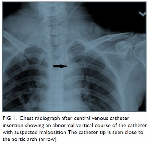
Figure 1. Chest radiograph after central venous catheter insertion showing an abnormal vertical course of the catheter with suspected malposition. The catheter tip is seen close to the aortic arch (arrow)
 

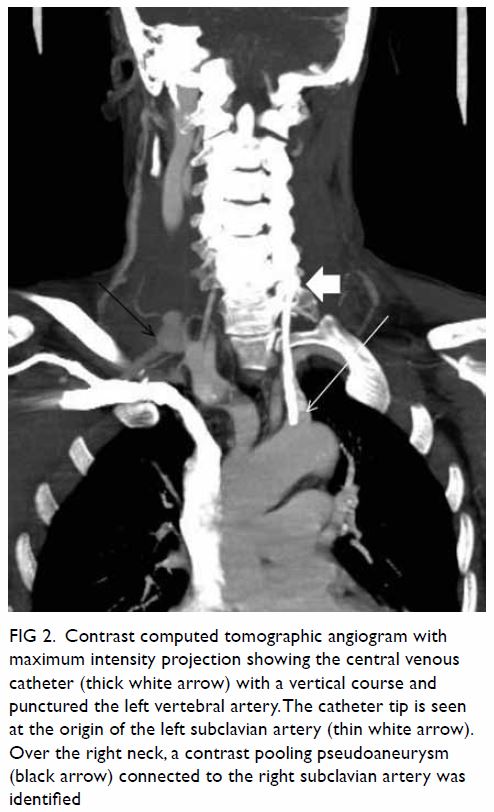
Figure 2. Contrast computed tomographic angiogram with maximum intensity projection showing the central venous catheter (thick white arrow) with a vertical course and punctured the left vertebral artery. The catheter tip is seen at the origin of the left subclavian artery (thin white arrow). Over the right neck, a contrast pooling pseudoaneurysm (black arrow) connected to the right subclavian artery was identified
 

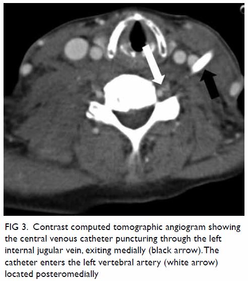
Figure 3. Contrast computed tomographic angiogram showing the central venous catheter puncturing through the left internal jugular vein, exiting medially (black arrow). The catheter enters the left vertebral artery (white arrow) located posteromedially
 
The opinion of a vascular surgeon was sought and the catheter was removed under general anaesthesia with repair of the vertebral artery. Oozing was noted from the left IJV exit site. Haemostasis was controlled by direct pressure onto the IJV. Urgent right subclavian angiogram (Fig 4) was performed by interventional radiologists, confirming the presence of a pseudoaneurysm, which was successfully embolised with coils.
 

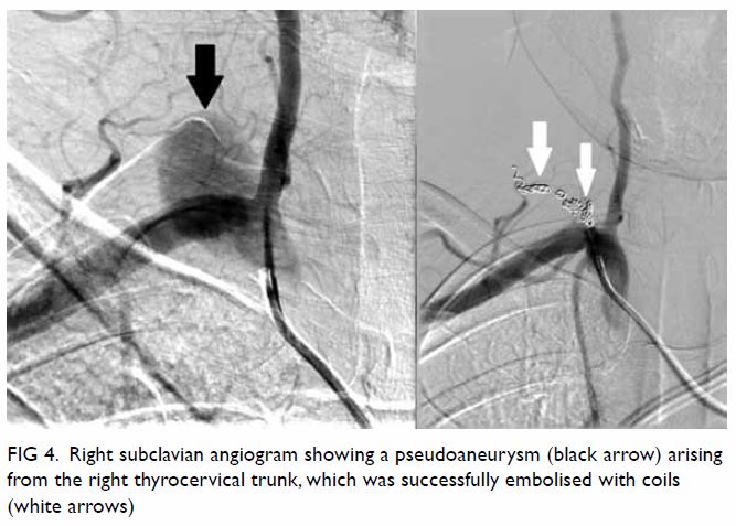
Figure 4. Right subclavian angiogram showing a pseudoaneurysm (black arrow) arising from the right thyrocervical trunk, which was successfully embolised with coils (white arrows)
 
Postoperatively, the patient progressed well, with her peritonitis controlled by intravenous antibiotics. A new CVC for temporary haemodialysis was inserted via her right IJV by interventional radiologists under fluoroscopic and real-time ultrasound guidance.
 
This case concurs with a previous report that the incidence of mechanical complications after multiple attempts is higher than after one attempt.2 Real-time ultrasound guided venepuncture of the IJV has a higher first insertion attempt success rate, and decreased rate of arterial puncture as compared with the anatomic landmark approach; this technique is currently recommended by the Association of Anaesthetists of Great Britain and Ireland for all CVC insertions.3 Credentialing of ultrasound-guided CVC insertion should be advocated in Hong Kong, with adequate training provided by accredited trainers.
 
Author contributions
All authors have made substantial contributions to the concept of this pictorial medicine; acquisition and interpretation of data, drafting of the article, and critical revision for important intellectual content.
 
Funding/support
This research received no specific grant from any funding agency in the public, commercial, or not-for-profit sectors.
 
Declaration
All authors have disclosed no conflicts of interest. All authors had full access to the data, contributed to the study, approved the final version for publication, and take responsibility for its accuracy and integrity.
 
References
1. McGee DC, Gould MK. Preventing complications of central venous catheterization. N Engl J Med 2003;348:1123-33. Crossref
2. Schummer W, Schummer C, Rose N, Niesen WD, Sakka SG. Mechanical complications and malpositions of central venous cannulations by experienced operators. A prospective study of 1794 catheterizations in critically ill patients. Intensive Care Med 2007;33:1055-9. Crossref
3. Bodenham A, Babu S, Bennett J, et al. Association of Anaesthetists of Great Britain and Ireland: Safe Vascular Access 2016. Anaesthesia 2016;71:573-85. Crossref

Emphysematous epididymo-orchitis: an uncommon but life-threatening cause of scrotal pain

DOI: 10.12809/hkmj176876
© Hong Kong Academy of Medicine. CC BY-NC-ND 4.0
 
PICTORIAL MEDICINE
Emphysematous epididymo-orchitis: an uncommon but life-threatening cause of scrotal pain
HW Lau, MB, ChB, FRCR1; CH Yu, MB, ChB1; SM Yu, FRCR, FHKCR2; LF Lee, MB, ChB3
1 Department of Radiology, Tuen Mun Hospital, Tuen Mun, Hong Kong
2 Department of Radiology, United Christian Hospital, Kwun Tong, Hong Kong
3 Division of Urology, Department of Surgery, United Christian Hospital, Kwun Tong, Hong Kong
 
Corresponding author: Dr HW Lau (jackylauhw@gmail.com)
 
 Full paper in PDF
 
An 80-year-old man presented to the emergency department in August 2016 with a 2-day history of painful scrotal swelling and fever. There was no history of trauma. He had a history of hypertension, diabetes mellitus and bullous pemphigoid, and was taking long-term steroid therapy. Upon presentation, the patient had a fever of 38.8°C and fast atrial fibrillation with a heart rate of 170 beats per minute. Blood pressure was 149/104 mm Hg. Physical examination of the patient revealed scrotal skin erythema and a firm left scrotal swelling that was tender on palpation. No cough impulse could be elicited. His white cell count was 21.8 × 10⁹ /L and blood glucose level was 21.6 mmol/L. Ultrasound of the left hemiscrotum showed a fluid collection with hyperechoic interfaces and ring-down artefact suggestive of the presence of gas, obscuring the underlying structures (Fig 1). Subsequent non-contrast computed tomographic scan of the pelvis confirmed that the mottled gas and fluid density were confined to the left hemiscrotum (Fig 2a). The left testis and epididymis were poorly delineated. There was no gas density in the subcutaneous layer of the scrotum or perineum or in the intraperitoneal cavity. There was also no evidence of indirect inguinal hernia (Fig 2b). A diagnosis of emphysematous epididymo-orchitis was made. Emergency surgery was arranged a few hours after imaging. Intra-operatively, gas and pus were seen within the left tunica vaginalis. The left epididymis was necrotic and almost destroyed but the left testis was relatively spared with friable tissue and patches of necrosis (Fig 3). Drainage of pus and left orchidectomy were performed. Pus culture revealed Escherichia coli. The patient subsequently developed septic shock and died 3 days after the operation.
 

Figure 1. Ultrasound image of the left hemiscrotum showing a fluid collection with hyperechoic interfaces (white arrows) and ring-down artefact (black arrow) suggestive of the presence of gas. Underlying structures including the testis and epididymis were obscured
 

Figure 2. Axial computed tomography of the pelvis showing (a) mottled gas densities confined to the left hemiscrotum (white arrow) with no evidence of gas in the subcutaneous layer of the scrotum or perineum, virtually excluding the possibility of Fournier’s gangrene; and (b) absence of bowel loop in the left inguinal canal (white arrow) excluded the possibility of incarcerated indirect inguinal hernia
 

Figure 3. Intra-operative photo showing a grossly inflamed and slightly necrotic epididymis (white arrow). The testis (black arrow) was relatively spared with friable tissue and patches of necrosis
 
Emphysematous cholecystitis, pyelonephritis, and cystitis are not uncommonly seen in patients with poorly controlled diabetes mellitus. It is nonetheless rare to see gas-forming infection of the epididymis and testis although this is also reported to be associated with diabetes mellitus.1 2 3 Long-term use of steroid in our patient may have been an additional risk factor due to immunosuppression. Based on the clinical presentation and ultrasound findings of our patient, the main differential diagnoses included Fournier’s gangrene and incarcerated indirect inguinal hernia. Computed tomographic scan was very helpful in delineating the definitive diagnosis. Gas pockets confined to the scrotal sac without involvement of the subcutaneous layer of the perineum virtually excluded the possibility of Fournier’s gangrene. Incarcerated indirect inguinal hernia was excluded due to the absence of bowel loops in the scrotal sac.
 
When a diabetic patient presents with acute scrotal pain and features of infection, careful examination during the initial ultrasound scan for the presence of gas within the scrotum is essential as this will alter the subsequent management plan as it implies a much poorer prognosis than non–gas-forming epididymo-orchitis.
 
In conclusion, emphysematous epididymo-orchitis is an uncommon but life-threatening disease. Ultrasound and computed tomographic scan are essential to identify this entity for early treatment.
 
Author contributions
All authors have made substantial contributions to the concept or design of this study; acquisition of data; analysis or interpretation of data; drafting of the article; and critical revision for important intellectual content.
 
Funding/support
This research received no specific grant from any funding agency in the public, commercial, or not-for-profit sectors.
 
Declaration
All authors have disclosed no conflicts of interest. All authors had full access to the data, contributed to the study, approved the final version for publication, and take responsibility for its accuracy and integrity.
 
References
1. Mathur A, Manish A, Maletha M, Luthra NB. Emphysematous epididymo-orchitis: a rare entity. Indian J Urol 2011;27:399-400. Crossref
2. Mandava A, Rao RP, Kumar DA, Naga Prasad IS. Imaging in emphysematous epididymo-orchitis: a rare cause of acute scrotum. Indian J Radiol Imaging 2014;24:306-9. Crossref
3. Hegde RG, Balani A, Merchant SA, Joshi AR. Synchronous infection of the aorta and the testis: emphysematous epididymo-orchitis, abdominal aortic mycotic aneurysm, and testicular artery pseudoaneurysm diagnosed by use of MDCT. Jpn J Radiol 2014;32:425-30. Crossref

Pancreatic pseudocyst rupture into the portal vein diagnosed by magnetic resonance imaging

DOI: 10.12809/hkmj164980
© Hong Kong Academy of Medicine. CC BY-NC-ND 4.0
 
PICTORIAL MEDICINE
Pancreatic pseudocyst rupture into the portal vein diagnosed by magnetic resonance imaging
HC Lee, FRCR, FHKCR1; KH Tse, FRCR, FHKCR2
1 Department of Radiology, United Christian Hospital, Kwun Tong, Hong Kong
2 Department of Radiology, Princess Margaret Hospital, Laichikok, Hong Kong
 
Corresponding author: Dr HC Lee (lhc874@ha.org.hk)
 
 Full paper in PDF
 
A 38-year-old man presented to the United Christian Hospital, Hong Kong, with acute epigastric pain in October 2014. He was a chronic drinker and had experienced intermittent abdominal pain for 6 months. His serum amylase level was elevated (454 IU/L), and a diagnosis of acute-on-chronic pancreatitis was made. The patient was treated conservatively.
 
Magnetic resonance cholangiopancreatography performed 3 months after hospital discharge showed a 5.8-cm-diameter unilocular cystic mass over the pancreatic head (Fig 1). The main and right portal veins showed a signal intensity identical to that of the cystic pancreatic lesion on all phases, without any contrast enhancement (Fig 2). There was communication between the main portal vein and the cystic mass (Fig 3). The presence of multiple collateral veins in the hepatic hilum was consistent with cavernous transformation (Fig 2c). Features were suggestive of a pancreatic head pseudocyst that had ruptured into the main portal vein.
 

Figure 1. Coronal T2-weighted magnetic resonance image showing a cystic lesion at the pancreatic head (arrow)
 

Figure 2. Axial magnetic resonance images: (a) T1-weighted pre-contrast image and (b) T1-weighted post-contrast arterial phase image showing low signal in the main and right portal veins without contrast enhancement (arrows); (c) T1-weighted post-contrast portal venous phase image showing low signal in the main and right portal veins without contrast enhancement (arrow), and with collateral veins at the hepatic hilum (arrowhead) suggesting cavernous transformation; and (d) T2-weighted image showing high signal intensity within the right and main portal veins (arrow)
 

Figure 3. Two-dimensional slab magnetic resonance cholangiopancreatogram showing a pancreatic head cystic lesion with communication (arrow) with the main portal vein, both as high signal intensity (a normal portal vein with flowing blood should be of low signal intensity); the non-dilated biliary tree (arrowheads) and the gallbladder are also visible
 
The patient presented again 1 month later with recurrent pancreatitis. Contrast computed tomography (CT) showed that the pancreatic pseudocyst had enlarged, to 7.6 cm in diameter (Fig 4). Pancreatic cystojejunostomy and cholecystectomy were performed. Intra-operatively, a 10-cm cystic lesion at the pancreatic head was found, and 200 mL of clear fluid was aspirated. Intra-operative ultrasonography showed the lack of flow in the main portal vein.
 

Figure 4. Reformatted coronal computed tomographic image in portal venous phase showing fluid attenuation in the main and right portal veins (PV), absence of contrast enhancement, and communication between the portal veins and the pancreatic cystic lesion (arrow)
 
The patient had a few more episodes of recurrent pancreatitis thereafter. The last CT examination, performed 2 years after surgery, showed a reduction in the size of the pseudocyst, to 2 cm. The patient remains on regular follow-up.
 
Discussion
Rupture of a pancreatic pseudocyst into the portal vein is an uncommon complication; only a handful of cases have been reported in the literature.1 It has been postulated that portal vein thrombosis occurs first, followed by erosion of the portal vein by pancreatic enzymes present in the pseudocyst, and subsequent lysis of the thrombus and filling of the portal vein with fluid.1 2 It has also been reported that rupture of the pseudocyst into the portal vein may be the initial event, followed by the development of portal vein thrombosis.3 4
 
Previously reported cases have used various diagnostic techniques. Invasive methods include endoscopic retrograde cholangiopancreatography and portography with surgery. Non-invasive methods include ultrasonography, CT, and magnetic resonance imaging (MRI). In all reported cases in which MRI was performed, the signal intensity of fluid in the portal vein matched that of the pancreatic pseudocyst.1 2 3 Direct communication between the portal vein and the pancreatic pseudocyst was clearly seen in most cases. The presence of residual thrombus or concomitant existence of complete thrombosis of the portal vein has also been reported.1
 
There is no well-established treatment protocol. Options include conservative management, endoscopic or percutaneous procedures, or surgery. The patient’s clinical condition and symptoms, patency of the portal vein, communication between the pseudocyst and pancreatic duct, size of pseudocyst, and any other complicating factors should be considered in treatment planning.3
 
In summary, rupture of a pancreatic pseudocyst into the portal vein is an uncommon complication. On MRI, demonstration of fluid signal in the portal vein that matches the signal intensity of a pancreatic pseudocyst allows the diagnosis to be confidently made, obviating the need for more invasive investigations.
 
Declaration
The authors have no conflicts of interest to disclose.
 
References
1. Dayal M, Sharma R, Madhusudhan KS, et al. MRI diagnosis of rupture of pancreatic pseudocyst into portal vein: case report and review of literature. Ann Gastroenterol 2014;27:173-6.
2. Riddell A, Jhaveri K, Haider M. Pseudocyst rupture into the portal vein diagnosed with MRI. Br J Radiol 2005;78:265-8. Crossref
3. Ng TS, Rochefort H, Czaplicki C, et al. Massive pancreatic pseudocyst with portal vein fistula: case report and proposed treatment algorithm. Pancreatology 2015;15:88-93. Crossref
4. Dawson BC, Kasa D, Mazer MA. Pancreatic pseudocyst rupture into the portal Vein. South Med J 2009;102:728-32. Crossref

Characteristic imaging features of clonorchiasis

DOI: 10.12809/hkmj166198
© Hong Kong Academy of Medicine. CC BY-NC-ND 4.0
 
PICTORIAL MEDICINE
Characteristic imaging features of clonorchiasis
WK Lo, MB, BS, FRCR1; SM Yu, FRCR, FHKAM (Radiology)2; James CS Chan, FRCR, FHKAM (Radiology)2
1 Department of Diagnostic and Interventional Radiology, Kwong Wah Hospital, Yaumatei, Hong Kong
2 Department of Radiology and Organ Imaging, United Christian Hospital, Kwun Tong, Hong Kong
 
Corresponding author: Dr WK Lo (waikglo@gmail.com)
 
 Full paper in PDF
 
A 43-year-old Chinese man presented to United Christian Hospital, Hong Kong, in January 2016, with a 2-year history of vague right upper quadrant pain. Blood test results were unremarkable except for an episode of transient eosinophilia. Ultrasonography showed mildly dilated intrahepatic ducts. Magnetic resonance cholangiopancreatography showed diffuse and uniform mild dilatation of peripheral intrahepatic bile ducts (Fig 1) consistent with the ‘too many intrahepatic ducts’ sign (Fig 2). Notably, there was no central extrahepatic bile duct dilatation. Subsequently requested stool microscopy was positive for Clonorchis sinensis ova. The patient was given a course of praziquantel.


Figure 1. Axial T2-weighted magnetic resonance image showing dilatation of the peripheral intrahepatic ducts (arrows)


Figure 2. Reformatted magnetic resonance cholangiographic images showing the ‘too many intrahepatic ducts’ sign in a patient with clonorchiasis (a), compared with a healthy individual (b)
 
Clonorchiasis is a foodborne zoonosis caused by consumption of raw or undercooked freshwater fish infested by the liver fluke C sinensis. C sinensis larvae penetrate the scales of freshwater fish and encyst in subcutaneous tissues. Cysts, when consumed by a definitive host, hatch in the intestine and migrate to the biliary tree, where the adult flukes establish residence. Clonorchiasis is endemic in many parts of Asia, including China, Korea, and Vietnam.1 Clonorchiasis and its complications are not commonly seen in affluent populations, such as those in Hong Kong. However, the increasing mobility of people around the region means that clinicians should be vigilant of the condition and alert to its symptoms. Knowledge of the characteristic imaging features of clonorchiasis would prompt stool microscopy and allow early diagnosis.
 
Within the biliary tree, adult C sinensis flukes reside in small- or medium-sized peripheral intrahepatic ducts. The organisms cause dilatation of the intrahepatic ducts. In heavy infestations, the extrahepatic ducts, the gallbladder, or even the pancreatic duct can also be involved. The dilated peripheral ducts can be visualised by direct cholangiography, or to a better extent by magnetic resonance cholangiopancreatography (owing to the independence of rate and volume of the contrast injection), as the ‘too many intrahepatic ducts’ sign.2 Occasionally, linear or elliptical filling defects representing the flukes can be seen.3
 
Light infestations with C sinensis can be asymptomatic. Heavier infestations can result in fever, jaundice, right upper quadrant pain, and eosinophilia. Untreated, chronic clonorchiasis induces chronic inflammation of the bile ducts. Recurrent pyogenic cholangitis, cholelithiasis, pancreatitis, and cholangiocarcinoma are the main complications.4
 
Infestations are diagnosed by observation of C sinensis ova during stool microscopy. Immunological and molecular techniques of diagnosis are still at the experimental stage.1 Praziquantel is the only drug treatment for clonorchiasis that is recommended by the World Health Organization.1
 
In summary, clinicians in Asia should be vigilant of clonorchiasis. In modern clinical practice, magnetic resonance cholangiopancreatography is often requested for non-invasive evaluation of the biliary tree. Knowledge and recognition of the characteristic imaging features of clonorchiasis would prompt timely investigation by stool microscopy. Early diagnosis and treatment of clonorchiasis can prevent complications such as recurrent pyogenic cholangitis and cholangiocarcinoma.
 
Declaration
The authors have no conflicts of interest to disclose.
 
References
1. World Health Organization. Foodborne trematode infections—clonorchiasis. Available from: http://www.who.int/foodborne_trematode_infections/clonorchiasis/en/. Accessed on 27 Nov 2016.
2. Choi D, Hong ST. Imaging diagnosis of clonorchiasis. Korean J Parasitol 2007;45:77. Crossref
3. Lim JH. Radiologic findings of clonorchiasis. AJR Am J Roentgenol 1990;155:1001-8. Crossref
4. Choi BI, Han JK, Hong ST, Lee KH. Clonorchiasis and cholangiocarcinoma: etiologic relationship and imaging diagnosis. Clin Microbiol Rev 2004;17:540-52. Crossref

Cutaneous manifestation mimicking Stevens-Johnson syndrome in a critically ill patient: looks similar but totally different

DOI: 10.12809/hkmj165051
© Hong Kong Academy of Medicine. CC BY-NC-ND 4.0
 
PICTORIAL MEDICINE
Cutaneous manifestation mimicking Stevens-Johnson syndrome in a critically ill patient: looks similar but totally different
Jo A Lim, MD, MRCP, D Derm1; SE Chong, MD, MMed2,3; Huda Zainal Abidin, MD, MMed3; Mohd H Hassan, MBBS, MMed3
1 Department of Internal Medicine, Hospital Sultan Abdul Halim, 08000 Sungai Petani, Kedah, Malaysia
2 Regenerative Medicine Cluster, Advanced Medical and Dental Institute, Universiti Sains Malaysia, Bertam, 13200 Kepala Batas, Penang, Malaysia
3 Department of Anesthesiology and Intensive Care, School of Medical Sciences, Universiti Sains Malaysia, 16150 Kota Bharu, Kelantan, Malaysia
 
Corresponding author: Dr SE Chong (sechong@usm.my)
 
 Full paper in PDF
 
Case
A 60-year-old gentleman was intubated due to severe leptospirosis and multi-organ failure: acute kidney injury and liver failure with disseminated intravascular coagulation (DIC). He was ventilated in the intensive care unit for 7 days. He was treated with ceftriaxone, pantoprazole, fentanyl, midazolam, and required frequent fluid challenge and inotropic support.
 
On the day of extubation, the patient was noted to have ulcers over the angles of the mouth with crusted blood and seropurulent discharge (Fig 1). He also had diffuse erythema and desquamation over the tips of the fingers (Fig 2), and a large purpuric patch over the lateral aspect of both thighs (Fig 3) with generalised scaly dry skin over the body.
 

Figure 1. Localised oral and lip ulcers confined to previous endotracheal tube placement site, as opposed to the diffuse oral and lip ulcers seen in Stevens-Johnson syndrome
 

Figure 2. Erythematous fingers with skin desquamation over the fingertips
 

Figure 3. Purpuric patch with scaly skin rash over lateral thigh
 
He was treated as Stevens-Johnson syndrome (SJS). Antibiotic therapy was stopped and intravenous hydrocortisone was started but his ulcers continued to worsen. A dermatological opinion was arranged and revealed that the oral and tongue mucosa erosions were confined to the site of previous endotracheal tube placement rather than being the diffuse oral and lips erosions of SJS. Nikolsky sign was negative. The conjunctiva was clear, and there was involvement of the nasal, urethral or anal mucosa.
 
In view of the confined area of mucosa involvement, he was diagnosed with medical device–related pressure ulcers. The purpura and ecchymosis were due to the underlying coagulopathy secondary to septic shock with DIC. Potential infective causes, eg vegetating herpes simplex, staphylococcal scalded skin syndrome, were excluded by negative wound culture. There were also no features of SJS on skin biopsy.
 
The steroid was stopped immediately and antibiotics resumed. Albumin level was optimised. After 2 weeks of oral care, the patient’s skin condition improved (Fig 4) and he finally attained full recovery.
 

Figure 4. Ulcers improved 4 days after stopping hydrocortisone and restarting antibiotics
 
Discussion
Tracheal intubation is one of the best methods of airway protection1 but serious complications may occur, especially in critically ill patients.2
 
Stevens-Johnson syndrome is part of a spectrum of severe cutaneous adverse reactions that affect skin and mucous membranes. It is commonly caused by certain medications or infections. Skin lesions may be purpuric macule spots, erythema, or sometimes violaceous target-like lesions. Mucous membrane erosions and ulcers almost always appear in the eyes, mouth and lips, but may also occur in the upper airway, genitourinary and gastrointestinal tract. Assessment is often difficult when mucous membrane involvement is not yet apparent.3
 
Hypotensive episodes during septic shock may lead to reduced perfusion pressure to the skin and mucosa. This may cause pressure point areas to develop pressure sores.4 Hypoalbuminaemia and DIC may cause further deterioration of the ulcers. Pressure sores are one of the most common complications among patients undergoing mechanical ventilation in a poorly managed setting.5
 
Physicians should always be extra cautious when diagnosing SJS. The consensus definition should always be consulted with referral to a dermatologist or performance of a skin biopsy if there is any doubt.3 An incorrect diagnosis of SJS may lead to a totally inappropriate spectrum of treatment such as cessation of appropriate life-saving antibiotics and the commencement of steroid therapy that will increase the risk of infection and impair wound healing.
 
References
1. International Liaison Committee on Resuscitation. 2005 International Consensus on Cardiopulmonary Resuscitation and Emergency Cardiovascular Care Science with Treatment Recommendations. Part 4: Advanced life support. Resuscitation 2005;67:213-47.
2. von Goedecke A, Herff H, Paal P, Dörges V, Wenzel V. Field airway management disasters. Anesth Analg 2007;104:481-3. Crossref
3. Mockenhaupt M. Severe drug-induced skin reactions: clinical pattern, diagnostics and therapy. J Dtsch Dermatol Ges 2009;7:142-60. Crossref
4. Black JM, Edsberg LE, Baharestani MM, et al. Pressure ulcers: avoidable or unavoidable? Results of the National Pressure Ulcer Advisory Panel Consensus Conference. Ostomy Wound Manage 2011;57:24-37.
5. Tang WM, Tong CK, Yu WC, Tong KL, Buckley TA. Outcome of adult critically ill patients mechanically ventilated on general medical wards. Hong Kong Med J 2012;18:284-90.

Is it an orbital foreign body?

DOI: 10.12809/hkmj164952
© Hong Kong Academy of Medicine. CC BY-NC-ND 4.0
 
PICTORIAL MEDICINE
Is it an orbital foreign body?
Mohamed Shaheeda, FRCSEd, MPH; Stacey C Lam, MB, ChB; Noel CY Chan, FRCSEd, FCOphthHK; Hunter KL Yuen, FRCSEd, FCOphthHK
Department of Ophthalmology and Visual Sciences, The Chinese University of Hong Kong, Shatin, Hong Kong
 
Corresponding author: Dr Stacey C Lam (staceylam@gmail.com)
 
 Full paper in PDF
 
A 58-year-old man presented with left globe rupture following blunt orbital trauma. Visual acuity was light perception only. There was lid swelling, ecchymosis, and a deformed globe with scleral laceration. Computed tomography (CT) of the orbit revealed a 3.7-mm hyperdensity at the superomedial aspect of the left globe, interpreted by the radiologist as a possible intra-orbital foreign body (Fig).
 

Figure. (a) Coronal computed tomographic scan in soft tissue window showing bilateral hyperdensities at the superomedial aspect of both orbits at the level of the trochlear apparatus (arrows). There is also a left collapsed globe with intra-ocular haemorrhage and gas (arrowheads). (b) Axial computed tomographic scan in soft tissue window showing hyperdensity at the anteromedial left orbit (arrow)
 
Thorough history for the mechanism of trauma, physical examination for a wound site and thin cut CT sections were reviewed. History revealed blunt orbital trauma by a metallic rod rather than a sharp penetrating injury. No cutaneous entry site was evident. Computed tomography revealed bilateral hyperdensities at the level of the trochlea, more pronounced on the left than the right, with no metallic streak artefacts. All of the above supported a diagnosis of trochlear calcification rather than an intra-orbital foreign body, and the patient subsequently underwent emergency repair of scleral laceration without orbital exploration.
 
Discussion
The trochlea is a cartilaginous pulley at the superomedial aspect of the orbit through which the superior oblique muscle tendon passes freely. It has a synovium-lined space, and like other synovial joints of the body, the trochlea can develop calcifications in the cartilage, tendon, or within the bursa-like cleft.1 The reported prevalence of incidental trochlear calcifications on CT is 3-16%, with over 50% being unilateral.2 3 No clear cause has been identified, but prior studies have postulated degenerative, inflammatory, metabolic or traumatic aetiologies.4 Since trochlear calcification is asymptomatic, most cases go unnoticed by radiologists as well as ophthalmologists. In the presence of co-existing orbital trauma, it can be misdiagnosed as an intraorbital foreign body. Surgical exploration around the trochlear region may cause damage and scarring leading to diplopia.
 
Differentiation of the two entities requires history taking, physical examination, and proper imaging. A foreign body is present in one in six cases of orbital trauma, with metallic objects and glass being the most common.5 A review of the history to determine mechanism of injury is useful but may be unreliable. Examination of the skin and conjunctiva, particularly the fornices, may help detect subtle penetrating injuries. Radiological assessment includes plain films, ultrasound, CT, and magnetic resonance imaging. Plain films are limited to detection of metallic foreign bodies, and ultrasound has limited use because foreign bodies can be masked by surrounding highly reflective structures such as bone. Computed tomography is an excellent means to identify high-density objects such as metal or glass, but not organic matter. Magnetic resonance imaging is contra-indicated in metallic foreign bodies, but may be useful for organic foreign matter. Features of trochlear calcification may include symmetrical presentation and typical site at the trochlear apparatus. Its morphology has been described as comma, dot, and inverted U shape.2 In contrast, orbital foreign bodies are often unilateral with no specific size, shape, or location. Metallic foreign bodies may also generate streak artefacts.
 
Orbital calcifications can be incidental or pathological. It is important to recognise trochlear calcifications as distinct from foreign bodies so as to avoid unnecessary surgical exploration in the presence of orbital trauma.
 
References
1. Sobel RK, Goldstein SM. Trochlear calcification: A common entity. Orbit 2012;31:94-6. Crossref
2. Xiao TL, Kalariya NM, Yan ZH, et al. Trochlear calcification and intraorbital foreign body in ocular trauma patients. Chin J Traumatol 2009;12:210-3.
3. Shriver EM, McKeown CA, Johnson TE. Trochlear calcification mimicking an orbital foreign body. Ophthal Plast Reconstr Surg 2011;27:143-4. Crossref
4. Buch K, Nadgir RN, Tannenbaum AD, Ozonoff A, Fujita A, Sakai O. Clinical significance of trochlear calcifications in the orbit. AJNR Am J Neuroradiol 2014;35:573-7. Crossref
5. Nasr AM, Haik BG, Fleming JC, Al-Hussain HM, Karcioglu ZA. Penetrating orbital injury with organic foreign bodies. Ophthalmology 1999;106:523-32. Crossref

Birt-Hogg-Dubé syndrome: a rare cause of familial spontaneous pneumothorax

DOI: 10.12809/hkmj165022
© Hong Kong Academy of Medicine. CC BY-NC-ND 4.0
 
PICTORIAL MEDICINE
Birt-Hogg-Dubé syndrome: a rare cause of familial spontaneous pneumothorax
HM Luk, FHKAM (Paediatrics); Tony MF Tong, MSc; Ivan FM Lo, FHKAM (Paediatrics)
Clinical Genetic Service, Department of Health, 3/F Cheung Sha Wan Jockey Club Clinic, 2 Kwong Lee Road, Shamshuipo, Hong Kong
 
Corresponding author: Dr HM Luk (luksite@gmail.com)
 
 Full paper in PDF
 
A family with a strong history of pneumothorax was referred to our genetic clinic for assessment. There were three siblings who had all developed spontaneous pneumothorax at the age of 30, 58, and 59 years. All were non-smokers with no pre-existing pulmonary disease. High-resolution computed tomography of the thorax for all showed multiple thin-walled pulmonary cysts of variable size on both sides, mainly located at the basal and peripheral lung regions (Fig). Lung biopsy was not informative. Physical examination revealed multiple smooth, dome-shaped papules over the face and ears in one of siblings (Fig b). There were no other features of tuberous sclerosis or history of renal disease in the family. Based on the dermatological findings and diffuse multicystic lung disease, Birt-Hogg-Dubé (BHD) syndrome was suspected. FLCN gene analysis revealed a heterozygous FLCN{NM_144997.5}:c.1285dupC mutation in all affected members. The diagnosis of BHD syndrome was substantiated. Renal imaging was arranged for surveillance of potential renal cell carcinoma.
 

Figure. (a) Chest X-ray showing bilateral multiple pulmonary cysts over the basal region (arrows). (b) Clinical photo of fibrofolliculomas over pinna (arrows). (c and d) High-resolution computed tomography of the thorax showing lower-zone predominant pulmonary cysts of variable shapes and sizes (circles). The walls are thin, sharply demarcated and do not enhance with contrast (arrowhead)
 
The BHD syndrome is a rare autosomal dominant disease characterised by three major organ manifestations1:
(1) Fibrofolliculomas and other benign skin tumours such as trichodiscomas and acrochordons; these skin lesions are predominantly located on the facial, cervical, and upper truncal regions as smooth, dome-shaped, and white to flesh-coloured papules.
(2) Increased susceptibility to renal cell carcinoma of mixed histologies; the most frequent subtype is a hybrid oncocytic tumour with features of renal oncocytoma and chromophobe renal cell carcinoma.
(3) Multiple bilateral pulmonary cysts and spontaneous pneumothorax.
 
The clinical features of BHD syndrome are heterogeneous with wide intra-familial and inter-familial variation. It is caused by mutations of the FLCN gene. Any combination of the cutaneous, renal, and pulmonary features mentioned above present in an individual or multiple family members should alert the clinician to the possibility of BHD syndrome.
 
Bilateral multiple pulmonary cysts are a highly penetrant feature in BHD syndrome. As a result, the risk of pneumothorax in BHD patients is 50 times higher than that of the general population.2 Approximately 80% to 90% of BHD patients develop lung cysts, usually after early mid-adulthood. The BHD-associated lung cysts tend to be located at the basilar and mediastinal regions of the lungs, in contrast to the typically apical location in primary spontaneous pneumothorax and emphysema. Radiologically, the BHD-associated lung cysts are usually irregularly shaped, variable in size and number, and with sharply demarcated thin walls that do not enhance on computed tomographic imaging.
 
Fibrofolliculomas are present in more than 80% of patients with BHD syndrome and typically appear after the age of 20 years. They are dome-shaped, white to flesh-coloured, non-painful and non-pruritic papules located on the facial, cervical, and upper truncal regions.
 
The most threatening complication of BHD syndrome is renal cell carcinoma. It occurs in approximately 15% of BHD patients by the age of 70 years.3 Therefore regular surveillance is mandatory.
 
Physicians should be alert to the possibility of BHD syndrome in a patient who presents with diffuse cystic lung disease, particularly in the presence of a positive family history. Early referral to a clinical genetic service and multidisciplinary management is recommended. Early diagnosis and regular renal surveillance aim to greatly reduce renal cell carcinoma–associated morbidity and mortality.
 
References
1. Menko FH, van Steensel MA, Giraud S, et al. Birt-Hogg-Dubé syndrome: diagnosis and management. Lancet Oncol 2009;10:1199-206. Crossref
2. Gupta N, Seyama K, McCormack FX. Pulmonary manifestations of Birt-Hogg-Dubé syndrome. Fam Cancer 2013;12:387-96. Crossref
3. Stamatakis L, Metwalli AR, Middelton LA, Marston Linehan W. Diagnosis and management of BHD-associated kidney cancer. Fam Cancer 2013;12:397-402. Crossref

A child with giant tumefactive perivascular spaces

DOI: 10.12809/hkmj164918
© Hong Kong Academy of Medicine. CC BY-NC-ND 4.0
 
PICTORIAL MEDICINE
A child with giant tumefactive perivascular spaces
Neeraj R Mahboobani, MB, BS, FRCR; LF Cheng, FRCR, FHKAM (Radiology); Johnny KF Ma, FRCR, FHKAM (Radiology)
Department of Radiology, Princess Margaret Hospital, Laichikok, Hong Kong
 
Corresponding author: Dr Neeraj R Mahboobani (neeraj.mahboobani@gmail.com)
 
 Full paper in PDF
 
A 4-year-old girl was admitted with a 2-day history of a high temperature up to 41°C. She developed sudden-onset generalised tonic-clonic seizure. She had no travel history. Physical examination showed neck stiffness. The working diagnosis was meningitis. Non-contrast computed tomographic (CT) brain prior to lumbar puncture revealed clustered well-defined cystic lesions with no calcifications measuring up to 1.5 cm in the white matter of the right frontal and parietal lobes (Fig 1).
 

Figure 1. Non-contrast computed tomography of the brain on the day of admission showing clustered well-defined cystic lesions with no calcifications measuring up to 1.5 cm in the white matter of the right frontal and parietal lobes (arrows)
 
The child subsequently developed status epilepticus. The neurosurgical unit was consulted and brain abscesses were suspected. In view of her clinical deterioration, burr hole and aspiration of the cystic lesions were planned after magnetic resonance imaging (MRI). The signal intensity of the lesions was identical to that of cerebrospinal fluid (CSF) on all MRI sequences (Figs 2 and 3). There was no rim enhancement around the lesions. Minimal fluid-attenuated inversion recovery (FLAIR) hyperintense signals were noted in the white matter adjacent to the lesions. There was no restricted diffusion on diffusion-weighted imaging.
 

Figure 2. Magnetic resonance imaging (MRI) on day 2 following admission at the level of the right frontal lobe lesions
(a) T1-weighted, (b) T2-weighted, (c) fluid-attenuated inversion recovery (FLAIR) imaging showing the lesions to be identical to cerebrospinal fluid signal on all MRI sequences. Minimal FLAIR hyperintense signals seen around the cysts are suggestive of gliosis (arrows). (d) No enhancement on post-gadolinium scan is shown (arrow). (e) Diffusion-weighted imaging and (f) apparent diffusion coefficient map showing no restricted diffusion (arrows)


Figure 3. Magnetic resonance imaging (MRI) on day 2 following admission at the level of the right parietal lobe lesions
(a) T1-weighted, (b) T2-weighted, (c) fluid-attenuated inversion recovery (FLAIR) imaging showing the lesions to be identical to the cerebrospinal fluid signal on all MRI sequences. Minimal FLAIR hyperintense signals seen around the cysts are suggestive of gliosis (arrows). (d) No enhancement on post-gadolinium scan is shown (arrows). (e) Diffusion-weighted imaging and (f) apparent diffusion coefficient map showing no restricted diffusion (arrows)
 
Imaging features were not compatible with brain abscess so surgical aspiration was withheld. Cerebral hydatid disease was considered a possible diagnosis based on imaging and the child was commenced on oral albendazole 15 mg/kg/day. This diagnosis was subsequently disproved given the lack of an exposure history, absence of Echinococcus granulosus antibody in serum, and absence of cysts in the liver and spleen on ultrasonography.
 
Influenza A virus antigen was subsequently detected in nasopharyngeal aspirate. The child was prescribed antiviral treatment with consequent cessation of seizures and clinical improvement. The clinical diagnosis was influenza virus–associated encephalopathy.
 
Follow-up CT and MRI 3 weeks later showed the non-enhancing cystic lesions to be unchanged in size and signal characteristics. The lesions were classified as giant tumefactive perivascular spaces (GTPVS).
 
The perivascular spaces (PVS) of the brain are pial-lined, interstitial fluid-filled cystic spaces. They accompany penetrating arteries as they enter the brain parenchyma. These are normal structures that can be found in all ages.1 Most PVS are small in size and usually measure less than 5 mm.1 Larger (>1 cm) PVS have been reported and these are called GTPVS, which can occur as single or multiple clustered cysts.2 Such GTPVS in cerebral white matter can have mild FLAIR hyperintensities in the surrounding white matter1 that can be due to gliosis.2
 
Of note, GTPVS are often incidental findings on imaging. They occur in characteristic locations alongside penetrating vessels in the mesencephalothalamic area, cerebral white matter, and cerebellar dentate nuclei.2 They follow the CSF signal on MRI and do not enhance.
 
Sometimes, GTPVS can be misinterpreted as other pathological processes.1 2 3 Lack of a solid component and complete suppression on FLAIR differentiates them from cystic neoplasms.1 Nil to minimal peri-lesional FLAIR signals help to differentiate them from cystic infarction and mucopolysaccharidosis. Parasitic cysts, in particular hydatid cysts, can have identical imaging features and necessitate clinical correlation for exclusion as in this case. Neuroepithelial cysts also have identical imaging features but can only be differentiated from GTPVS by histopathology.1 Knowledge of this entity, its imaging features, and characteristic locations is essential to avoid unnecessary medical treatment and surgical biopsy/intervention.
 
References
1. Kwee RM, Kwee TC. Virchow-Robin spaces at MR imaging. Radiographics 2007;21:1071-86. Crossref
2. Salzman KL, Osborn AG, House P, et al. Giant tumefactive perivascular spaces. AJNR Am J Neuroradiol 2005;26:298-305.
3. Sankararaman S, Velayuthan S, Ambekar S, Gonzalez-Toledo E. Giant tumefactive perivascular spaces: A further case. J Pediatr Neurosci 2013;8:108-10. Crossref

Urothelial carcinoma with paraneoplastic leukocytosis

DOI: 10.12809/hkmj164956
© Hong Kong Academy of Medicine. CC BY-NC-ND 4.0
 
PICTORIAL MEDICINE
Urothelial carcinoma with paraneoplastic leukocytosis
CW Hsu, MD, PhD; HY Su, MD
Department of Emergency Medicine, E-Da Hospital and I-Shou University, Kaohsiung, Taiwan
 
Previously presented at the 8th Asian Conference on Emergency Medicine, Taiwan, 7-10 November 2015.
 
Corresponding author: Dr HY Su (hys927@hotmail.com)
 
 Full paper in PDF
 
An 88-year-old male with no underlying disease presented to our emergency department in March 2015 with a 2-week history of right flank pain but no fever or urinary tract symptoms. He was conscious, alert, and well-oriented. His blood pressure was 145/89 mm Hg, with a heart rate of 82 beats/min and respiratory rate of 16 breaths/min on arrival. Physical examination revealed obvious right flank knocking tenderness. Laboratory testing revealed white blood cell (WBC) count of 34.7 x 10⁹ /L (reference range [RR], 4.5-11.0 x 10⁹ /L), haemoglobin level of 95 g/L (RR, 135-175 g/L), platelet count of 496 x 109 /L (RR, 150-450 x 109 /L), C-reactive protein level of 24.3 mg/L (RR, 0-10 mg/L), and serum calcium level of 3.31 mmol/L (RR, 2.18-2.58 mmol/L). Urinalysis demonstrated no pyuria, but 50 to 75 red blood cells per high-power field. Prothrombin time, activated partial thromboplastin time, and fibrinogen were within normal limits. An abdominal plain film revealed a mass lesion with a concealing right lower psoas shadow (Fig 1). Subsequent abdominal computed tomographic (CT) scan demonstrated a cyst-like lesion with irregular surface, involving adjacent tissues in the retroperitoneal cavity (Fig 2).
 

Figure 1. A plain abdominal film revealing a mass lesion (arrows) with obliteration of right lower psoas shadow (arrowheads)
 

Figure 2. Abdominal computed tomographic scans demonstrating a cyst-like lesion with irregular surface (asterisk) that involves the adjacent psoas muscle, vertebral body (arrowhead), ureter, and great vessels (arrow) in the retroperitoneal cavity
 
A CT-guided biopsy was performed because the discrepancy between clinical presentation and laboratory tests and image study made it difficult for physicians to discriminate psoas muscle abscess from malignancy. Ultimate pathology of biopsy specimens revealed a high-grade urothelial carcinoma with tumour necrosis. The TNM staging was stage IV (cT4N0M0). Blood and urine cultures were all negative. Follow-up 1 month later revealed a WBC count of 34.7 x 10⁹ /L.
 
Urothelial carcinoma originates in the urinary system. Paraneoplastic leukocytosis, defined by a WBC count of >20.0 x 10⁹ /L on more than two occasions 30 days apart, occurs in 0.6% of urothelial carcinoma cases.1 The aetiology of paraneoplastic leukocytosis is considered to be related to the production of granulocyte colony-stimulating factor by tumour cells.2 3 Hypercalcaemia, anaemia and thrombocytosis, as seen in this case, are also frequently seen in paraneoplastic syndrome. It is associated with advanced urothelial cancer and indicates a poor prognosis.1 3 Muscle invasion is also frequently found in cases of urothelial cancer with paraneoplastic leukocytosis.1 The cyst-like pattern of urothelial cancer on abdominal CT scan in combination with the paraneoplastic leukocytosis can mislead physicians into making an incorrect diagnosis, such as pyogenic psoas muscle abscess. We advise physicians to always be aware of urothelial cancer with paraneoplastic leukocytosis while managing a cyst-like lesion in the retroperitoneal cavity.
 
References
1. Izard JP, Gore JL, Mostaghel EA, Wright JL, Yu EY. Persistent, unexplained leukocytosis is a paraneoplastic syndrome associated with a poor prognosis in patients with urothelial carcinoma. Clin Genitourin Cancer 2015;13:e253-8. Crossref
2. Ito N, Matsuda T, Kakehi Y, Takeuchi E, Takahashi T, Yoshida O. Bladder cancer producing granulocyte colony-stimulating factor. N Engl J Med 1990;323:1709-10. Crossref
3. Kumar AK, Satyan MT, Holzbeierlein J, Mirza M, Van Veldhuizen P. Leukemoid reaction and autocrine growth of bladder cancer induced by paraneoplastic production of granulocyte colony-stimulating factor—a potential neoplastic marker: a case report and review of the literature. J Med Case Rep 2014;8:147. Crossref

Extraluminal location of a Foley catheter balloon

DOI: 10.12809/hkmj164938
© Hong Kong Academy of Medicine. CC BY-NC-ND 4.0
 
PICTORIAL MEDICINE
Extraluminal location of a Foley catheter balloon
CL Cho, FRCS Ed (Urol), FHKAM (Surgery); Wayne KW Chan, FRCS Ed (Urol), FHKAM (Surgery); Ringo WH Chu, FRCS Ed (Urol), FHKAM (Surgery); IC Law, FRCS Ed (Urol), FHKAM (Surgery)
Division of Urology, Department of Surgery, Kwong Wah Hospital, Yaumatei, Hong Kong
 
Corresponding author: Dr CL Cho (chochaklam@yahoo.com.hk, ccl296@ha.org.hk)
 
 Full paper in PDF
 
Indwelling urinary catheters are generally safe but may be associated with complications. Although intraperitoneal or extraperitoneal perforation is rare, the condition can be life-threatening.1 Abdominopelvic computed tomography (CT) is commonly performed in hospitalised patients. In many cases the urinary bladder is catheterised and included in the scan; CT scan is a reliable method to evaluate many pathologies of the urinary bladder.2 The apparent extraluminal position of a Foley catheter tip or balloon can be misleading, however.3 We present a case in which a Foley catheter balloon was inflated in a bladder diverticulum mimicking an extraluminal location on a CT scan.
 
Case
A 68-year-old man was admitted with abdominal distension and suprapubic pain in April 2016. He was a visitor to Hong Kong and had a history of recurrent acute urinary retention. A urethral Foley catheter had been inserted in his home country a week before presentation and he travelled to Hong Kong with the catheter in situ. A urological assessment was scheduled on his return home. He had a history of open left nephrectomy performed over 20 years ago for urinary stone disease but otherwise had good medical history.
 
On presentation he was afebrile and normotensive. The abdomen was grossly distended with suprapubic tenderness and a general surgeon was consulted. Digital rectal examination revealed a grossly enlarged non-suspicious prostate. Clear urine and good urine outputs from the Foley catheter were noted. Laboratory tests showed an elevated white blood cell count of 14.5 x 10⁹ /L and normal creatinine level of 125 µmol/L. A nasogastric tube was inserted with a working diagnosis of intestinal obstruction. An urgent contrast CT scan revealed grossly dilated small bowel loops with free fluid in the pelvis and paracolic gutters. The wall of the urinary bladder was thickened with a large prostate and intravesical prostatic protrusion (Fig 1). The tip of the Foley catheter appeared to be at an extravesical location (Fig 2).
 

Figure 1. Coronal computed tomographic scan demonstrating the thickened urinary bladder wall (arrow) and large intravesical prostatic protrusion (arrowhead)
 

Figure 2. Axial computed tomographic scan of the pelvis demonstrating (a) the Foley catheter through the thickened urinary bladder wall (arrow) and extraluminal location of balloon (arrowhead); and (b) the extraluminal location of balloon and Foley catheter tip (arrowhead)
 
A urological opinion was sought and the patient underwent exploratory laparotomy. Intra-operatively, the tip of the Foley catheter and balloon were noted inside a large 5-cm bladder diverticulum at the dome of the urinary bladder. There was a 5-mm concealed perforation at the bladder diverticulum with surrounding dusky tissue covered by slough. Bladder diverticulectomy was performed. The urinary bladder was closed in a two-layer fashion and confirmed water-tight. No bowel injury was evident and an extensive washout was performed. A pelvic drain and 18F Foley catheter were inserted.
 
Postoperatively, the patient progressed well. Diet was resumed on postoperative day 3 and the drain was removed. There were no wound complications and the patient was fit for discharge on day 5. He returned to his home country with the Foley catheter in situ. We advised maintenance of bladder drainage until surgery for benign prostate hyperplasia could be performed.
 
This case concurs with a previous report that extraluminal location of a Foley catheter balloon on imaging can be misleading.3 Exploratory laparotomy based on the radiological findings alone may not be appropriate, especially when the clinical suspicion of bladder perforation is low. Further studies including cystogram should be considered in case of doubt.
 
References
1. White SA, Thompson MM, Boyle JR, Bell PR. Extraperitoneal bladder perforation caused by an indwelling urinary catheter. Br J Surg 1994;81:1212. Crossref
2. Caoili EM, Cohan RH, Korobkin M, et al. Urinary tract abnormalities: initial experience with multi-detector row CT urography. Radiology 2002;222:353-60. Crossref
3. Abadi S, Brook OR, Solomonov E, Fischer D. Misleading positioning of a Foley catheter balloon. Br J Radiol 2006;79:175-6. Crossref

Pages