Use of colposcopy in a patient with recurrent genital ulcers

DOI: 10.12809/hkmj154536
© Hong Kong Academy of Medicine. CC BY-NC-ND 4.0
 
PICTORIAL MEDICINE
Use of colposcopy in a patient with recurrent genital ulcers
Tomoko Matsuzono, MRCOG, FHKAM (Obstetrics and Gynaecology); WH Li, FHKCOG, FHKAM (Obstetrics and Gynaecology)
Department of Obstetrics and Gynaecology, Queen Elizabeth Hospital, Jordan, Hong Kong
Corresponding author: Dr Tomoko Matsuzono (tomoko821@gmail.com)
 
 Full paper in PDF
 
Case
The patient was a 40-year-old female with a 5-month history of recurrent painful labial ulcers in December 2013. Investigations by a private physician revealed a negative VDRL (Venereal Disease Research Laboratory) test and vulval swab culture grew commensals only. The ulcers, however, failed to heal following treatment with antibiotics.
 
Vulval biopsy was performed at her first visit and histopathology report revealed only a non-specific ulcer with negative stains for fungus, bacteria, acid-fast bacilli, and herpes simplex virus. Papanicolaou smear showed atypical squamous cells of unknown significance (ASCUS).
 
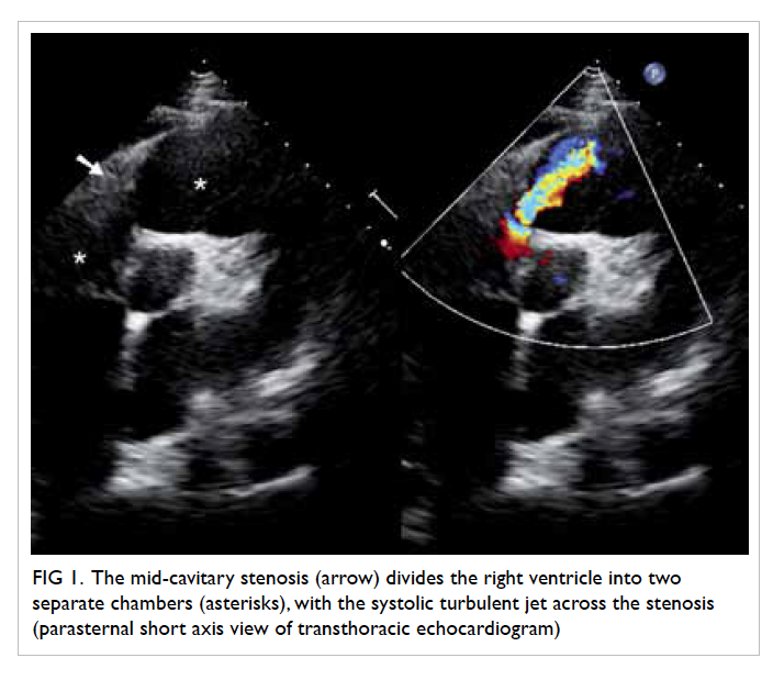
In the absence of any obvious pathology for the non-healing ulcer, colposcopy was arranged: four ulcers were identified within the vagina, scattered 
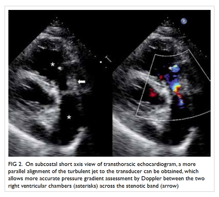
over the right and left vaginal walls (Fig 1). A left lower vulval ulcer measuring 3 cm with slightly raised edges was also seen (Fig 2). Biopsies were taken over the cervix, vaginal, and vulval ulcers. Histopathology of these biopsies confirmed the presence of cervicitis and ulcers, with no evidence of malignancy.
 

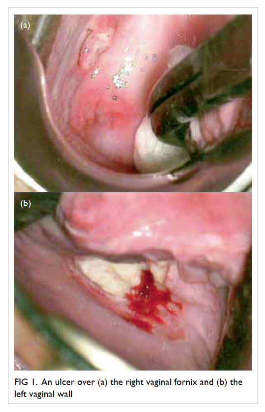
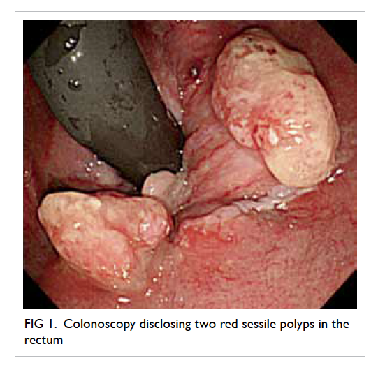
Figure 1. An ulcer over (a) the right vaginal fornix and (b) the left vaginal wall
 

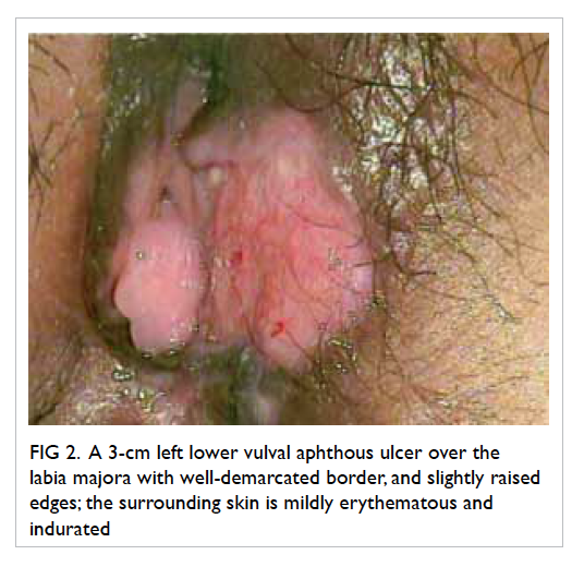
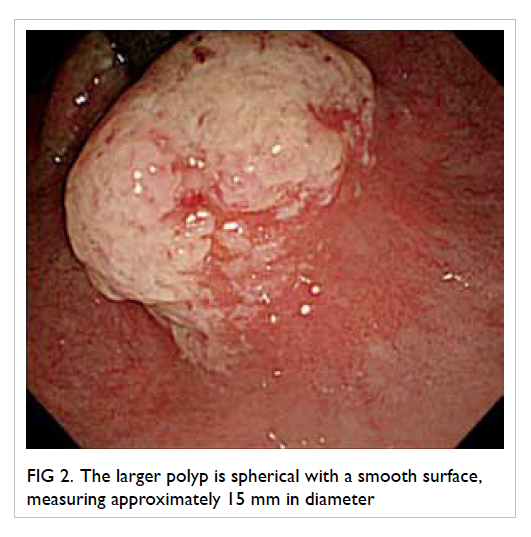
Figure 2. A 3-cm left lower vulval aphthous ulcer over the labia majora with well-demarcated border, and slightly raised edges; the surrounding skin is mildly erythematous and indurated
 
Upon systemic review, the patient revealed that she had been suffering from recurrent mouth ulcers and bruising over both shins. Subsequent examination revealed multiple aphthous mouth ulcers, as well as erythema nodosum over both shins.
 
Based on the clinical signs, a diagnosis of Behçet’s disease was made. The patient was commenced on prednisolone, and all ulcers had healed at subsequent follow-up.
 
Discussion
Behçet’s disease is a rare cause of genital ulceration. It is a multisystem vasculitic disorder and the diagnosis is based on clinical criteria.1 2 3 4 Our patient fulfilled the criteria of recurrent oral and genital ulcerations as well as a cutaneous manifestation.
 
Although there is little published evidence to support the value of colposcopy and biopsy in the diagnosis of Behçet’s disease, colposcopy enables a more detailed evaluation of the genital tract, and can be used to exclude the possibility of malignancy. Histopathological results in Behçet’s ulcer are typically non-specific with chronic active inflammation.
 
Papanicolaou smear in our patient showed ASCUS, but histopathology result for the cervix was normal. In a prospective study by Özdemir et al,2 abnormal cervical cytology, acetowhite epithelium, and iodine-negative epithelium on colposcopy were more common in Behçet’s patients. Nonetheless, the majority of ASCUS results revealed a normal finding for cervical histopathology. It was suggested that this was due to the benign inflammatory changes in the cervical epithelium.2
 
As in this case, diagnosis for recurrent genital ulcers can be difficult although when information about oral ulcers became available, a diagnosis of Behçet’s became more obvious. A complete history and physical examination that pays particular attention to signs or symptoms of an underlying associated systemic condition are essential when evaluating patients with recurrent genital ulcers.
 
References
1. Patel S, Prime K. Recurrent vulval ulceration: could it be Behçet’s disease? Int J STD AIDS 2012;23:683-4. Crossref
2. Özdemir S, Özdemir M, Celik C, Balevi A, Toy H, Kamış U. Evaluation of patients with Behçet’s disease by cervical cytology and colposcopic examination. Arch Gynecol Obstet 2012;285:1363-8. Crossref
3. Keogan MT. Clinical Immunology Review Series: an approach to the patient with recurrent orogenital ulceration, including Behçet’s syndrome. Clin Exp Immunol 2009;156:1-11. Crossref
4. Bandow GD. Diagnosis and management of vulvar ulcers. Dermatol Clin 2010;28:753-63. Crossref

Acute basilar artery occlusion: an easily missed uncommon but devastating emergency

DOI: 10.12809/hkmj154530
© Hong Kong Academy of Medicine. CC BY-NC-ND 4.0
 
PICTORIAL MEDICINE
Acute basilar artery occlusion: an easily missed uncommon but devastating emergency
WP Chu, FRCR, FHKAM (Radiology); WC Wong, FRCR, FHKAM (Radiology); Bill A Lo, FRCR, FHKAM (Radiology); KK Lai, FRCR, FHKAM (Radiology)
Department of Radiology, Tseung Kwan O Hospital, Tseung Kwan O, Hong Kong
Corresponding author: Dr WP Chu (drvictorchu@yahoo.com)
 
 Full paper in PDF
 
A 55-year-old man was admitted in August 2014 as an emergency with sudden onset of vertigo, dizziness, and left-sided weakness. Initial Glasgow Coma Scale score was 15/15. Physical examination revealed left hemiparesis and an upgoing left plantar response. Both pupils were reactive with the left one slightly smaller than the right. Muscle power was grade 4 over 5 for the left upper and lower limbs. Urgent computed tomography (CT) examination of the brain revealed a hyperdense basilar artery, which was initially unnoticed (Fig 1). Subsequently, the patient’s level of consciousness rapidly decreased and intubation was required. Urgent magnetic resonance angiography (MRA) identified loss of flow-related signals along the basilar artery (Fig 2). Diffusion-weighted imaging (DWI) found restricted diffusion at the pons and bilateral cerebellar hemispheres (Fig 3). The brainstem appeared normal on the T2-weighted images and there was loss of flow void at the basilar artery (Fig 4). These neuroimaging findings were consistent with acute occlusion of the basilar artery, cytotoxic oedema at the brainstem and bilateral cerebellum. The patient died a week later despite intensive medical intervention.
 

Figure 1. (a) An axial non-contrast computed tomography (CT) brain with usual viewing window (width 90; centre 30) shows hyperdense basilar artery (red arrow) and normal attenuation of the M1 segment of the left middle cerebral artery (MCA; white arrowhead). (b) The same CT image is magnified with its window adjusted (width 54; centre 36). Hyperdense basilar artery (red arrow) is better delineated with greater contrast with the left MCA (white arrowhead). (c) A magnified non-contrast CT brain with narrowed window shows the Hounsfield unit of the basilar artery measured 57 (red arrow) while that of the left middle cerebral artery measured 37 (white arrowhead)
 

Figure 2. Time-of-flight magnetic resonance angiography shows absence of flow-related signal along the basilar artery (red arrow), highly suggestive of occlusion
 

Figure 3. (a) Diffusion-weighted imaging (b value=1000 s/mm2) shows hyperintense lesions at the pons (arrow) and bilateral cerebellum (arrowheads). (b) The apparent diffusion coefficient map shows low signals at the corresponding sites. Together with the clinical course and the magnetic resonance angiography findings, the overall picture is consistent with acute infarcts at the posterior circulation
 

Figure 4. An axial T2-weighted turbo spin echo image of the brainstem shows loss of flow void at the basilar artery (red arrow)
Note the normal flow void (dark) bilateral cavernous portion of the internal carotid arteries (white arrowheads). No abnormal signal was detected at the pons (green arrowhead)
 
Acute basilar occlusion is a true neurological emergency. Early diagnosis and treatment are essential to prevent brainstem infarct and death. It is uncommon and accounts for 1% of all strokes.1 Nonetheless, when present, a hyperdense basilar artery is evident on non-contrast CT images in approximately 65% of patients2 and enables the diagnosis to be confirmed. Hyperdensity at the occluded basilar artery is due to an intraluminal blood clot and is analogous to the ‘hyperdense middle cerebral artery sign’ of acute thromboembolism of middle cerebral artery.
 
A very high index of suspicion is required because CT findings can be subtle. Diagnosis requires careful scrutiny of the basilar artery and the posterior circulation. Hyperdense basilar artery may be the only sign before development of an established infarct. Pitfalls to diagnosis include vascular wall calcification secondary to atherosclerosis, partial volume averaging, haematocrit elevation, and vessel dilation. Meticulous evaluation of the CT images of thin collimation and narrowed window, careful comparison of the density of the basilar artery with other intracranial vessels and previous CT images, if available, will be helpful. A blood clot within the basilar artery will present as a hyperdense intraluminal filling defect. Vascular calcification may present as rim or curvilinear peripheral hyperdensity. In patients with hemo-concentration, there should be generalised increased attenuations of the intracerebral vasculature instead of focal abnormality. Both magnetic resonance imaging (MRI) and MRA can demonstrate the extent of vascular occlusion and the secondary changes including cytotoxic oedema for patients with diagnostic uncertainty. Limited sequences, including time-of-flight MRA and DWI, may be performed within 15 minutes. Of note, DWI is the most sensitive MRI technique to detect cytotoxic oedema before radiological changes are evident on other MRI sequences. Close collaboration between the neurologists, the neuro-interventional radiologists, and neurosurgeons is essential for the management of such patients. Treatment options include intravenous thrombolysis, catheter-directed intra-arterial thrombolysis, and endovascular mechanical thrombectomy. The best approach, however, needs to be defined by future large-scale studies.
 
References
1. Goldmakher GV, Camargo EC, Furie KL, et al. Hyperdense basilar artery sign on unenhanced CT predicts thrombus and outcome in acute posterior circulation stroke. Stroke 2009;40:134-9. Crossref
2. Mattle HP, Arnold M, Lindsberg PJ, Schonewille WJ, Schroth G. Basilar artery occlusion. Lancet Neurol 2011;10:1002-14. Crossref

Bisphosphonate-associated atypical femur fracture in a 90-year-old Caucasian woman

DOI: 10.12809/hkmj144384
© Hong Kong Academy of Medicine. CC BY-NC-ND 4.0
 
PICTORIAL MEDICINE
Bisphosphonate-associated atypical femur fracture in a 90-year-old Caucasian woman
KG Gopinath, MD, FRACP; PK Shibu, FRACP, MRCP
Aged and Extended Care Services, The Queen Elizabeth Hospital, Woodville SA 5011, University of Adelaide, SA 5000, Australia
Corresponding author: Dr KG Gopinath (gops95@yahoo.com)
 
 Full paper in PDF
 
A 90-year-old Caucasian female was admitted to the hospital following a fall preceded by left thigh pain for 2 weeks in May 2013. Her medical history included postmenopausal osteoporosis, depression, ischaemic heart disease, and atrial fibrillation. She was on alendronate for 11 years, as well as aspirin, bisoprolol, digoxin, calcium, vitamin D, frusemide, metformin, paracetamol, pantoprazole, and sertraline for around 14 years prior to hospital admission.
 
The Figure shows an atypical diaphyseal fracture of the femur commonly associated with long-term bisphosphonate therapy. These fractures usually occur in patients taking bisphosphonates for more than 5 years although it is known to occur with shorter duration of usage and in bisphosphonate-naïve patients (10%).1 This patient fulfilled all the ASBMR (American Society for Bone and Mineral Research) task force major and minor criteria for atypical fractures.2 The mandatory major criterion is fracture located along the femoral diaphysis from just distal to the lesser trochanter to just proximal to the supracondylar flare. In addition, at least four of five major features must be present: (1) The fracture is associated with minimal or no trauma, as in a fall from a standing height or less. (2) The fracture line originates at the lateral cortex and is substantially transverse in its orientation, although it may become oblique as it progresses medially across the femur. (3) Complete fractures extend through both cortices and may be associated with a medial spike; incomplete fractures involve only the lateral cortex. (4) The fracture is non-comminuted or minimally comminuted. (5) There is localised periosteal or endosteal thickening of the lateral cortex at the fracture site (“beaking” or “flaring”). The minor features not essential for diagnosis include: generalised increase in cortical thickness of the femoral diaphysis, unilateral or bilateral prodromal symptoms such as dull or aching pain in the groin or thigh, bilateral incomplete or complete femoral diaphysis fracture, and delayed fracture healing. Differential diagnoses in these patients include femoral fractures with subtrochanteric extension, pathological fractures associated with tumours, and periprosthetic fractures.2
 

Figure. Complete transverse, non-comminuted diaphyseal fracture of the left femur (arrow) is shown. Cortical thickening associated with bowing (arrowhead) is noted in the right femur
 
Management strategies include cessation of bisphosphonates, protected weight-bearing, prophylactic intramedullary rod insertion, and use of anabolic bone agents like teriparatide or strontium.3 It is unclear whether a drug holiday is useful to prevent these fractures.4 Our patient was treated with intramedullary nailing and commenced on strontium after cessation of bisphosphonates. Greater awareness of this condition would prevent misdiagnosis especially in frail older patients and facilitate proper management.
 
Declaration
Dr PK Shibu has received educational grants and honorarium from Novartis Pty Australia for Osteoporosis and Fracture Liaison related clinical research projects or lectures in the past and received honorarium from Amgen Ltd for presenting at educational meetings in the past.
 
References
1. Dell RM, Adams AL, Greene DF, et al. Incidence of atypical nontraumatic diaphyseal fractures of the femur. J Bone Miner Res 2012;27:2544-50. Crossref
2. Shane E, Burr D, Abrahamsen B, et al. Atypical subtrochanteric and diaphyseal femoral fractures: second report of a task force of the American Society for Bone and Mineral Research. J Bone Miner Res 2014;29:1-23. Crossref
3. Carvalho NN, Voss LA, Almeida MO, Salgado CL, Bandeira F. Atypical femoral fractures during prolonged use of bisphosphonates: short-term responses to strontium ranelate and teriparatide. J Clin Endocrinol Metab 2011;96:2675-80. Crossref
4. Diab DL, Watts NB. Bisphosphonate drug holiday: who, when and how long. Ther Adv Musculoskelet Dis 2013;5:107-11. Crossref

Acute tumour bleeding in a patient with tuberous sclerosis and bilateral renal angiomyolipomata

DOI: 10.12809/hkmj144320
© Hong Kong Academy of Medicine. CC BY-NC-ND 4.0
 
PICTORIAL MEDICINE
Acute tumour bleeding in a patient with tuberous sclerosis and bilateral renal angiomyolipomata
Raymond WM Kan, MB, BS, MRCSEd; CF Kan, MB, BS, FHKAM (Surgery); WH Au, MB, BS, FHKAM (Surgery)
Department of Surgery, Queen Elizabeth Hospital, Jordan, Hong Kong
Corresponding author: Dr Raymond WM Kan (kwm.raymond@yahoo.com)
 
 Full paper in PDF
 
A 20-year-old woman presented to our hospital with tuberous sclerosis in January 2007. She had bilateral enlarging renal angiomyolipomata. She presented with a 1-day history of acute-onset severe right loin pain. She had no recent history of haematuria and her haemodynamics were stable. Figure a shows bilateral fat-containing renal masses, compatible with bilateral renal angiomyolipomata. The left kidney (black arrows) was displaced and compressed by a large renal angiomyolipoma in front of it. In the right kidney, there were multiple fat-containing renal angiomyolipomata. There was a rim of haematoma surrounding the right kidney (white arrows), indicating a recent haemorrhage. Figure b shows a crescent-shaped hyperdensity (white dotted arrows), indicating active tumour bleeding. On the left side, although there was no active bleeding, the thick-calibre artery supplying the renal angiomyolipoma (black dotted arrow) showed the vascular nature of this tumour. This patient was later successfully treated by transcatheter superselective arterial embolisation.
 

Figure
 
Brain magnetic resonance imaging of the same patient (Fig c) showed an enhancing subependymal nodule at the left lateral ventricle (white arrow). This lesion was suggestive of a giant cell astrocytoma, which is typically associated with tuberous sclerosis complex.
 

Cor triatriatum: a rare cause of embolisation

DOI: 10.12809/hkmj144431
© Hong Kong Academy of Medicine. CC BY-NC-ND 4.0
 
PICTORIAL MEDICINE
Cor triatriatum: a rare cause of embolisation
KF Leung, FRCP (Edin, Glasg), FHKAM (Medicine); Alexson TK Lau, MRCP (UK), FHKCP
Department of Medicine, United Christian Hospital, Kwun Tong, Hong Kong
Corresponding author: Dr KF Leung (leungkwokfai@yahoo.com.hk)
 Full paper in PDF
 
Three consecutive video clips showing the presence of cor triatriatum:
(1) The presence of cor triatriatum in the left atrium is shown by a portable echocardiogram machine at the apical four-chamber view.
(2) Flow accentuation across the fenestration in the cor triatriatum is shown by a standard echocardiogram machine at a modified apical four-chamber view.
(3) The presence of cor triatriatum is shown by transoesophageal echocardiogram at midoesophageal long-axis view.
 
Transthoracic echocardiogram was arranged for a 42-year-old woman who had been diagnosed with ischaemic stroke in April 2013. While no thrombus was found, a membranous structure with two moderately sized fenestrations near the anterior and lateral border of the left atrium was noted by portable transthoracic echocardiogram (Fig a, Video [1]). Flow accentuation with gradient up to 7 mm Hg across the fenestrations and spontaneous echo contrast were also documented by formal transthoracic echocardiogram (Fig b, Video [2]) and transoesophageal echocardiogram (Fig c, Video [3]). Computed tomography of the heart with contrast was arranged and the findings concurred with the echocardiogram findings (Fig d). In addition to cardiac structural abnormalities, the patient was noted to have atrial fibrillation. The overall picture was compatible with cor triatriatum, atrial fibrillation, and history of embolic stroke. She refused both anticoagulation and surgical excision of the cor triatriatum membrane at the time of diagnosis. However, 2 months later, the patient developed acute ischaemia of the right arm. Computed tomography revealed a 4-cm filling defect at the right proximal brachial artery. Echocardiogram did not reveal any intra-cardiac thrombus. Surgical embolectomy was successful in restoring the distal pulses. Excision of the cor triatriatum along with the modified Cox Maze III procedure and left atrial appendage plication were subsequently performed.
 

Figure. (a) Cor triatriatum shown by portable echocardiogram at apical four-chamber view. (b) Flow accentuation across cor triatriatum at a modified four-chamber view. (c) Flow accentuation across cor triatriatum by transoesophageal echo at mid-oesophageal long-axis view. (d) Cor triatriatum shown on computed tomography
 
Cor triatriatum is an uncommon congenital anomaly. The left atrium is subdivided into a proximal and a distal chamber by a fenestrated fibromuscular membrane. The reported incidence is around 0.1% of all congenital cardiac diseases.1 The most commonly associated structural abnormalities in adults include secundum atrial septal defect, mitral regurgitation, and left superior vena cava with unroofed coronary sinus.2 Patients present with symptoms comparable to mitral stenosis due to their similar haemodynamic effects with dyspnoea, orthopnoea, and haemoptysis. Cardioembolic stroke is another recognised complication and echocardiographic features of embolic stroke, including left atrial thrombus and spontaneous echo contrast, are often found.3 Transthoracic echocardiogram is the initial investigation of choice due to its accessibility. Transoesophageal echocardiogram is needed to define the structure precisely and to screen for other co-existing congenital abnormalities. Computed tomography of the heart can supplement the echocardiographic findings before definitive treatment.4 Open surgical resection of the accessory membrane is indicated in patients with obstructive symptoms. The procedure is performed through median sternotomy and atriotomy according to the morphology of the membranous defect and co-existing abnormalities. The operative result is excellent when patients present early and there are no co-existing cardiac anomalies.5
 
References
1. Niwayama G. Cor triatriatum. Am Heart J 1960;59:291-317. Crossref
2. Reddy TD, Valderrama E, Bierman FZ. Images in cardiology. Atrioventricular septal defect with cor triatriatum. Heart 2002;87:215. Crossref
3. Park KJ, Park IK, Sir JJ, et al. Adult cor triatriatum presenting as cardioembolic stroke. Intern Med 2009;48:1149-52. Crossref
4. Su CS, Tsai IC, Lin WW, Lee T, Ting CT, Liang KW. Usefulness of multidetector-row computed tomography in evaluating adult cor triatriatum. Tex Heart Inst J 2008;35:349-51.
5. Rodefeld MD, Brown JW, Heimansohn DA, et al. Cor triatriatum: clinical presentation and surgical results in 12 patients. Ann Thorac Surg 1990;50:562-8. Crossref

An uncommon complication of infective endocarditis

DOI: 10.12809/hkmj144248
© Hong Kong Academy of Medicine. CC BY-NC-ND 4.0
 
PICTORIAL MEDICINE
An uncommon complication of infective endocarditis
KW Lam, MB, BS, FHKAM (Medicine); KW Au Yeung, MB, BS, FHKAM (Anaesthesiology); KY Lai, MB, BS, FHKAM (Medicine)
Intensive Care Unit, Queen Elizabeth Hospital, Jordan, Hong Kong
Corresponding author: Dr KW Lam (lamkw1@ha.org.hk)
 Full paper in PDF
 
A 20-year-old man presented to us with confusion and generalised skin rash in April 2010. On examination, he was febrile and in shock. He was detected with a mitral regurgitation murmur and mild neck stiffness. His white cell count was elevated and platelet count was low. His liver function was mildly impaired. Chest X-ray showed acute pulmonary oedema. Computed tomography (CT) scan of brain showed multiple haematomas in his left occipital lobe and right parietal lobe, and subarachnoid haemorrhage (Fig 1a). Blood culture showed methicillin-sensitive Staphylococcus aureus (MSSA).
 

Figure 1. Complications of infective endocarditis developed by the patient: (a) cerebral haemorrhage in the left occipital lobe (arrows), (b) saddle embolism (arrow)
 
Transthoracic echocardiogram showed a huge vegetation measuring 4 cm x 2.22 cm attached to the base of anterior leaflet of mitral valve, resulting in perforation of the base of leaflet with severe mitral regurgitation (Fig 2a). The diagnosis was infective endocarditis due to MSSA, complicated by ruptured chordae tendineae with severe acute mitral regurgitation and multiple, septic cerebral emboli. The source of infection was suspected to be the skin. He was treated with high-dose intravenous cloxacillin and gentamicin. His heart failure was treated with frusemide.
 

Figure 2. Echocardiographic findings in the vegetation (a) before and (b) after embolisation
 
Three weeks later, he complained of sudden onset of right lower limb pain and numbness. His CT angiogram showed a large saddle embolus in the lower abdominal aorta (Fig 1b). Emergent right-sided lower femoral and bilateral iliac artery embolectomy was performed.
 
On postoperative echocardiogram, the size of the vegetation was found to be decreased to 1.5 cm in diameter (Fig 2b). He then underwent mitral valvular replacement about 1 week later.
 
Discussion
The incidence of community-acquired native-valve endocarditis in western countries ranges from 1.7 to 6.2 cases per 100 000 person-years, with a male-to-female ratio of 1.7:1.1 Recently, the incidence of S aureus infective endocarditis is on the rise. Infective endocarditis due to S aureus is more common among young adults, especially the intravenous injection drug users.1 Usually, the tricuspid valve is involved.2
 
Extra cardiac complications of infective endocarditis usually include embolic events. The rate of embolic events has a relationship with the initiation of antibiotic therapy. In one study, after the commencement of appropriate antimicrobial treatment, the rate of embolism fell from 13 per 1000 patient-days during the first week of treatment to fewer than 1.2 per 1000 patient-days 2 weeks after treatment.3 A review involving 281 patients with suspected infective endocarditis demonstrated that the incidence of embolic events was greater with mitral than aortic valve vegetations (25% vs 10%).4 For mitral valve vegetations, the rate of embolism was higher if these were attached to the anterior leaflet rather than posterior leaflet. Some studies showed that the rate of embolism correlated with the size of vegetation, with the risk being higher if the diameter of the vegetation was greater than 1 cm.1
 
Up to 65% of embolic events of infective endocarditis are associated with neurological involvement. Such neurological complications account for 20% to 40% of all patients with infective endocarditis.5 Saddle embolus is a large embolus that straddles the arterial bifurcation and, thus, blocks both branches of the aorta. Saddle embolisation at the aortic bifurcation is an uncommon but serious complication. From the literature search, only eight cases were reported and all were caused by fungal endocarditis.6
 
References
1. Mylonakis E, Calderwood SB. Infective endocarditis in adults. N Engl J Med 2001;345:1318-30. Crossref
2. Hecht SR, Berger M. Right-sided endocarditis in intravenous drug users. Prognostic features in 102 episodes. Ann Intern Med 1992;117:560-6. Crossref
3. Heiro M, Nikoskelainen J, Engblom E, Kotilainen E, Marttila R, Kotilainen P. Neurologic manifestations of infective endocarditis: a 17-year experience in a teaching hospital in Finland. Arch Inern Med 2000;160:2781-7. Crossref
4. Rohmann S, Erbel R, Görge G, et al. Clinical relevance of vegetation localization by transoesophageal echocardiography in infective endocarditis. Eur Heart J 1992;13:446-52.
5. Røder BL, Wandall DA, Espersen F, Frimodt-Møller N, Skinhøj P, Rosdahl VT. Neurologic manifestations in Staphylococcus aureus endocarditis: a review of 260 bacteremic cases in nondrug addicts. Am J Med 1997;102:379-86. Crossref
6. Kawamoto T, Nakano S, Matsuda H, Hirose H, Kawashima Y. Candida endocarditis with saddle embolism: a successful surgical intervention. Ann Thorac Surg 1989;48:723-4. Crossref

Inflammatory myoglandular polyps of the rectum

DOI: 10.12809/hkmj134189
© Hong Kong Academy of Medicine. CC BY-NC-ND 4.0
 
PICTORIAL MEDICINE
Inflammatory myoglandular polyps of the rectum
Akira Hokama, MD#; Chiharu Kobashigawa, MD#; Jiro Fujita, MD
Department of Infectious, Respiratory, and Digestive Medicine, University of the Ryukyus, 207 Uehara, Nishihara, Okinawa 903-0215, Japan
# Currently at Department of Endoscopy, University of the Ryukyus, 207 Uehara, Nishihara, Okinawa 903-0215, Japan
 
Corresponding author: Dr Akira Hokama (hokama-a@med.u-ryukyu.ac.jp)
 
 Full paper in PDF
Case report
An 84-year-old woman with advanced pharyngeal cancer underwent colonoscopy for intermittent rectal bleeding in October 2012. Colonoscopy disclosed two red sessile polyps in the rectum (Fig 1). The larger one was spherical with a smooth surface, measuring approximately 15 mm in diameter (Fig 2). Biopsy showed hyperplastic glands and marked proliferation of smooth muscle cells in the lamina propria, consistent with a diagnosis of inflammatory myoglandular polyp (IMGP) [Fig 3]. The patient denied colonoscopic treatment and stays in a hospice.
 

Figure 1. Colonoscopy disclosing two red sessile polyps in the rectum
 

Figure 2. The larger polyp is spherical with a smooth surface, measuring approximately 15 mm in diameter
 

Figure 3. Biopsy shows hyperplastic glands and marked proliferation of smooth muscle cells in the lamina propria, consistent with a diagnosis of inflammatory myoglandular polyp (H&E; original magnification, x 100)
 
Inflammatory myoglandular polyp is a rare, non-neoplastic polyp of the colorectum with histological features of inflammatory granulation tissue in the lamina propria, proliferation of smooth muscle cells, and hyperplastic glands with variable cystic changes.1 Since Nakamura et al1 first documented IMGP in 1992, only 60 cases of IMGPs have been reported worldwide.2 As most IMGPs are located in the rectum and the sigmoid colon, a common symptom of the condition is haematochezia. Although the causes of IMGP are obscure, chronic trauma from the faecal stream and peristalsis may contribute to its pathogenesis.1 With prolonged irritation, small, sessile IMGPs can enlarge and become pedunculated. Characteristic features include hyperaemic surface with patchy mucous exudation and erosion. Inflammatory myoglandular polyp differs from other non-neoplastic polyps including inflammatory cap polyps, inflammatory cloacogenic polyps, juvenile polyps, inflammatory fibroid polyps, polyps secondary to mucosal prolapse syndrome, polypoid prolapsing mucosal folds of diverticular disease in terms of its clinical and histopathological features.2 Most IMGPs can be treated by endoscopic resection. Because IMGP follows a benign course, endoscopic resection might be unnecessary when biopsy confirms the histopathological diagnosis.3
 
References
1. Nakamura S, Kino I, Akagi T. Inflammatory myoglandular polyps of the colon and rectum. A clinicopathological study of 32 pedunculated polyps, distinct from other types of polyps. Am J Surg Pathol 1992;16:772-9. CrossRef
2. Meniconi RL, Caronna R, Benedetti M, et al. Inflammatory myoglandular polyp of the cecum: case report and review of literature. BMC Gastroenterol 2010;10:10. CrossRef
3. Hirasaki S, Okuda M, Kudo K, Suzuki S, Shirakawa A. Inflammatory myoglandular polyp causing hematochezia. World J Gastroenterol 2008;14:5353-5. CrossRef
 
Find HKMJ in MEDLINE:
 

Double-chambered right ventricle: a commonly overlooked diagnosis

DOI: 10.12809/hkmj134187
© Hong Kong Academy of Medicine. CC BY-NC-ND 4.0
 
PICTORIAL MEDICINE
Double-chambered right ventricle: a commonly overlooked diagnosis
Joe KT Lee, MRCP (UK), FHKAM (Medicine); KL Tsui, FRCP (Edin, Glasg), FHKAM (Medicine)
Department of Medicine, Pamela Youde Nethersole Eastern Hospital, Chai Wan, Hong Kong
 
Corresponding author: Dr Joe KT Lee (jktlee@gmail.com)
 
 Full paper in PDF
A 72-year-old woman presented with decreased exercise tolerance since 2007. Based on a transthoracic echocardiogram (TTE) in another hospital, the patient was diagnosed to have perimembranous ventricular septal defect (VSD) with pulmonary hypertension. Upon referral to our unit in 2013, a more meticulous TTE examination revealed a mid-cavitary stenosis in the right ventricle (RV) which was best appreciated in the parasternal short axis and the subcostal short axis view (Figs 1 and 2). The systolic pressure gradient measured by continuous-wave Doppler between the two RV chambers was markedly elevated to 80 mm Hg. There was also severe tricuspid regurgitation with a dilated RV. The findings were confirmed on a transoesophageal echocardiogram (TEE) and the diagnosis was revised as double-chambered right ventricle (DCRV) with perimembranous VSD (Fig 3). A right heart catheterization study showed a stenotic band over the right ventricular outflow tract (RVOT) [Fig 4]. The systolic pressure at the proximal RV chamber was markedly elevated to 75 mm Hg. However, we failed to manipulate the catheter across the stenotic band to measure the pressure gradient across the two chambers. The coronary angiographic results were normal. The patient was referred to cardiothoracic surgeons and open heart surgery was undertaken. After right ventriculotomy, the anomalous infundibular muscle bundle and a small perimembranous VSD were identified. The obstructive muscle bundle was resected and VSD was repaired. The tricuspid valve was repaired by means of annuloplasty. In postoperative TTE, the previously noted high pressure gradient across the RVOT was no longer present. The right ventricular systolic pressure normalised to 15 mm Hg.
 

Figure 1. The mid-cavitary stenosis (arrow) divides the right ventricle into two separate chambers (asterisks), with the systolic turbulent jet across the stenosis (parasternal short axis view of transthoracic echocardiogram)
 

Figure 2. On subcostal short axis view of transthoracic echocardiogram, a more parallel alignment of the turbulent jet to the transducer can be obtained, which allows more accurate pressure gradient assessment by Doppler between the two right ventricular chambers (asterisks) across the stenotic band (arrow)
 

Figure 3. The systolic turbulent jet across the mid-cavitary stenosis (arrow) in between the two right ventricle chambers (asterisks) [75° on transoesophageal echocardiogram]
 

Figure 4. The right-heart ventriculogram on postero-anterior projection reveals a stenotic band in the right ventricular outflow tract (arrows) and the two right ventricle chambers (asterisks)
 
Discussion
Double-chambered RV is characterised by the presence of an anomalous muscle bundle (AMB), which divides the RV into two separate chambers, namely, the proximal high-pressure and the distal low-pressure chambers. The AMB is considered a hypertrophied moderator band or the accentuated septoparietal trabeculation.1 2 These congenital anatomical substrates and other acquired haemodynamic factors lead to the development of DCRV.
 
Double-chambered RV is an uncommon condition and it is only seen in 0.5% to 2% of all cases of congenital heart disease.3 Most cases are diagnosed in childhood or adolescence before the age of 20 years. The occurrence in adults is rare and has only been described in case reports and small case series. About 80% to 90% of DCRV cases are associated with VSD, or sometimes with other congenital cardiac anomalies. Patients with DCRV usually present with shortness of breath and decreased exercise tolerance, but they may also have atypical symptoms such as chest pain, dizziness, and syncope.
 
The irregular shape and retrosternal position of RV, and its close proximity to the precordium impose diagnostic difficulty by TTE, especially in adults. Quite often, the mid-cavitary turbulent jet on TTE is mistaken as an intracardiac shunt, and the high RV systolic pressure is falsely interpreted as pulmonary hypertension. In a case series, only 15.6% of patients with DCRV could be correctly diagnosed by TTE.3 A high clinical suspicion and awareness of this clinical entity are required for precise diagnosis. The subcostal view of TTE may sometimes provide better visualisation of the RV and RVOT. It also allows better alignment of the turbulent jet for pressure gradient measurement. When TTE is not confirmative, TEE and cardiac catheterization serve as complementary tools. The use of cardiac magnetic resonance imaging as non-invasive assessment is now emerging as an alternative diagnostic modality for DCRV.
 
Surgical repair of RV by resection of the AMB is indicated in symptomatic patients, and it yields excellent long-term haemodynamic and functional results.4 Surgical treatment is also suggested in asymptomatic patients who have significantly elevated midventricular pressure gradient, that is, >40 mm Hg as the obstruction can progress rapidly over just a few years.5
 
References
1. Wong PC, Sanders SP, Jonas RA, et al. Pulmonary valve-moderator band distance and association with development of double-chambered right ventricle. Am J Cardiol 1991;68:1681-6. CrossRef
2. Alva C, Ho SY, Lincoln CR, et al. The nature of the obstructive muscular bundles in double-chambered right ventricle. J Thorac Cardiovasc Surg 1999;117:1180-9. CrossRef
3. Hoffman P, Wojcik AW, Rozanski J, et al. The role of echocardiography in diagnosing double chambered right ventricle in adults. Heart 2004;90:789-93. CrossRef
4. Telagh R, Alexi-Meskishvili V, Hetzer R, et al. Initial clinical manifestations and mid- and long-term results after surgical repair of double-chambered right ventricle in children and adults. Cardiol Young 2008;18:268-74. CrossRef
5. Oliver JM, Garrido A, Gonzalez A, et al. Rapid progression of midventricular obstruction in adults with double-chambered right ventricle. J Thorac Cardiovasc Surg 2003;126:711-7. CrossRef
 
Find HKMJ in MEDLINE:
 

Walker-Warburg syndrome: rare congenital muscular dystrophy associated with brain and eye abnormalities

DOI: 10.12809/hkmj134137
© Hong Kong Academy of Medicine. CC BY-NC-ND 4.0
 
PICTORIAL MEDICINE
Walker-Warburg syndrome: rare congenital muscular dystrophy associated with brain and eye abnormalities
CY Lee, FRCR, MB, ChB
Department of Radiology, Tuen Mun Hospital, Tuen Mun, Hong Kong
 
Corresponding author: Dr CY Lee (prodigycat@gmail.com)
 
 Full paper in PDF
Case report
A 7-month-old boy was found to have developmental delay, abnormal muscle tone, and abnormal eye movement in December 2012. Physical examination of the eyes revealed wandering gaze with convergent squint. Ophthalmology was consulted and bilateral retrolental masses were suspected. Blood tests revealed elevated serum creatine kinase level.
 
Computed tomography of orbits showed bilateral hyperdense retrolental tubular opacities with small retinal haemorrhage on the right (Fig 1). Computed tomography of brain also showed communicating hydrocephalus. Magnetic resonance imaging of orbits showed deformed bilateral globes, abnormal T1-weighted and T2-weighted hypo–to–iso-intense contrast-enhancing triangular bands with base near the optic disc and apex at the posterior surface of lens, compatible with bilateral persistent hyperplastic primary vitreous. T1- and T2-weighted hyperintensity at right vitreous body was compatible with previous haemorrhage.
 

Figure 1. Computed tomography of orbits shows bilateral, hyperdense retrolental tubular opacities (arrows), later confirmed to be bilateral, persistent hyperplastic primary vitreous on magnetic resonance imaging
 
Magnetic resonance imaging of the brain showed pachygyria, hydrocephalus, absent septum pellucidum, and hypoplasia of corpus callosum forming type II lissencephaly (Fig 2). Mega cisterna magna, hypoplastic pons, and cerebellar vermis were compatible with posterior cranial fossa malformation (Fig 3). Band-like structures in the bilateral periventricular regions with signal changes similar to grey matter were suggestive of band heterotopic grey matter.
 

Figure 2. T1-weighted axial magnetic resonance imaging of brain shows pachygyria, hydrocephalus, absent septum pellucidum, and hypoplasia of corpus callosum forming type II lissencephaly (arrows)
 

Figure 3. (a) T1-weighted sagittal and (b) axial magnetic resonance imaging of brain show mega cisterna magna, hypoplastic pons, and cerebellar vermis compatible with posterior cranial fossa malformation (arrows)
 
Radiological findings of type II lissencephaly, posterior fossa malformation and retinal anomaly, together with clinical findings of developmental delay, abnormal muscle tone, and elevated serum creatine kinase level were compatible with diagnosis of Walker-Warburg syndrome.
 
Discussion
Congenital muscular dystrophy (CMD) comprises a heterogeneous group of disorders. Walker-Warburg syndrome is one phenotype of CMD known to occur due to dystroglycanopathy,1 which is an autosomal recessive condition. The overall incidence is unknown but a survey in Northeastern Italy has reported an incidence rate of 1.2 per 100 000 live births.2
 
Walker-Warburg syndrome affects the brain, eye, and muscles with characteristic malformation. Diagnostic criteria for Walker-Warburg syndrome include type II lissencephaly, cerebellar malformation, retinal malformation, and CMD.3 Common associated anomalies include anterior chamber malformation of the eye and hydrocephalus. The less commonly observed anomalies include Dandy-Walker malformation, cleft lip and palate, congenital macrocephaly or microcephaly, posterior encephalocoele, ocular colobomas, congenital cataracts, and genital abnormalities. Neuroimaging findings other than lissencephaly include band heterotopia, cerebellar vermian hypoplasia, dysgenesis of corpus callosum, abnormal white matter changes, hypoplastic cerebral peduncles, intraventricular haemorrhage, cerebellar polymicrogyria, collicular fusion, and fusion of occipital poles.4 Laboratory investigations usually show elevated serum creatine kinase level, myopathic/dystrophic muscle pathology, and altered alpha-dystroglycan.2
 
Differentiation of Walker-Warburg syndrome from other dystroglycanopathies, for example, muscle-eye-brain disease or Fukuyama CMD, depends on the severity of clinical presentation including motor function and intellectual disability, and involvement of the central nervous system and eye.1 Walker-Warburg syndrome is believed to be the most severe form of dystroglycanopathy with most children dying before the age of 3 years.2 No specific treatment is available for this syndrome. Management is mainly supportive and preventive.
 
References
1. Sparks S, Quijano-Roy S, Harper A, et al. Congenital Muscular Dystrophy Overview. 2001 Jan 22 [Updated 2012 Aug 23]. In: Pagon RA, Adam MP, Ardinger HH, et al, editors. GeneReviews® [Internet]. Seattle (WA): University of Washington, Seattle; 1993-2014. Available from: http://www.ncbi.nlm.nih.gov/books/NBK1291/. Accessed Sep 2013.
2. Vajsar J, Schachter H. Walker-Warburg syndrome. Orphanet J Rare Dis 2006;1:29. CrossRef
3. Dobyns WB, Pagon RA, Armstrong D, et al. Diagnostic criteria for Walker-Warburg syndrome. Am J Med Genet 1989;32:195-210. CrossRef
4. Zaleski CG, Abdenour GE. Pediatric case of the day. Walker-Warburg syndrome (cerebro-ocular dysplasia-muscular dystrophy). Radiographics 1997;17:1319-23. CrossRef

Pneumonitis and extreme failure to thrive

DOI: 10.12809/hkmj134106
© Hong Kong Academy of Medicine. CC BY-NC-ND 4.0
 
PICTORIAL MEDICINE
Pneumonitis and extreme failure to thrive
KL Hon, MD, FCCM1; TF Leung, FRCPCH, FHKAM (Paediatrics)1; YS Yau, MRCP (UK), FHKAM (Paediatrics)2
1 Department of Paediatrics, The Chinese University of Hong Kong, Prince of Wales Hospital, Shatin, Hong Kong
2 Department of Paediatrics, Queen Elizabeth Hospital, Jordan, Hong Kong
 
Corresponding author: Prof KL Hon (ehon@cuhk.edu.hk)
 
 Full paper in PDF
Failure to thrive is an uncommon, challenging, but important basket of differential diagnoses to manage in the city of Hong Kong. Diagnosis may be non-organic or functional. During outbreaks of avian influenza in Mainland China and MERS/SARI (Middle East respiratory syndrome/severe acute respiratory illness) coronavirus infection in the Middle East in early 2013,1 a 27-month-old girl was brought by her parents to Hong Kong following a long period of hospitalisation and investigations in Mainland China for recurrent pneumonia, chronic diarrhoea, lymphadenopathy, oral candidiasis, and failure to thrive. Reportedly, no bacterial or viral pathogens had been found. Antenatal anti–human immunodeficiency virus (HIV) antibody testing was negative in the mother. There had been no adverse reaction to Bacille Calmette-Guérin and routine immunisations. The child had been exclusively breastfed and her growth and development were normal until 9 months of age. The child was admitted to the paediatric intensive care unit (ICU) of a Hong Kong hospital for management. She was noted to have profound failure to thrive with a body weight of only 5 kg (Fig 1). Chest X-ray showed diffuse pneumonitis (Fig 2). She required oxygen supplementation but mechanical ventilation was not needed.
 

Figure 1. Cachexic girl with pneumonitis
 

Figure 2. Chest X-ray showing fulminant pneumonitis
 
Which of the following investigations will most likely give the underlying diagnosis?
1. White cell counts and differentials for congenital neutrophil abnormality
2. Complement C3 and C4 levels for congenital complement deficits
3. Immunoglobulin A for immunoglobinopathy
4. H7N9, coronavirus, mycoplasma, and chlamydia serology for atypical pneumonia
5. HIV testing
 
After performing various investigations (Table), the child tested positive for HIV infection, confirming that she had acquired immunodeficiency syndrome (AIDS). Computed tomographic scan of thorax showed pneumonitis (Fig 3). Treatment for Pneumocystis jiroveci pneumonia and cytomegalovirus infection was commenced. Following stabilisation, the child was referred to a paediatric infectious disease specialist for continuation of care. Highly active antiretroviral therapy was started when opportunistic infections were under control. The child was last seen in June 2014; she was asymptomatic and had been thriving well.
 

Table. Some relevant investigations performed in the patient
 

Figure 3. Computed tomography thorax with contrast showing pneumonitis with diffuse ground-glass opacities and interseptal thickening, but no lung consolidation or hilar lymphadenopathy
 
The parents refused HIV testing but reported that the child had received blood products after onset of illness while in the Mainland hospital. Both were subsequently confirmed HIV positive. Nevertheless, HIV was most likely to be vertically (mother to child) transmitted.2 3 4 5 In many areas of the world, HIV/AIDS has become a chronic rather than an acutely fatal disease.3 Half of the infants born with HIV die before 2 years of age without treatment.2 3 4 5
 
Pneumonitis is usually caused by viruses or atypical pathogens. Since the atypical pneumonitis epidemic of coronavirus in 2003, Hong Kong and the rest of the world have heightened surveillance for outbreaks of atypical pneumonitis with novel pathogens such as avian or swine influenza, or coronavirus.1 In the cosmopolitan city of Hong Kong, paediatric HIV remains a relatively rare diagnosis. Nevertheless, due to the busy trafficking between Hong Kong and Mainland China, paediatricians in Hong Kong must be vigilant of such possibility in Hong Kong children of Mainland parents. Despite the misleading history by the parents about negative screening for HIV, paediatricians at the paediatric ICU were prompt to arrive at the definitive diagnosis by requesting HIV testing and considering HIV infection as a possibility to explain the combination of pneumonitis and extreme failure to thrive in this child.
 
This case is interesting and highlights the importance of excluding HIV infection in a child with dual symptoms of recurrent infections and failure to thrive, even when the mother had tested negative for HIV antibodies initially; the test may have been performed in a window period during pregnancy.
 
Differential diagnoses for failure to thrive include child abuse and neglect, cystic fibrosis (rare in Hong Kong), gastroesophageal reflux, growth failure, growth hormone deficiency, and HIV infection.6 7 The history and physical examination should guide any laboratory or ancillary testing. Most infants and children with growth failure related to environmental factors need very limited laboratory screenings. This child presented with recurrent infections and candidiasis. Approach to recurrent infections resulting in failure to thrive may include HIV testing, sweat test for cystic fibrosis (if history is relevant), metabolic and endocrinology screening, tuberculosis testing, and stool studies.
 
Basing on disease onset, this is most likely a case of vertical transmission of HIV. In infants, the onset of AIDS symptoms can take a few months; in contrast, it can be many years before adults develop symptoms of HIV. Thus, repeated HIV testing is very important to initiate timely treatment in the parents.
 
References
1. Hon KL. Severe respiratory syndromes: travel history matters. Travel Med Infect Dis 2013;11:285-7. CrossRef
2. Sepkowitz KA. AIDS—the first 20 years. N Engl J Med 2001;344:1764-72. CrossRef
3. Knoll B, Lassmann B, Temesgen Z. Current status of HIV infection: a review for non–HIV-treating physicians. Int J Dermatol 2007;46:1219-28. CrossRef
4. Coutsoudis A, Kwaan L, Thomson M. Prevention of vertical transmission of HIV-1 in resource-limited settings. Expert Rev Anti Infect Ther 2010;8:1163-75. CrossRef
5. Thorne C, Newell ML. HIV. Semin Fetal Neonatal Med 2007;12:174-81. CrossRef
6. Nangia S, Tiwari S. Failure to thrive. Indian J Pediatr 2013;80:585-9. CrossRef
7. Hendaus M, Al-Hammadi A. Failure to thrive in infants (review). Georgian Med News 2013;(214):48-54.

Pages