Trachyonychia in a patient with chronic myeloid leukaemia after imatinib mesylate

DOI: 10.12809/hkmj134084
© Hong Kong Academy of Medicine. CC BY-NC-ND 4.0
 
PICTORIAL MEDICINE
Trachyonychia in a patient with chronic myeloid leukaemia after imatinib mesylate
YM Lau, FHKCP, FHKAM (Medicine); YK Lam, FHKCP, FHKAM (Medicine); KH Leung, MRCP; SY Lin, FHKCP, FHKAM (Medicine)
Department of Medicine and Geriatrics, United Christian Hospital, Kwun Tong, Hong Kong
 
Corresponding author: Dr YM Lau (lym570@ha.org.hk)
 
 Full paper in PDF
An 86-year-old man presented with leukocytosis in December 2009. Bone marrow biopsy showed chronic myeloid leukaemia in chronic phase and cytogenetic studies showed t(9;22)(q34;q11.2) translocation. He was initially put on imatinib 300 mg daily; subsequently, this was increased to 400 mg 
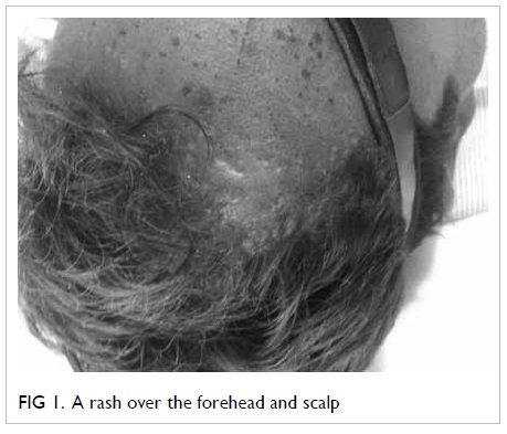
daily. He developed pruritic skin rash within 3 months of initiating imatinib. Initially, the skin condition improved with topical steroid. However, there was progressive development of white streaks and scaling of skin over the face, scalp, trunk, limbs, and trachyonychia with onycholysis of fingers and toes. There were no mucosal lesions. Skin biopsy findings were consistent with lichenoid drug reaction. Imatinib was stopped and changed to nilotinib. The skin and nail conditions progressively improved while the patient was on nilotinib.
 
Imatinib mesylate has been the standard treatment for chronic myeloid leukaemia for 10 years.1 Imatinib mesylate inhibits tyrosine kinases of bcr/abl, c-kit, and platelet-derived growth factor receptors, and cutaneous reactions are the commonest side-effects in patients receiving this drug.2 3 Trachyonychia results from disruption of the nail matrix cells, and can be induced by chemotherapeutic agents.4 Although paronychial inflammation is commonly induced by kinase inhibitors, trachyonychia is rarely reported. Cross-reactivity between different tyrosine kinases has rarely been reported.5 The absence of cross-reactivity between imatinib and nilotinib in this patient suggests that the mechanism of drug reaction is not related to the inhibition of tyrosine kinase.
 

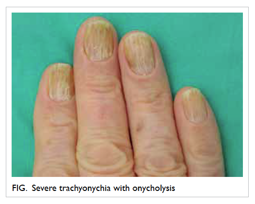
Figure. Severe trachyonychia with onycholysis
 
References
1. Peggs K, Mackinnon S. Imatinib mesylate—the new gold standard for treatment of chronic myeloid leukemia. N Engl J Med 2003;348:1048-50. CrossRef
2. Gardembas M, Rousselot P, Tulliez M, et al. Results of a prospective phase 2 study combining imatinib mesylate and cytarabine for the treatment of Philadelphia-positive patients with chronic myelogenous leukemia in chronic phase. Blood 2003;102:4298-305. CrossRef
3. Wahiduzzaman M, Pubalan M. Oral and cutaneous lichenoid reaction with nail changes secondary to imatinib: report of a case and literature review. Dermatol Online J 2008;14:14.
4. Chen W, Yu YS, Liu YH, Sheen JM, Hsiao CC. Nail changes associated with chemotherapy in children. J Eur Acad Dermatol Venereol 2007;21:186-90. CrossRef
5. Novitzky-Basso I, Craddock C. Cross-intolerance to imatinib, dasatinib and nilotinib therapy in a patient with chronic myeloid leukaemia. Eur J Haematol 2011;86:548-9. CrossRef

To scan or not to scan, to enhance or not to enhance? That is the question

DOI: 10.12809/hkmj134120
© Hong Kong Academy of Medicine. CC BY-NC-ND 4.0
 
PICTORIAL MEDICINE
To scan or not to scan, to enhance or not to enhance? That is the question
Lily Li, MRCS (Eng), MB/BChir1; Jonathan Costello, MRCPI, FCEM2
1 Department of Trauma and Orthopaedics, Lister Hospital, Stevenage, Hertfordshire SG1 4AB, United Kingdom
2 Emergency Department, Royal Free Hospital, London NW3 2QG, United Kingdom
 
Corresponding author: Dr Lily Li (xl228@doctors.org.uk)
 
 Full paper in PDF
A 12-year-old boy presented to the Emergency Department (ED) with reduced level of consciousness in February 2011. Collateral history established a pre-hospital witnessed seizure (requiring benzodiazepine administration) preceded by auditory hallucinations. Apart from uncomplicated malaria at the age of 5 years, there was no other medical history of relevance. Initial review was consistent with post-ictal presentation. An additional generalised seizure was witnessed in the ED within 30 minutes of admission requiring termination with additional intravenous benzodiazepine. In view of recurrent presentation, he was electively intubated and commenced on parenteral phenytoin. In addition, empirical acyclovir and ceftriaxone were administered. An unenhanced computed tomographic (CT) scan was normal (a). However, a subsequent enhanced scan revealed diffuse right parieto-occipital arteriovenous malformation (b). This case challenges the prevalent practice of non-performance of CT in new-onset seizure disorders and, if performed, the practice of performing solely non-enhanced CT scans in such presentations.
 

Should we perform polypectomy or not?

DOI: 10.12809/hkmj134090
© Hong Kong Academy of Medicine. CC BY-NC-ND 4.0
 
PICTORIAL MEDICINE
Should we perform polypectomy or not?
WY Mak, MB, BS, MRCP (UK); YT Hui, MRCP (UK), FHKAM (Medicine); Jodis TW Lam, FHKAM (Medicine), FRCP (Edin)
Department of Medicine, Queen Elizabeth Hospital, Jordan, Hong Kong
 
Corresponding author: Dr WY Mak (mwy612@ha.org.hk)
 
 Full paper in PDF
A 78-year-old woman, with a known history of rheumatoid arthritis, complained of dizziness and was found to have iron-deficiency anaemia with a haemoglobin level of 105 g/L in January 2013. She was on treatment with methotrexate, sulphasalazine, and non-steroidal anti-inflammatory medications. Oesophagogastroduodenoscopy performing for anaemia showed a linear ulcer over the anterior wall of the stomach. Subsequent colonoscopic examination was done to look for the cause of iron-deficiency anaemia and revealed a 1 cm–long everted umbilicated polypoid lesion in the ascending colon (Fig a). On close examination of the lesion using narrow band imaging (NBI), the mucosal pattern was normal with no endoscopic features of adenomatous polyp. Upon further air insufflation, the everted lesion invaginated and turned into a diverticulum (Fig b). Thus, a diagnosis of an inverted colonic diverticulum (ICD) was made.
 

Figure. (a) An inverted colonic diverticulum (white arrow) was found next to two small diverticula (black arrows) by narrow band imaging. (b) The inverted colonic diverticulum (white arrow) which was located next to the two small diverticula (black arrows) invaginated and became a diverticulum
 
Inverted colonic diverticulum is a rare condition. A retrospective analysis of colonic examinations showed that the prevalence was only 0.7%.1 The majority (approximately 75%) of ICDs were found in the sigmoid colon.1 Right-sided colonic ICD, as illustrated in our case, was not commonly seen. An ICD is typically described as a broad-based lesion with normal overlying mucosa lying within a bed of colonic diverticula. It can resemble an adenomatous polyp of variable size. It is essential to correctly diagnose this condition as inadvertent ‘polypectomy’ may potentially lead to bowel perforation. Currently, there are several endoscopic strategies that can help in distinguishing ICD from an adenomatous polyp. Firstly, gentle air insufflation may cause evertion of inverted diverticula. In some cases, a jet of water may be used to flatten the lesion.2 Secondly, probing the lesion gently with a biopsy forceps will show a soft lesion with easy indentation. Interestingly, it was recently suggested that the presence of Aurora rings can support the diagnosis of an ICD.3 Aurora rings are described as concentric rings surrounding the base of an ICD which can be demonstrated with the use of NBI or chromoendoscopy. If doubt exists, double-contrast barium enema or computed tomography colonography may help to distinguish between the two entities. Endoscopic ultrasound (EUS) has also been used to characterise such lesions. In a report, the diagnosis of sigmoid ICD was made by the EUS features of a thickened but normal-looking colonic mucosa in a polyp-like lesion.4 Most importantly, endoscopic removal and biopsy of ICD should be avoided as potentially fatal bowel perforation may occur.5
 
In conclusion, ICD is an uncommon but important clinical finding. Endoscopists should always be aware of the possibility of ICD during colonoscopic examination as inadvertent biopsy or resection of these lesions can lead to potentially serious complications.
 
References
1. Merino R, Kinney T, Santander R, et al. Inverted colonic diverticulum: an infrequent and dangerous endoscopic finding [abstract]. Gastrointest Endosc 2005;61:AB257. CrossRef
2. Cappell MS. The water jet deformation sign: a novel provocative colonoscopic maneuver to help diagnose an inverted colonic diverticulum. South Med J 2009;102:295-8. CrossRef
3. Share MD, Avila A, Dry SM, Share EJ. Aurora rings: a novel endoscopic finding to distinguish inverted colonic diverticula from colon polyps. Gastointest Endosc 2013;77:308-12. CrossRef
4. Yoshida M, Kawabata K, Kutsumi H, et al. Polypoid prolapsing mucosal folds associated with diverticular disease in the sigmoid colon: usefulness of colonoscopy and endoscopic ultrasonography for diagnosis. Gastrointest Endosc 1996;44:489-91. CrossRef
5. D’Ovidio V, Di Camillo M, Pimpo MT, Meo D, Vernia P, Caprilli R. An unusual complicated polypectomy and inverted colonic diverticula. Colorectal Dis 2010;12:491-2. CrossRef

Urinary bladder inguinal hernia: an uncommon cause of scrotal swelling

DOI: 10.12809/hkmj134057
© Hong Kong Academy of Medicine. CC BY-NC-ND 4.0
 
PICTORIAL MEDICINE
Urinary bladder inguinal hernia: an uncommon cause of scrotal swelling
HL She, MB, BS; KC Lam, MB, BS; KK Wong, MB, BS; Wendy WM Lam, MB, BS
Department of Radiology, Queen Mary Hospital, Pokfulam, Hong Kong
 
Corresponding author: Dr HL She (helenshe1025@gmail.com)
 
 Full paper in PDF
A 77-year-old man with benign prostate hypertrophy (BPH) presented to our hospital in October 2011 with a history of right groin swelling for several months. He was otherwise asymptomatic. Physical examination revealed a reducible right inguinal hernia. Ultrasound (USG) examination of the groins showed a fluid-filled lesion within the right scrotum. It had a beaked appearance at its cranial portion, which could be traced entering the right inguinal canal (Fig 1a). A tiny calcified focus was noted within this fluid-filled structure (Fig 1b). The normal right testis was displaced inferiorly (Fig 1c). Findings were suggestive of urinary bladder inguinal hernia with a bladder stone within. It was confirmed with non-contrast computed tomography (CT) of the abdomen and pelvis, which showed herniation of the urinary bladder along the inguinal canal and into the right scrotum, with a small bladder stone within (Fig 2).
 

Figure 1. (a) Ultrasonography demonstrates a fluid-filled structure with a beaked appearance within the right scrotum. (b) A tiny calcified focus within the fluid-filled structure in the right scrotum is seen (white arrow). (c) A normal right testis is displaced inferiorly by the fluid-filled structure (black arrow)
 

Figure 2. (a) Non-contrast computed tomography (CT) of the abdomen and pelvis in a reformatted image demonstrates herniation of the urinary bladder into the right scrotum (arrow). (b) Non-contrast CT axial view shows herniation of the urinary bladder into right scrotum (arrow) with a tiny bladder stone within (arrowhead)
 
Urinary bladder herniation is an uncommon condition, encountered in 1% to 4% of inguinal hernias. However, over the age of 50 years, the frequency increases to about 10%.1 Most patients are asymptomatic and usually found incidentally on imaging for workup of inguinal hernias, or even at the time of herniorrhaphy. Occasionally, patients may complain of urinary symptoms especially at an advanced stage, and may entail double-phase urination, that is, manually compressing the scrotum for complete bladder emptying. Predisposing factors include obesity, bladder outlet obstruction (eg due to BPH), and weakened abdominal musculature.2 Imaging modalities including USG, intravenous urogram, CT, and magnetic resonance imaging usually facilitate the diagnosis. Standard treatment entails surgical repair. It is important to be aware of this diagnosis, as apart from complications like urinary tract obstruction, urinary traction infection and urinary bladder infarction, unknowing herniorrhaphy may lead to bladder injury.1 3
 
References
1. Bisharat M, O’Donnell ME, Thompson T, et al. Complications of inguinoscrotal bladder hernias: a case series. Hernia 2009;13:81-4. CrossRef
2. Vindlacheruvu RR, Zayyan K, Burgess NA, Wharton SB, Dunn DC. Extensive bladder infarction in a strangulated inguinal hernia. Br J Urol 1996;77:926-7. CrossRef
3. Oruç MT, Akbulut Z, Ozozan O, Coşkun F. Urological findings in inguinal hernias: a case report and review of the literature. Hernia 2004;8:76-9. CrossRef

Diffuse xanthomatous eruption

DOI: 10.12809/hkmj134046
© Hong Kong Academy of Medicine. CC BY-NC-ND 4.0
 
PICTORIAL MEDICINE
Diffuse xanthomatous eruption
HF Cheng, MB, BS, MRCP (UK); William YM Tang, FRCP (Edin), FHKAM (Medicine); KC Lee, FRACPath, FHKAM (Pathology)
DERM 1 Skin Specialists Centre, Room 1102, Champion Building, 301-309 Nathan Road, Kowloon, Hong Kong
 
Corresponding author: Dr HF Cheng (chf@doctor.com)
 
 Full paper in PDF
A 37-year-old non-smoker with no history of drug allergy and history of childhood asthma presented with itchy rash over his back for 1 month, which progressed to involve his limbs and both axillae, in January 2013. The patient was not taking any medication apart from health supplements. He did not have any complaints of joint pain or fever. He was seen by a general practitioner who managed the rash as viral infection. Family history of hyperlipidaemia was negative. On examination, the patient was an obese man with body mass index of 32 kg/m2, blood pressure of 152/89 mm Hg, and pulse rate of 100 beats/min. There were widespread, reddish-yellow papular eruptions over both sides of the trunk and limbs, sparing the face, scalp, oral cavity, and ears (Fig 1). There were no scales, vesicles, pus formation, or erosions. The size of the lesions ranged from to 0.1 cm to 0.4 cm (Fig 2). There was no corneal arcus, regional lymphadenopathy, abdominal organomegaly or arthropathy. A skin biopsy of the lesion showed features of eruptive xanthoma (Figs 3 and 4). Fasting blood examination showed markedly elevated levels of total cholesterol (12.1 mmol/L), serum triglycerides (40.36 mmol/L), and plasma glucose (14.7 mmol/L). Thus, he was urgently referred to an endocrinologist.
 

Figure 1. Clinical photo showing diffuse eruption on the trunk
 

Figure 2. Close-up view demonstrating the colour, surface contour, and configuration of the lesion. Note also the absence of epidermal change
 

Figure 3. A wedge-shaped dermal lesion consists of histiocytes and lymphocytic infiltration (H&E; original magnification, x 40)
 

Figure 4. Characteristic lace-like granular material between collagen in the dermis is shown. Note the presence of foamy histiocytes (arrows) with admixed lymphocytes (arrowhead). Neutrophils are not conspicuous in this case (H&E; original magnification, x 200)
 
Discussion
Eruptive xanthoma is a benign lesion and patients usually consult because of itchiness or for cosmetic reasons. Morbidity arises from metabolic complications such as acute pancreatitis or myocardial infarction. The macroscopic lesions arise from phagocytosis in the dermis of plasma lipoproteins that leak from capillaries.1 Laboratory workup is mandatory to exclude diabetes, nephrotic syndrome, or hypothyroidism. Screening of family members is essential as genetic factors may contribute in the development of the condition.2 Eruptive xanthoma can occur in individuals with normal lipid levels.3 Under these circumstances, it is prudent to exclude occult malignancy (eg lymphoproliferative disorders and monoclonal paraproteinaemia) or infections (eg human immunodeficiency virus infection).4 Solitary lesions necessitate enquiry about previous local trauma, dermatoses, or surgical operation for Köbner phenomenon might have happened.
 
Differential diagnoses include eruptive xanthogranuloma, xanthoma disseminatum, and Langerhans cell histiocytosis (LCH). Xanthogranuloma usually arises in the head-and-neck regions of children. It is mostly a solitary, small-sized papule or nodule. Ophthalmologic evaluation is indicated if ocular involvement is suspected. Histopathology shows collection of lipidised histiocytes, inflammatory infiltrates, and Touton giant cells in the dermis. Known as non-LCH, xanthoma disseminatum is a non-familial histiocytic disorder. Mucocutaneous as well as systemic involvement has been reported. It shares similar histopathological features with eruptive xanthogranuloma but eosinophils may be absent, and Touton giant cells may be inconspicuous. Treatment, by far, is unsatisfactory. Langerhans cell histiocytosis comprises a spectrum of disorders with varied clinical manifestations including cutaneous involvement. Confirmation of diagnosis rests on histopathology. Within the lesion is dense infiltration by abnormal Langerhans cells which are characterised by their folded nuclei. Presence of a mixed inflammatory infiltrate with eosinophils in the background forms the classical picture of LCH. The Langerhans cells in LCH differ from the typical Langerhans cells by the lack of dendritic cell processes, and this feature is best demonstrated by CD1a immunostaining. All these differential diagnoses lack the characteristic deposits of lace-like material between collagen seen in eruptive xanthoma.
 
Gradual resolution of the cutaneous lesions is usually expected upon normalisation of lipid level in patients with eruptive xanthoma. En-bloc surgical excision or carbon dioxide laser vaporisation is equally practical, depending on the extent of the disease. The use of carbon dioxide laser has been reported in a skin phototype VI patient with xanthoma disseminatum with cosmetically acceptable post-inflammatory hyperpigmentation.5 Finally, referral to a physician is needed in patients with concomitant metabolic syndrome.
 
References
1. Massengale WT, Nesbitt LT Jr. Xanthomas. In: Bolognia JL, Jorizzo JL, Rapini RP, editors. Dermatology. Vol 2. 2nd ed. London, England: Mosby Elsevier; 2008: 1411-9.
2. Pickens S, Farber G, Mosadegh M. Eruptive xanthoma: a case report. Cutis 2012;89:141-4.
3. Williford PM, White WL, Jorizzo JL, Greer K. The spectrum of normolipemic plane xanthoma. Am J Dermatopathol 1993;15:572-5. CrossRef
4. Ramsay HM, Garraido MC, Smith AG. Normolipaemic xanthomas in association with human immunodeficiency virus infection. Br J Dermatol 2000;142:571-3. CrossRef
5. Carpo BG, Grevelink SV, Brady S, Gellis S, Grevelink JM. Treatment of cutaneous lesions of xanthoma disseminatum with a CO2 laser. Dermatol Surg 1999;25:751-4. CrossRef

Tumour-induced osteomalacia

DOI: 10.12809/hkmj133981
© Hong Kong Academy of Medicine. CC BY-NC-ND 4.0
 
PICTORIAL MEDICINE
Tumour-induced osteomalacia
Anjali A Bhatt, MD1; Suma S Mathews, MS, DLO2; Anusha Kumari, MD3; Thomas V Paul, MD, PhD1
1 Department of Endocrinology, Diabetes and Metabolism, Christian Medical College, Vellore 632 004, India
2 Department of ENT, Christian Medical College, Vellore 632 004, India
3 Department of General Pathology, Christian Medical College, Vellore 632 004, India
 
Corresponding author: Dr Thomas V Paul (thomasvpaul@yahoo.com)
 
 Full paper in PDF
Tumour-induced osteomalacia (also known as oncogenic osteomalacia) is an uncommon condition. The fibroblast growth factor 23 (FGF-23), a polypeptide secreted by mesenchymal tumours, causes phosphaturia, which in turn results in defective mineralisation. In addition, FGF-23 causes suppression of the enzyme 1α-hydroxylase located in the proximal convoluted tubules of kidneys and responsible for final activation of vitamin D (from 25-hydroxyvitamin D to 1,25-dihydroxyvitamin D).1 Once the tumour causing the osteomalacia is found, its excision usually results in complete remission of the bone disorder.2 Herein we report on a patient who presented to us with the features of oncogenic osteomalacia.
 
A 32-year-old woman born of non-consanguineous parents complained of gradually increasing proximal muscle weakness of both lower limbs over 1 year in February 2012. She also had pain in both hips while walking, which restricted her daily activities. She had no history of any chronic gastro-intestinal illness, and was not taking medication which might affect bone metabolism. Nor was there a family history of any similar illness. Clinical examination revealed severe proximal muscle weakness in both lower limbs. Active movements like external rotation and abduction at the both hips were painful. Otherwise the examination was unremarkable.
 
Biochemical evaluation revealed hypophosphataemia with phosphaturia (Table). Radiology of the hips showed bilateral femoral neck pseudofractures (Fig 1). These abnormalities favoured a diagnosis of hypophosphataemic osteomalacia. Further workup yielded a high FGF-23 level and computed tomography of the paranasal sinuses showed a soft tissue tumour in the left ethmoidal sinus (Fig 2).
 

Table. Biochemical parameters
 

Figure 1. Pseudofractures seen in both femoral neck (arrows)
 

Figure 2. Heterogeneously enhancing soft tissue lesion in left posterior ethmoid sinus (arrow)
 
The patient was treated with phosphate supplements and underwent complete excision of the left ethmoidal sinus mass. Histopathological examination confirmed the diagnosis of a phosphaturic mesenchymal tumour. The patient remained stable after surgery. Two months later she was asymptomatic, by which time her muscle weakness had resolved markedly. Respective serial serum phosphate concentrations were 0.90, 1.00, and 1.35 mmol/L at 1, 2 and 4 weeks after surgery. After surgery, her plasma FGF-23 levels were undetectable.
 
Phosphaturic mesenchymal tumours are rare mesenchymal tumours and mostly comprised of a single histological entity with mixed connective tissue (designated PMT-MCT). Such tumours can occur in the soft tissue or bone.3 Most PMT-MCTs are histologically and clinically benign with rare instances of malignancy. Microscopically, PMT-MCTs are variable in appearance, usually being composed of small, bland round-to-spindle cells embedded in a vascular to myxochondroid matrix with variable amounts of mature adipose tissue (Fig 3). This matrix calcifies in an unusual ‘grungy’ fashion, inciting an osteoclast-rich and fibrohistiocytic response. A very prominent feature of PMT-MCT is its elaborate intrinsic microvasculature. Malignant PMT-MCTs resemble undifferentiated pleomorphic sarcomas or fibrosarcomas.3 After complete excision of the tumour, most patients improve dramatically4 and become symptomatically, biochemically, and radiologically better. Such results should prompt the physicians to search for such treatable and potentially curable causes, whenever they encounter hypophosphataemic osteomalacia.
 

Figure 3. High-power view showing distinctive grungy pattern of matrix calcification with surrounding bland small round-to-spindle cells (H&E; original magnification, x 40)
 
References
1. Chokyu I, Ishibashi K, Goto T, Ohata K. Oncogenic osteomalacia associated with mesenchymal tumor in the middle cranial fossa: a case report. J Med Case Rep 2012;6:181. CrossRef
2. Komínek P, Stárek I, Geierová M, Matoušek P, Zeleník K. Phosphaturic mesenchymal tumour of the sinonasal area: case report and review of the literature. Head Neck Oncol 2011;3:16. CrossRef
3. Folpe AL, Fanburg-Smith JC, Billings SD, et al. Most osteomalacia-associated mesenchymal tumors are a single histopathologic entity: an analysis of 32 cases and a comprehensive review of the literature. Am J Surg Pathol 2004;28:1-30. CrossRef
4. Chong WH, Molinolo AA, Chen CC, Collins MT. Tumor-induced osteomalacia. Endocr Relat Cancer 2011;18:R53-77. CrossRef

Fever with vesicular rash in an adult

Hong Kong Med J 2014;20:264.e3–4 | Number 3, June 2014
DOI: 10.12809/hkmj/134018
© Hong Kong Academy of Medicine. CC BY-NC-ND 4.0
 
PICTORIAL MEDICINE
Fever with vesicular rash in an adult
Prashant Nasa, MD, FNB1; Ankur Gupta, MD, DM2; Shakti Jain, MD3; Kishore Mangal, MD, IDCCM1
1 Department of Critical Care Medicine, Max Super Speciality Hospital, Shalimar Bagh, New Delhi 110088, India
2 Department of Nephrology, Max Super Speciality Hospital, Shalimar Bagh, New Delhi 110088, India
3 Department of Microbiology, Max Super Speciality Hospital, Shalimar Bagh, New Delhi 110088, India
 
Corresponding author: Dr Prashant Nasa (dr.prashantnasa@hotmail.com)
A 30 year-old man was admitted with fever and myalgia for 4 days, rashes over abdomen and forehead for 2 days (Fig 1), and breathing difficulty for 1 day in January 2013. There was a history of similar rashes affecting his wife 7 days earlier. On examination, he was tachypnoeic (respiratory rate, 24 breaths/min), conscious, and oriented. He had a papulo-vesicular rash on the forehead, scalp, and trunk but the extremities were spared. Laboratory investigations revealed thrombocytopenia (platelet counts, 30 x 109/L), deranged liver function tests (serum bilirubin 4.6 mg/dL, direct bilirubin 2.9 mg/dL, aspartate transaminase 1316 IU/L, alanine transaminase 989 IU/L, alkaline phosphates 116 IU/L). The chest X-ray showed no gross abnormality and the smear of skin scrapings showed multinucleated epithelial giant cell (Tzanck cells), abundant erythrocytes, and sporadic leukocytes (Fig 2). Aerobic bacterial culture of a swabbed vesicle fluid was sterile. The history and characteristic pattern of the vesicular rash on the scalp and abdomen with no history of childhood immunisation against chickenpox clinched the diagnosis of chickenpox. The diagnosis is primarily clinical. Confirmation requires either viral culture of the vesicular fluid or direct fluorescent antibody testing of serum,1 but these tests are not readily available. Smear examination showing Tzanck cells in vesicle fluid may be positive in chicken pox, but also occurs in herpes simplex, herpes zoster, cytomegalovirus infection, and even pemphigus vulgaris.1 The differential diagnosis in our case was severe Chicken pox, disseminated herpes simplex infection, disseminated herpes zoster infection, drug eruptions, and pityriasis lichenoides et varioliformis acuta. However the clinical history (including family and vaccination history) favoured severe chickenpox.
 

Figure 1. A rash over the forehead and scalp
 

Figure 2. A Giemsa stain of skin scrapings from the base of a vesicle showing a multinucleated epithelial giant cell (Tzanck cell) [white arrow], abundant erythrocytes (black arrowhead), and sporadic leukocytes (black arrow) [Giemsa stain; original magnification, x 200]
 
Chickenpox is rarely fatal, although it is generally more severe in adults than children; those who are pregnant or immunocompromised have more severe forms of the disease.2 Serious manifestations of varicella-zoster virus (VZV) infection include pneumonia, hepatitis, and encephalitis.2 3 The standard treatment for severe adult VZV infection with or without involvement of vital organs entails early recourse to intravenous antivirals such as acyclovir. Whether severe or not, meticulous skin care, hygiene, and droplet precautions should be implemented to prevent cross-transmission to others till the patient’s lesions desquamate.
 
References
1. Herpesviruses: Varicella zoster virus (VZV). In: Shors T. Understanding viruses. 2nd ed. Burlington, MA: Jones & Bartlett; 2011: 459.
2. Maggi U, Russo R, Conte G, et al. Fulminant multiorgan failure due to varicella zoster virus and HHV6 in an immunocompetent adult patient, and anhepatia. Transplant Proc 2011;43:1184-6. CrossRef
3. Mohsen AH, McKendrick M. Varicella pneumonia in adults. Eur Respir J 2003;21:886-91. CrossRef

Pulmonary tuberculosis complicating asbestosis

Hong Kong Med J 2014;20:265.e3–5 | Number 3, June 2014
DOI: 10.12809/hkmj134019
© Hong Kong Academy of Medicine. CC BY-NC-ND 4.0
 
PICTORIAL MEDICINE
Pulmonary tuberculosis complicating asbestosis
YF Shea, MRCP (UK), FHKAM (Medicine)1; Janice JK Ip, MB, BS, FRCR2
1 Department of Medicine, Queen Mary Hospital, The University of Hong Kong, Pokfulam, Hong Kong
2 Department of Radiology, Queen Mary Hospital, The University of Hong Kong, Pokfulam, Hong Kong
 
Corresponding author: Dr YF Shea (elphashea@gmail.com)
An 87-year-old man who previously worked in shipyard with asbestosis was admitted in November 2012 because of fever of unknown origin. He presented with fever on-and-off for 2 months and cough. On physical examination, there was no cervical lymphadenopathy or hepatosplenomegaly and the chest was clear. Complete blood picture, and liver and renal function tests remained unremarkable. Chest X-ray (CXR) and computed tomography (CT) of the thorax yielded calcified pleural plaques, diaphragmatic calcification, diffuse centrilobular nodules, and interstitial septal thickening (Fig 1). Sputum and urine cultures were negative. Further investigations included smears and cultures for acid-fast bacilli and testing for Mycobacterium tuberculosis (MTB) by polymerase chain reaction of sputum, urine, and bronchoalveolar larvage samples, all of which were negative. Searches for aspergillus antigen, cryptococcal antigen, the Weil-Felix test, the Widal test, nasopharyngeal aspirate for influenza and mycoplasma, urine examination for legionella antigen, automminue profiling, and tests for tumour markers, human immunodeficiency virus, and sputum cytology were all non-contributory. The patient’s C-reactive protein was elevated to 3.39 mg/dL (reference range, <0.76 mg/dL). His fever had persisted on-and-off for 2 months despite multiple courses of broad-spectrum antibiotics. Positron emission tomography (PET)–CT yielded pronounced micronodules, especially over both upper lobes (Fig 1) and patchy ground glass opacities over both lower lobes with increased 18-fluorodeoxyglucose (18FDG) uptake (maximum standardised uptake values of up to 4; Figs 2 and 3). The absence of pulmonary masses, mediastinal or hilar lymphadenopathy or nodular thickening of interlobular septa and any bronchovascular bundle made malignancy or lymphangitis carcinomatosis unlikely. Based on the radiology, the patient was diagnosed to have pulmonary MTB for which anti-MTB treatment was initiated. He developed sudden cardiac arrest 1 day later, and failed resuscitation. Sputum and urine sample culture results available after the patient’s demise grew MTB.
 

Figure 1. (a) Chest X-ray and (b) computed tomography of the thorax showing calcified pleural plaques and diaphragmatic calcifications bilaterally (arrows)
 

Figure 2. Computed tomography thorax (lung window) of the upper lobes taken (a) 2 months earlier and 2 months later are shown; an increase in micronodules (arrows) was evident in (b) as compared with (a)
 

Figure 3. (a) Computed tomography thorax showing a background of calcified pleural plaques and an increase in patchy ground glass opacities over both lower lobes (arrows). (b) Positron emission tomography–computed tomography showing the parenchymal pattern of increased 18fluorodeoxyglucose uptake (maximum standardised uptake values of up to 4) over the both lower lobes (arrows)
 
Asbestosis is caused by the inhalation of asbestos which was once used as an electrical and thermal insulator. Asbestos causes fibrosis of the pleura and lung, as well as malignant mesothelioma, and PET-CT is a well-known means of picking up this complication as a linear area of intense 18FDG uptake surrounding the lungs.1 Fever of unknown origin can be due to infection, malignancy, autoimmune conditions, or drugs. Tuberculosis was one of the most common infectious causes. One of the diagnostic difficulties in our patient was the presence of interstitial lung disease making the interpretation of CXR and CT images challenging. Soussan et al2 has classified the PET appearance of pulmonary MTB into lung and lymphatic patterns and demonstrated improved diagnostic accuracy after taking account of the other specific CT changes such as upper lobe consolidation with cavitations or multiple ill-defined micronodules surrounding a cavity. Findings pertaining to our patient fitted well into previously described lung pattern of increased 18FDG uptake in MTB. These included predominant lung parenchymal involvement and together with an interval excess of micronodules, especially over the both upper lobes, which led us to make a radiological diagnosis of MTB. The increased in 18FDG uptake is caused by local accumulation of inflammatory cells.3 Thus, PET-CT is useful in identifying an active pulmonary tuberculoma in the absence of initial microbiological proof. Monitoring the response to anti-MTB treatment can also provide early evidence of drug-resistant MTB.3 4 The earlier performance of PET-CT and institution of anti-MTB treatment may have changed the clinical outcome of our patient.
 
References
1. Alavi A, Gupta N, Alberini JL, et al. Positron emission tomography imaging in nonmalignant thoracic disorders. Semin Nucl Med 2002;32:293-321. CrossRef
2. Soussan M, Brillet PY, Mekinian A, et al. Patterns of pulmonary tuberculosis on FDG-PET/CT. Eur J Radiol 2012;81:2872-6. CrossRef
3. Kosterink JG. Positron emission tomography in the diagnosis and treatment management of tuberculosis. Curr Pharm Des 2011;17:2875-80. CrossRef
4. Demura Y, Tsuchida T, Uesaka D, et al. Usefulness of 18F-fluorodeoxyglucose positron emission tomography for diagnosing disease activity and monitoring therapeutic response in patients with pulmonary mycobacteriosis. Eur J Nucl Med Mol Imaging 2009;36:632-9. CrossRef

Amelanotic melanoma masquerading as a pyogenic granuloma: caution warranted

Hong Kong Med J 2014;20:265.e1–2 | Number 3, June 2014
DOI: 10.12809/hkmj133987
© Hong Kong Academy of Medicine. CC BY-NC-ND 4.0
 
PICTORIAL MEDICINE
Amelanotic melanoma masquerading as a pyogenic granuloma: caution warranted
Noah LW So, MB, BS; CF Chan, MB, BS, FHKAM (Orthopaedic Surgery); Kenneth WY Ho, MB, BS, FHKAM (Orthopaedic Surgery); YL Lam, MB, ChB, FHKAM (Orthopaedic Surgery)
Department of Orthopaedics and Traumatology, Queen Mary Hospital, 102 Pokfulam Road, Hong Kong
 
Corresponding author: Dr Noah LW So (noahlwso@gmail.com)
Case
In April 2012, a 69-year-old Chinese woman complained of a non-healing raised lesion over the lateral aspect of her left fifth toe which had developed 2 years earlier. It appeared to have started as a minor abrasion after wearing ill-fitting shoes. A painless swelling then developed over that site with occasional friction-induced bleeding. She attended several podiatry treatment sessions and tried applying topical medications such as silver nitrate. The lesion did not resolve, and increased in size and eventually developed into a pink nodule. Apart from hypercholesterolaemia, the patient was an otherwise healthy non-smoker and non-drinker who enjoyed sunbathing on the beach in her youth, often spending half a day under the sun.
 
On physical examination, a 1 cm x 1 cm round and dull red nodule with superficial ulceration was noted on the lateral aspect of the left fifth toe. There were no signs of infection and no contact bleeding was evident (Fig). Given the history of minor trauma, tendency to bleed with minor friction and the appearance of the lesion, pyogenic granuloma was the main differential diagnosis. Owing to failure of repeated conservative treatment, excision of the lesion was performed.
 

Figure. Left fifth toe before excision showing the malignant melanoma (arrow): (a) lateral view and (b) plantar view (oblique angle)
 
Histology revealed it to be a malignant melanoma; no melanin pigment was detected but the tumour cells were positive for melanocytic markers. A staging positron emission tomography–computed tomography showed no metastasis. A wide resection involving ray amputation of the 4th and 5th toes was performed to ensure a clear resection margin.
 
Discussion
Amelanotic melanoma is reported to account for about 1.8% to 8.1% of all melanomas.1 Cheung et al2 defined amelanotic melanoma as being unsuspected clinically and with melanin in less than 5% of the tumour cells. Because the diagnosis was not suspected clinically, he noted that excisional biopsies were seldom performed, and that if undertaken, transection of the melanoma was common. In their database of 1170 patients, McClain et al3 found that red melanomas accounted for 3.9% of all such malignancies and up to 70% of those that were amelanotic. Regarding prognosis, their review found no significant difference between red amelanotic melanomas and pigmented melanomas in terms of disease-free survival, overall survival, metastasis, and recurrence. In other words, the mortality of red amelanotic melanomas appeared comparable to that of pigmented melanoma.
 
In our patient, the most notable risk factor for developing melanoma was prolonged sunlight exposure but such a history may be missed if the diagnosis is not suspected. Amelanotic melanomas appear clinically indistinguishable from pyogenic granulomas. We therefore caution all our readers to the possibility of amelanotic melanoma presenting as seemingly benign red lesions. Whilst uncommon in China (age-standardised incidence of 0.2 per 100 000 population and year),4 melanomas are highly malignant; both observation without investigation and excision without undertaking histology can delay the diagnosis with potentially disastrous consequences. The mnemonic ‘ABCD’ summarises features suggesting melanoma: Asymmetry, irregular Border, uneven Colour, Diameter of >6 mm.4 The mnemonic RRR (Red, Raised lesion, Recent change3) is proposed additionally for the diagnosis of red amelanotic melanomas. We recommend that for such red lesions of uncertain nature, referral to specialists experienced in managing melanomas is warranted.
 
References
1. Koch SE, Lange JR. Amelanotic melanoma: the great masquerader. J Am Acad Dermatol 2000;42:731-4. CrossRef
2. Cheung WL, Patel RR, Leonard A, Firoz B, Meehan SA. Amelanotic melanoma: a detailed morphologic analysis with clinicopathologic correlation of 75 cases. J Cutan Pathol 2012;39;33-9. CrossRef
3. McClain SE, Mayo KB, Shada AL, Smolkin ME, Patterson JW, Slingluff CL Jr. Amelanotic melanomas presenting as red skin lesions: a diagnostic challenge with potentially lethal consequences. Int J Dermatol 2012;51:420-6. CrossRef
4. LeBoit PE, Burg G, Weedon D, Sarasin A, editors. Pathology and genetics of skin tumours (IARC WHO Classification of Tumours). Lyon: IARC Press; 2006.

More than skin deep: Paget’s disease of the perineum

Hong Kong Med J 2014;20:264.e1–2 | Number 3, June 2014
DOI: 10.12809/hkmj133983
© Hong Kong Academy of Medicine. CC BY-NC-ND 4.0
 
PICTORIAL MEDICINE
More than skin deep: Paget’s disease of the perineum
JW Li, MB, BS, MRCP; CM Ng, MRCP, FHKAM
Department of Medicine, Queen Elizabeth Hospital, 30 Gascoigne Road, Kowloon, Hong Kong
 
Corresponding author: Dr JW Li (lijohnwing@gmail.com)
In July 2012, an extensive eczematous lesion was found incidentally over the external genitalia in a 77-year-old man who was being evaluated for lower limb cellulitis. The affected area had been present for 10 years; it was erythematous, macerated, and itchy. He had consulted countless doctors, but the itchiness was intense and the area of involvement was enlarging despite application of topical creams. On physical examination, the perineum was weepy with excoriation, crusts, and a serous discharge. The erythematous skin was indurated and had a well-defined margin. The penis shaft was swollen with palpable groin lymph nodes (Fig 1). Blood tests showed eosinophilia (1.1 x 10⁹/L) and an elevated erythrocyte sedimentation rate (58 mm/h).
 

Figure 1. Clinical picture of extramammary Paget’s disease of the perineum, showing its weepy nature and the sharp margin around the affected area
 
A clinical suspicion of extramammary Paget’s disease (EMPD) was suggested by the dermatologist. Skin biopsy of the right suprapubic region confirmed the diagnosis (Fig 2), while another biopsy of the left groin showed poorly differentiated invasive carcinoma, consistent with invasive EMPD. Urine for cytology, as part of the malignancy screening, showed atypical cells. Computed tomography of the abdomen revealed left ureteric obstruction with enhanced mural thickening of left ureter, of which transitional cell carcinoma could not be excluded. He was referred to a urologist for further assessment. Radiotherapy to the affected skin lesions was given in view of the extensive involvement.
 

Figure 2. The skin biopsy taken from the right suprapubic area shows many nests of cohesive pleomorphic malignant cells invading the epidermis. Immunohistochemical study demonstrates that the tumour cells are cytokeratin (CAM5.2) positive (not shown), confirming their epithelial nature (H&E; original magnification, x 200)
 
Extramammary Paget’s disease is a rare intraepithelial adenocarcinoma of the skin rich in apocrine glands, first described by Crocker in 1888.1 The disease involves the epidermis, but can potentially metastasise via the lymphatic system.2 Its clinical and histological features are similar to Paget’s disease of the nipple. The penis, scrotum, and vulva are frequently involved in sites. The erythematous area has a sharp margin and scaly surface. Intense itchiness is common and may result in erosion, excoriation, and lichenification.2
 
Our patient had a typical presentation with longstanding, distressing, and difficult-to-treat, eczema-like perineal lesion with a well-defined border. Delay in diagnosis, up to a decade,3 is notoriously common, because EMPD often mimics benign dermatological diseases such as eczema, contact dermatitis and fungal infection, and even malignant skin conditions like superficial melanoma, basal cell carcinoma, and Bowen’s disease.
 
A clinical diagnosis should be confirmed by skin biopsy. Immunohistochemistry shows the cells are positive for CEA, CAM 5.2, as well as keratins CK7 and 8. Once EMPD is diagnosed, thorough search for malignancy is mandatory, since up to 37% of patients have associated malignancies, in which case the condition is referred to as secondary EMPD.4 A recent study among Chinese patients showed that 8.3% out of 48 patients with EMPD developed malignancy.3 Location of skin involvement in EMPD predicts the type of cancer; perianal lesions are associated with anal and colorectal cancer, whereas penoscrotal lesions tend to be associated with urogenital malignancies.4 The positivity of Cytokeratin 20 in immunostaining is also highly associated with internal malignancies.5
 
Treatment is mainly surgical resection with reconstruction. Radiotherapy is an adjunctive therapy or reserved for frail elderly patients.2 Photodynamic therapy and topical imiquimod have been used. Despite these treatments, the local recurrence rate still ranges from 21% to 61%,3 with a median time to recurrence of 2 years.
 
Acknowledgement
We would like to thank Dr Shun-chin Ng, Dermatologist, Department of Health, Hong Kong, for his clinical care and manuscript advice. We thank Dr Wing-hung Lau, Department of Pathology, Queen Elizabeth Hospital, for provision of histological diagnosis and picture.
 
References
1. Crocker HR. Paget's disease affecting the scrotum and penis. Trans Pathol Soc Lond 1888;40:187-91.
2. Burns T, Breathnach S, Cox N, Griffiths C, editors. Rook's textbook of dermatology. 8th ed. Oxford, UK: Wiley-Blackwell; 2010.
3. Chan JY, Li GK, Chung JH, Chow VL. Extramammary Paget's disease: 20 years of experience in Chinese population. Int J Surg Oncol 2012;2012:416418.
4. Sarmiento JM, Wolff BG, Burgart LJ, Frizelle FA, Ilstrup DM. Paget's disease of the perianal region—an aggressive disease? Dis Colon Rectum 1997;40:1187-94. CrossRef
5. Liegl B, Liegl S, Gogg-Kamerer M, Tessaro B, Horn LC, Moinfar F. Mammary and extramammary Paget's disease: an immunohistochemical study of 83 cases. Histopathology 2007;50:439-47. CrossRef

Pages