Virilisation in a menopausal woman with a previous kidney transplant

DOI: 10.12809/hkmj164901
© Hong Kong Academy of Medicine. CC BY-NC-ND 4.0
 
PICTORIAL MEDICINE
Virilisation in a menopausal woman with a previous kidney transplant
TM Fung, FRCOG, FHKAM (Obstetrics and Gynaecology)1; WC Wong, FHKCPath, FHKAM (Pathology)2; KW Chan, FHKCP, FHKAM (Medicine)3; KS Fung, FHKCP, FHKAM (Medicine)3
1 Department of Obstetrics and Gynaecology, Princess Margaret Hospital, Laichikok, Hong Kong
2 Department of Clinical Pathology, Pamela Youde Nethersole Eastern Hospital, Chai Wan, Hong Kong
3 Department of Medicine and Geriatrics, Princess Margaret Hospital, Laichikok, Hong Kong
 
Corresponding author: Dr TM Fung (fungtm@ha.org.hk)
 
 
 Full paper in PDF
 
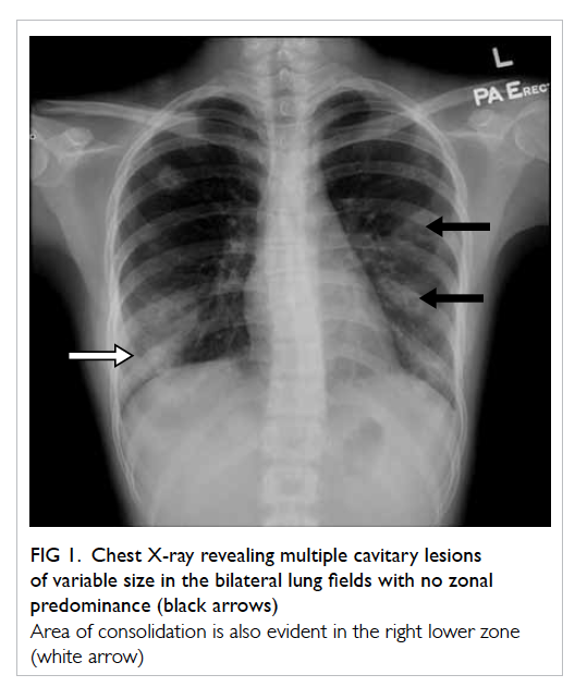
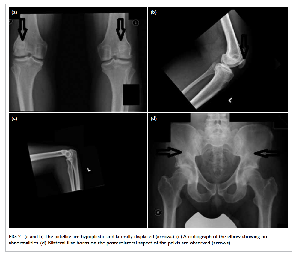
A 53-year-old woman, menopausal for 4 years and with a previous kidney transplant presented with a 3-year history of excessive facial and body hair that 
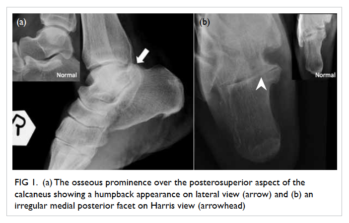
required daily shaving in January 2008 (Fig 1). She had had no postmenopausal bleeding or gynaecological disease. She was prescribed immunosuppressants and corticosteroids but no hormones. There was no acne or hoarseness of voice. Examination revealed frontal balding and excessive hair over her chin, both shins, and lower abdomen with mild clitoromegaly. Serum testosterone was 19 nmol/L (reference range, 0.35-2.65 nmol/L); 17-hydroxyprogesterone (17-OHP) and dehydroepiandrosterone sulfate (DHEAS) levels were normal. Adrenocorticotropic hormone and cortisol levels were normal after overnight dexamethasone suppression test. Her follicle-stimulating hormone, luteinising hormone, thyroid-stimulating hormone, and prolactin levels were also normal. Serum oestradiol was in the premenopausal range (298 nmol/L). Transvaginal ultrasound showed a normal uterus with endometrial thickness of 5 mm and no adnexal masses.
 

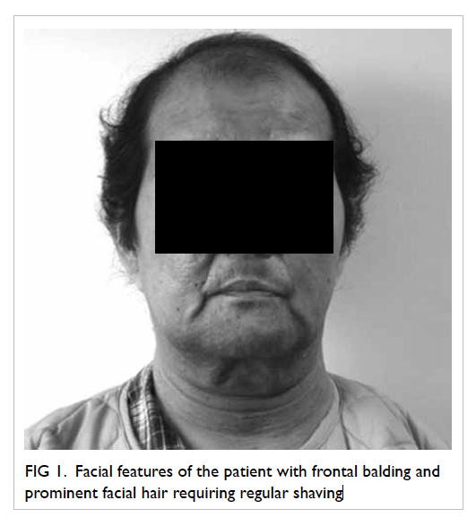
Figure 1. Facial features of the patient with frontal balding and prominent facial hair requiring regular shaving
 
Questions:
1. What are the differential diagnoses?
2. What further investigations are helpful?
3. How should the patient be managed?
 
Answers:
1. Androgen-secreting tumours of the ovary or adrenal gland.
Hirsutism involves excessive terminal hair growth in a masculine distribution in women. It can occur as a side-effect of immunosuppressants, eg cyclosporin with gingival hyperplasia and hirsutism. Nonetheless virilisation is uncommon. Cyclosporin should be changed to tacrolimus when hirsutism occurs.
Virilisation (development of male characteristics in women) occurs in less than 1% of patients with hirsutism. When it occurs (temporal hair recession and clitoromegaly in this patient) androgen-secreting tumours of the ovaries or adrenals should be suspected, particularly when onset is sudden with rapid progression.
2. Patients with virilisation should have serum testosterone, 17-OHP, and DHEAS checked. Serum testosterone level of >200 ng/dL (ie 6.94 nmol/L or 3 times normal) and/or DHEAS level of >700 µg/dL (ie 24.3 nmol/L) are indicative of virilising tumours.1 2 Computed tomography (CT) of the adrenals and pelvis should be considered. Ultrasonography of the pelvis may be difficult and not diagnostic in menopausal women because the ovaries are small and steroid-secreting tumours can be <1 cm in diameter.3 In this patient, normal levels of DHEAS and 17-OHP made adrenal tumour and congenital adrenal hyperplasia unlikely. Ovarian virilising tumours have to be suspected. Preoperative CT pelvis showed a 1-cm cyst in her right ovary.
3. Endometrial aspirate was performed, there was no hyperplasia or malignancy. She underwent laparotomy and bilateral salpingo-oophorectomy. There was a 1-cm yellowish tumour in the right ovary. The left ovary appeared normal. Hysterectomy was not done as the uterus was densely adhered to the bowel (history of 
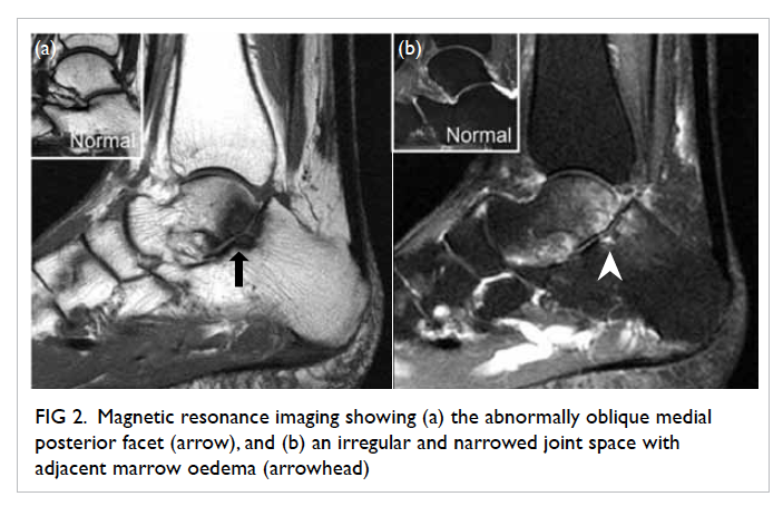
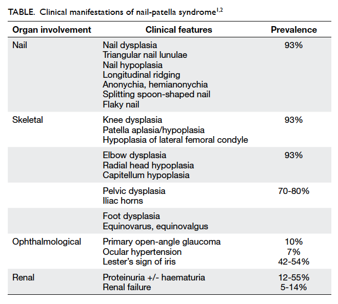
peritoneal dialysis). Histology confirmed stromal luteoma and hyperthecosis in both ovaries without malignancy (Fig 2). After surgery, her testosterone level normalised. Hair re-grew over her frontal region and her body hair reduced.
 

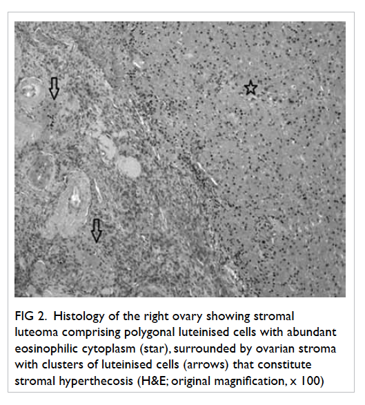
Figure 2. Histology of the right ovary showing stromal luteoma comprising polygonal luteinised cells with abundant eosinophilic cytoplasm (star), surrounded by ovarian stroma with clusters of luteinised cells (arrows) that constitute stromal hyperthecosis (H&E; original magnification, x 100)
 
Ovarian steroid cell tumours account for 0.1% to 0.2% of all ovarian tumours, and are composed of cells resembling steroid-secreting cells.4 Steroid cell tumours of small size and confined to the ovarian stroma are conventionally designated stromal luteomas and are usually associated with stromal hyperthecosis in adjacent stroma. They can have oestrogenic and/or androgenic manifestations with postmenopausal bleeding or virilisation, and are associated with endometrial hyperplasia or carcinoma.5 Hysterectomy and bilateral salpingo-oophorectomy is advised as some of these tumours may have malignant potential.
 
References
1. Somani N, Harrison S, Bergfeld WF. The clinical evaluation of hirsutism. Dermatol Ther 2008;21:376-91. Crossref
2. Hunter MH, Carek PJ. Evaluation and treatment of women with hirsutism. Am Fam Physician 2003;67:2565-72.
3. Outwater EK, Wagner BJ, Mannion C, McLarney JK, Kim B. Sex cord–stromal and steroid cell tumors of the ovary. Radiographics 1998;18:1523-46. Crossref
4. Hayes MC, Scully RE. Stromal luteoma of the ovary: a clinicopathological analysis of 25 cases. Int J Gynecol Pathol 1987;6:313-21. Crossref
5. Yamada S, Tanimoto A, Wang KY, Shimajiri S, Sasaguri Y. Stromal luteoma and nodular hyperthecosis of the bilateral ovaries associated with atypical endometrial hyperplasia of the uterus. Pathol Int 2009;59:831-3. Crossref

Posterior facet talocalcaneal non-osseous coalition: an uncommon but easily missed cause of hindfoot pain

DOI: 10.12809/hkmj164889
© Hong Kong Academy of Medicine. CC BY-NC-ND 4.0
 
PICTORIAL MEDICINE
Posterior facet talocalcaneal non-osseous coalition: an uncommon but easily missed cause of hindfoot pain
Arnold YH Tsang, MB, BS, FRCR; YY Cheuk, FHKCR, FHKAM (Radiology); Andrea WS Au-Yeung, FHKCR, FHKAM (Radiology)
Department of Diagnostic and Interventional Radiology, Kwong Wah Hospital, Yaumatei, Hong Kong
 
Corresponding author: Dr Arnold YH Tsang (arnoldtsang@gmail.com)
 
 
 Full paper in PDF
 
A 50-year-old female presented with chronic hindfoot pain in July 2015. She was treated for plantar fasciitis but the pain remained unresolved. Radiographs were initially interpreted as degenerative changes only. She was referred for further imaging. Review of the radiographs showed an osseous prominence over the posterosuperior aspect of the calcaneus, constituting the abnormal shape of the posterior facet of the subtalar joint with a humpback appearance on lateral view (Fig 1a). Joint space of the medial posterior facet was irregular and narrowed on Harris view (Fig 1b). Magnetic resonance imaging (MRI) showed the medial posterior facet of the subtalar joint to be abnormally oblique on sagittal plane, and the involved joint space was narrowed and irregular with adjacent marrow oedema (Fig 2). No bony bridging was seen. The middle facet was not involved. Included tendons appeared unremarkable and plantar fascia was not thickened. Features were suggestive of fibrocartilaginous coalition at the medial aspect of the posterior facet of the subtalar joint, also referred to as posteromedial talocalcaneal coalition. Computed tomography (CT) was also performed for surgical planning, and demonstrated clearly the irregular medial posterior facet, uninvolved middle facet and lateral posterior facet (Fig 3).
 

Figure 1. (a) The osseous prominence over the posterosuperior aspect of the calcaneus showing a humpback appearance on lateral view (arrow) and (b) an irregular medial posterior facet on Harris view (arrowhead)
 

Figure 2. Magnetic resonance imaging showing (a) the abnormally oblique medial posterior facet (arrow), and (b) an irregular and narrowed joint space with adjacent marrow oedema (arrowhead)
 

Figure 3. Computed tomography showing the irregular medial posterior facet, uninvolved middle facet (arrowhead) and lateral posterior facet (arrow)
 
Tarsal coalition can be osseous or fibrocartilaginous. Calcaneonavicular and talocalcaneal coalition accounts for 90% of hindfoot coalition,1 2 of which 50% are bilateral.1 The talocalcaneal joint, also referred to as the subtalar joint, consists of anterior, middle, and posterior facets. Talocalcaneal coalition usually involves the middle facet at the level of the sustentaculum tali. The incidence of middle facet coalition is less than 1%.1 3 Involvement of the posterior facet is even rarer, and is an easily missed cause of hindfoot pain. Continuous C sign is the classic sign well described for coalition over the middle facet. For posterior facet coalition, the humpback sign as described above is the radiographic finding to be recognised.1 3 4 The medial aspect of the posterior facet is more commonly involved, and Harris view can better demonstrate the medial joint that will be irregular and narrowed in non-osseous coalition. These findings are easily misinterpreted as degenerative changes.
 
Cross-sectional imaging including CT and MRI are valuable to confirm the diagnosis, define the location and extent of the segmentation anomaly, look for associated complications, and aid preoperative planning. In particular, CT is useful in determining the presence of any small bony bridging and is important for surgical planning; MRI is able to provide information about the degree of marrow oedema that may correlate with level of pain. It should be remembered that any unexplained marrow oedema around the subtalar joint, which is an atypical site for simple degenerative changes, should raise the suspicion of coalition. Osseous coalition is less likely to be missed on cross-sectional imaging as it will appear grossly abnormal with bony fusion across the involved facet (Fig 4). Potential complications of posterior facet coalition include peroneus muscle spasms, sinus tarsi syndrome, tarsal tunnel compression giving rise to a distended posterior tibial vein, calcaneal stress fracture, and premature osteoarthritis.1 4 Early recognition of non-osseous posterior facet talocalcaneal coalition is important, particularly in young patients, as surgical treatment can reduce complications later in life.
 

Figure 4. (a) Radiograph and (b) T1-weighted magnetic resonance imaging of another patient in our institution with osseous posterior facet coalition (arrows)
 
References
1. Staser J, Karmazyn B, Lubicky J. Radiographic diagnosis of posterior facet talocalcaneal coalition. Pediatr Radiol 2007;37:79-81. Crossref
2. Newman JS, Newberg AH. Congenital tarsal coalition: multimodality evaluation with emphasis on CT and MR imaging. Radiographics 2000;20:321-32. Crossref
3. McNally EG. Posteromedial subtalar coalition: imaging appearances in three cases. Skeletal Radiol 1999;28:691-5. Crossref
4. Moe DC, Choi JJ, Davis KW. Posterior subtalar facet coalition with calcaneal stress fracture. AJR Am J Roentgenol 2006;186:259-64. Crossref

Neurocysticercosis in a young Indian male

DOI: 10.12809/hkmj164815
© Hong Kong Academy of Medicine. CC BY-NC-ND 4.0
 
PICTORIAL MEDICINE
Neurocysticercosis in a young Indian male
Eugene PL Ng, MB, ChB; Peter YM Woo, MB, BS, FHKAM (Surgery); Alain KS Wong, MB, ChB, FHKAM (Surgery); KY Chan, MB, ChB, FHKAM (Surgery)
Department of Neurosurgery, Kwong Wah Hospital, Yaumatei, Hong Kong
 
Corresponding author: Dr Eugene PL Ng (npl566@ha.org.hk)
 
 Full paper in PDF
 
A 33-year-old Indian male was hospitalised for a 3-day history of headache and left lower limb weakness in December 2014. He had experienced no fever or seizures. He had visited New Delhi, India, the year before. Physical examination revealed the patient to be fully conscious with left lower limb monoparesis. There was no sensory deficit. Computed tomography (CT) revealed a superior parietal intra-axial lesion with a calcified focus (Fig 1a). Magnetic resonance imaging (MRI) delineated a 2 x 1.5 x 1.5 cm circumscribed hypointense cystic lesion with a contrast-enhancing wall and an eccentric intracystic signal with perilesional oedema (Figs 1b to 1d). Differential diagnoses included neurocysticercosis, brain abscess, brain metastasis, and malignant glioma. Craniotomy for excision was performed in view of the possibility of malignancy and the surgically accessible superficial location of the lesion. Intra-operatively, a firm capsular mass containing thick opaque material was seen (Fig 2a). Histology revealed a cysticercus within a fibrous capsule, compatible with the diagnosis of neurocysticercosis (Figs 2b and 2c). A 2-week course of oral albendazole was commenced and the patient was discharged with full recovery.
 

Figure 1. Images of the patient
(a) Computed tomography demonstrating an intra-axial right parietal lesion with a calcific focus (axial, white arrow). (b) Hole-with-dot sign: gadolinium contrast T1-weighted coronal magnetic resonance imaging demonstrating a ring-enhancing hypointense cystic lesion with an eccentric intracystic enhancing signal suggestive of a scolex at the colloidal vesicular stage of infection (white arrowhead, inset). (c) T3-weighted coronal image showing a hyperintense cyst (grey arrow) with a hypointense scolex (inset) and marked perilesional oedema. (d) Fluid-attenuated inversion recovery image depicting a hyperintense lesion (axial, grey arrowhead) with corresponding hypointense scolex (inset)
 

Figure 2. Intra-operative photographs and photomicrographs of the patient
(a) Intra-operative photographs of resected neurocysticercus with a fibrous wall containing thick opaque material. (b) The typical colloid cyst membrane (white arrow; H&E; original magnification, x 40). (c) Taenia solium scolex comprises four suckers (black arrow) and a double row of hooks (grey arrow) for host intestinal wall attachment (H&E; original magnification, x 100)
 
Neurocysticercosis is the most common parasitic infection of the central nervous system (CNS) caused by the larval form of Taenia solium. The peak age of incidence is between 25 and 35 years1 and the condition is endemic to the Indian subcontinent, coastal North Africa, sub-Saharan Africa, Latin America, and China. The main mode of transmission is by faecal-oral ingestion of tapeworm embryos.1 2 Consumption of contaminated poorly cooked pork is a less-frequent alternative source of infection since pigs are intermediate hosts.2 3 Within 72 hours of ingestion, larvae known as oncospheres are released and pass through the intestinal wall into the circulation, subsequently depositing in the CNS, retina, and skeletal muscle as cysticerci.1 2 The parasite can remain viable in the brain for several years after which it undergoes calcific degeneration.3 In endemic areas, the most common presentation is epilepsy, responsible for 30% of cases.1 Focal neurological deficits may occur including cranial nerve palsy due to basal meningitis. Obstructive hydrocephalus develops when lesions occupy the fourth ventricle.1 2
 
Characteristic radiological features include dystrophic calcification on CT imaging, cyst wall contrast enhancement on T1-weighted MRI and identification of the pathognomonic scolex, an eccentric focus of enhancement representing the tapeworm’s head, best delineated with fluid-attenuated inversion recovery sequences (hole-with-dot sign).3 4 Brain abscess or metastases are important differential diagnoses as they are also similarly located at the grey-white matter junction of the middle cerebral artery distribution, associated with disproportionately significant perilesional cerebral oedema and classically exhibit heterogeneous contrast enhancement. Malignant glioma was less likely in our patient since they are morphologically more infiltrative, and the present lesion was well-circumscribed. For this patient, the major distinguishing feature that supported a diagnosis of neurocysticercosis was the presence of dystrophic calcification on CT and, in retrospect, the presence of a scolex on MRI. Absolute criteria for a definitive diagnosis are either histological parasitic proof, imaging demonstration of a scolex, or subretinal parasites on fundoscopy (Table5). Serological enzyme-linked immunoelectrotransfer blot detection of anti-cysticercus antibodies or cerebrospinal fluid enzyme-linked immunosorbent assays are adjunctive investigations.1 6 Management of active neurocysticercosis includes antiepileptic drug administration, anti-inflammatory glucocorticoid therapy, and definitive antiparasitic therapy with albendazole (15 mg/kg per day) or praziquantel (50 mg/kg per day) for 2 to 4 weeks.1 Surgical excision is reserved for cysts that cause mass effect, hydrocephalus, or if the diagnosis is unclear.2
 

Table. Diagnostic criteria of neurocysticercosis5
 
In an era of increasing migration and international travel, patients from developing countries who present with seizures, raised intracranial pressure symptoms, or focal neurological deficit should be suspected of having neurocysticercosis when characteristic imaging findings are identified. In probable cases, a trial of antiparasitic therapy is recommended with serial scans arranged to monitor treatment response.
 
References
1. Garcia HH, Nash TE, Del Brutto OH. Clinical symptoms, diagnosis, and treatment of neurocysticercosis. Lancet Neurol 2014;13:1202-15. Crossref
2. Zymberg ST. Neurocysticercosis. World Neurosurg 2013;79(2 Suppl):S24.e5-8.
3. Dhesi B, Karia SJ, Adab N, Nair S. Imaging in neurocysticercosis. Pract Neurol 2015;15:135-7. Crossref
4. Lerner A, Shiroishi MS, Zee CS, Law M, Go JL. Imaging of neurocysticercosis. Neuroimaging Clin N Am 2012;22:659-76. Crossref
5. Del Brutto OH, Rajshekhar V, White AC Jr, et al. Proposed diagnostic criteria for neurocysticercosis. Neurology 2001;57:177-83. Crossref
6. Gekeler F, Eichenlaub S, Mendoza EG, Sotelo J, Hoelscher M, Löscher T. Sensitivity and specificity of ELISA and immunoblot for diagnosing neurocysticercosis. Eur J Clin Microbiol Infect Dis 2002;21:227-9. Crossref

Crowned dens syndrome: an uncommon cause of cord compression

DOI: 10.12809/hkmj164864
© Hong Kong Academy of Medicine. CC BY-NC-ND 4.0
 
PICTORIAL MEDICINE
Crowned dens syndrome: an uncommon cause of cord compression
Cindy SY Fung, MB, ChB, FRCR; Godfrey KF Tam, FRCR, FHKCR
Department of Radiology, North District Hospital, Sheung Shui, Hong Kong
 
Corresponding author: Dr Cindy SY Fung (sycindy@gmail.com)
 
 Full paper in PDF
 
A 65-year-old man presented to our hospital in June 2015 with a 2-week history of neck pain and progressive weakness in four limbs. There was no recent trauma history. He had a history of cervical myelopathy with decompression performed in 2011. On physical examination, an old scar on his neck was unremarkable with no signs of infection. Neurological examination revealed generalised weakness in all four limbs, more marked in bilateral upper limbs. All limbs were hypertonic with hyperreflexia. There was no sensory loss. His C-reactive protein level was elevated to 39.6 mg/L, white blood cell count was also elevated to 14.8 x 10⁹ /L. His cervical radiograph showed indistinct dens (Fig 1). No abnormal soft tissue thickening was seen. Screws of the previous posterior cervical decompression were in-situ. Computed tomography was performed and revealed erosion of the dens and some mildly hyperdense periodontoid soft tissue (Fig 2). Further study with magnetic resonance imaging showed T1 intermediate, T2 heterogeneously hypointense periodontoid soft tissue with patchy enhancement (Fig 3). The cervicomedullary junction was moderately compressed with internal T2 hyperintense cord signal, signifying cord oedema or myelomalacia. Radiograph of other joints found chondrocalcinosis in the triangular fibrocartilage of the right wrist, which is also a common manifestation of calcium pyrophosphate dihydrate (CPPD) crystal deposition disease (Fig 4). Overall features were compatible with crowned dens syndrome.
 

Figure 1. Lateral cervical spine radiograph demonstrating indistinct dens (arrow)
 

Figure 2. Sagittal reformatting of cervical spine computed tomographic scan revealing erosion of dens and hyperdense periodontoid soft tissue (arrow)
 

Figure 3. Sagittal (a) T1-weighted and (b) T2-weighted magnetic resonance images showing T1 intermediate, T2 heterogenously hypointense periodontoid mass with cord compression
 

Figure 4. Radiograph of other joints found chondrocalcinosis of the triangular fibrocartilage in the right wrist, compatible with calcium pyrophosphate dihydrate crystal deposition disease
 
Crowned dens syndrome was first described by Bouvet et al in 1985.1 It is a rare entity that presents clinically with severe upper neck pain and radiologically a crown-like density surrounding the odontoid process caused by deposition of CPPD crystals, which is now more commonly described, or hydroxyapatite (HA).1 It is more common in elderly patients with no history of trauma. Increased inflammatory indicators, such as an elevated C-reactive protein, are usually seen.2 Diagnosis is not easy as crowned dens syndrome can mimic a wide range of diseases such as meningitis, osteomyelitis, degenerative cervical spine disease, ankylosing spondylitis, gout, rheumatoid arthritis, temporal arteritis, metastatic bone disease, and spinal tumours.3 Computed tomography is the gold standard for identifying crowned dens syndrome, as it is able to depict the shape and site of calcification and any bone erosions. Radiography of other joints (wrist, knee, pubic symphysis) may help to ascertain whether the disease is due to CPPD or HA crystals, and is therefore recommended for routine patient management. Magnetic resonance imaging is indicated for the study of neurological complications as in our patient.4 Prednisolone and non-steroidal anti-inflammatory drugs in combination are the recommended treatment for symptom relief.2
 
Crowned dens syndrome is an under-recognised disease. Familiarity with the clinical and radiological features will help doctors provide prompt and effective treatment.
 
References
1. Bouvet JP, le Parc JM, Michalski B, Benlahrache C, Auquier L. Acute neck pain due to calcifications surrounding the odontoid process: the crowned dens syndrome. Arthritis Rheum 1985;28:1417-20. Crossref
2. Goto S, Umehara J, Aizawa T, Kokubun S. Crowned dens syndrome. J Bone Joint Surg Am 2007;89:2732-6. Crossref
3. Wu DW, Reginato AJ, Torriani M, Robinson DR, Reginato AM. The crowned dens syndrome as a cause of neck pain: report of two new cases and review of the literature. Arthritis Rheum 2005;53:133-7. Crossref
4. Scutellari PN, Galeotti R, Leprotti S, Ridolfi M, Franciosi R, Antinolfi G. The crowned dens syndrome. Evaluation with CT imaging. Radio Med 2007;112:195-207. Crossref

Median arcuate ligament syndrome

DOI: 10.12809/hkmj154821
© Hong Kong Academy of Medicine. CC BY-NC-ND 4.0
 
PICTORIAL MEDICINE
Median arcuate ligament syndrome
FH Ng, MB, ChB, FRCR; Ophelia KH Wai, MB, ChB, FRCR; Agnes WY Wong, MB, ChB, FRCR; SM Yu, MB, ChB, FRCR
Department of Radiology and Organ Imaging, United Christian Hospital, Kwun Tong, Kowloon, Hong Kong
 
Corresponding author: Dr FH Ng (nfh667@ha.org.hk)
 
 
 Full paper in PDF
 
Case
A middle-aged man admitted with abdominal pain and anaemia in December 2015. A computed tomographic (CT) angiogram demonstrated a superior indentation with focal narrowing in the proximal celiac axis (Fig 1). Conventional superior mesenteric arteriogram demonstrated prominent collaterals, and retrograde flow of contrast from the superior mesenteric artery (SMA) to the hepatic arteries (Fig 2). It was likely related to chronic compression of the proximal celiac artery. Low insertion of the median arcuate ligament (MAL) can be found in normal asymptomatic people. In this case, prominent collaterals and the retrograde flow from the SMA supported the diagnosis and may have explained his symptoms. In symptomatic cases, surgical division of the median arcuate ligament is the mainstay of treatment.
 

Figure 1. (a) Reformatted computed tomographic (CT) image showing the characteristic focal narrowing of the celiac artery with a hooked appearance (arrow) which is compressed by the median arcuate ligament (MAL). (b) The CT axial image again demonstrating the compression by the MAL
 

Figure 2. (a) Celiac angiogram showing post-stenotic prominent splenic artery and collaterals. (b) Angiogram of superior mesenteric artery (SMA) showing retrograde flow to celiac axis from the SMA via the pancreaticoduodenal arcade
 
Discussion
The MAL is a fibrous arch that unites the diaphragmatic crura on either side of the aortic hiatus. The crura pass superior and anterior to surround the aortic opening and to join the central tendon of the diaphragm. The ligament usually passes superior to the origin of the celiac axis. The insertion of the ligament may be low and therefore crossover the proximal portion of the celiac axis, causing a characteristic indentation. If it is significantly compressed on the celiac axis, this will compromise vascular flow and produce symptoms.
 
The MAL syndrome was first described in 1963 by Harjola1 and in 1965 by Dunbar et al.2 The definition of the syndrome relies on a combination of both clinical and radiographic features. Clinically, they described a classical triad of chronic postprandial abdominal pain, epigastric bruit, and weight loss.3 4 Extrinsic compression of the celiac trunk by the MAL occurs in 10% to 24% of patients.1 Usually, patients are asymptomatic and the classical triad is not always present, presumably due to collateral supply from the superior mesenteric circulation.1 2 The disease typically occurs in young patients and is more common in thin women who may present with epigastric pain and weight loss.1 The abdominal pain may be associated with eating, but not always.1 On physical examination, an abdominal bruit that varies with respiration may be audible in the mid-epigastric region. Symptoms are thought to arise from compression of the celiac axis with consequent compromised blood flow.
 
The diagnosis of celiac artery compression is traditionally made following conventional angiography. The use of thin-section multidetector CT and three-dimensional imaging techniques has greatly improved the ability to non-invasively obtain detailed images of the mesenteric vessels. Compression of the celiac axis by the MAL produces characteristic findings visible on CT angiography. Computed tomographic angiography can play a role in the diagnosis of this condition by demonstrating the characteristic focal narrowing of the celiac artery (Fig 1) with a hooked appearance that distinguishes this condition from other causes of celiac artery narrowing, such as atherosclerotic disease. Indentation of the origin of coeliac artery is exacerbated during the expiratory phase. Repeating CT on inspiration can often distinguish clinically significant narrowing from transient compression seen only during expiration in some patients, and is how most abdominal CT studies are performed.1
 
The majority of affected patients have no symptoms, thus radiographic finding of celiac axis compression alone may not be significant, unless it is correlated with clinical symptoms. Severe compression occurs in approximately 1% of patients.1 Severe stenosis will result in post-stenotic dilatation, and in some cases, the celiac axis will be fed by the SMA via the pancreaticoduodenal arcade. This was evident on the angiogram of our patient with prominent collaterals and retrograde flow from the SMA (Fig 2). His CT angiogram showed a characteristic hooked focal narrowing at the superior proximal celiac artery (Fig 1).
 
The surgical management of MAL syndrome is controversial.1 2 Surgical treatment in severe cases is advocated, particularly in cases with post-stenotic dilatation and collateral vessels (Fig 2), by division of the ligament. A laparoscopic approach is being increasingly adopted. Celiac angioplasty and stenting by endovascular means may be a future hot topic.2
 
References
1. Harjola PT. A rare obstruction of the coeliac artery: report of a case. Ann Chir Gynaecol Fenn 1963;52:547-50.
2. Dunbar JD, Molnar W, Beman FF, Marable SA. Compression of the celiac artery and abdominal angina. Am J Roentgenol Radium Ther Nucl Med 1965;95:731-44. Crossref
3. Horton KM, Talamini MA, Fishman EK. Median arcuate ligament syndrome: evaluation with CT angiography. Radiographics 2005;25:1177-82. Crossref
4. Duffy AJ, Panait L, Eisenberg D, Bell RL, Roberts KE, Sumpio B. Management of median arcuate ligament syndrome: a new paradigm. Ann Vasc Surg 2009;23:778-84. Crossref

Lemierre’s syndrome: an often forgotten but potentially life-threatening disease

DOI: 10.12809/hkmj154696
© Hong Kong Academy of Medicine. CC BY-NC-ND 4.0
 
PICTORIAL MEDICINE
Lemierre’s syndrome: an often forgotten but potentially life-threatening disease
C Lee, MB, BS, FRCR; Lorraine HY Sinn, MB, BS, FRCR; Sonia HY Lam, MB, BS, FRCR; WM Lam, MB, BS, FRCR
Department of Radiology, Queen Mary Hospital, Pokfulam, Hong Kong
 
Corresponding author: Dr C Lee (leechunbruce@gmail.com)
 
An earlier version of this paper was presented as a poster at the European Society of Thoracic Radiology held in Barcelona, Spain on 4-6 June 2015.
 
 Full paper in PDF
 
A 20-year-old Chinese female with good past health presented to the emergency department in October 2014 with a few days history of fever, dizziness, and headache. She was hypotensive upon presentation with blood pressure of 73/48 mm Hg and pulse rate of 90 beats/min but responded to fluid resuscitation. Physical examination revealed no other significant findings. Her white cell count was 4.4 x 10⁹ /L on admission but increased to 13.4 x 10⁹ five days later. Neutrophil predominance was observed. Subsequent gradual decline and normalisation of the white cell count was noted after initiation of intravenous antibiotics.
 
Computed tomographic (CT) abdomen and pelvis was initially requested based on the clinical suspicion of intra-abdominal sepsis or gynaecological pathologies but yielded no remarkable findings. Chest X-ray (CXR) was also performed for preliminary assessment and was initially unremarkable. Follow-up serial CXRs, however, showed an increasing number of cavitary lesions in both lungs with no zonal predominance (Fig 1).
 

Figure 1. Chest X-ray revealing multiple cavitary lesions of variable size in the bilateral lung fields with no zonal predominance (black arrows)
Area of consolidation is also evident in the right lower zone (white arrow)
 
Non-contrast CT thorax was deemed necessary for further characterisation of these lesions and confirmed multiple cavitary lesions of variable sizes involving all the lung lobes (Fig 2). Thick irregular walls with fluid content and perifocal consolidative changes were noted in some of these lesions, which are compatible with an infective process. Further imaging workup was then arranged to identify any potential source of the septic emboli but echocardiogram did not reveal any heart valve vegetation.
 

Figure 2. Computed tomographic thorax demonstrating multiple cavitary lesions of variable size involving all the lung lobes
The right lower zone lesion shows perifocal consolidative changes (white arrow) in some of these lesions, compatible with an infective process
 
On further questioning, the patient reported mild left neck pain. Doppler ultrasound of the neck was then performed based on the clinical suspicion of Lemierre’s syndrome. A markedly narrowed lumen with thickened wall and dampened flow signal was noted across the left internal jugular vein suspicious of venous thrombosis (Fig 3). This was subsequently confirmed on contrast CT neck and thorax (Fig 4).
 

Figure 3. Markedly narrowed lumen (white arrow) with thickened wall and dampened flow signal is noted across the left internal jugular vein suspicious of venous thrombosis
 

Figure 4. Reformatted computed tomographic image in an oblique sagittal projection showing a filling defect in the mid portion of the left internal jugular vein (IJV) [white arrow], compatible with jugular venous thrombosis
 
Mycoplasma-Fusobacterium polymerase chain reaction study confirmed the causative organism as Fusobacterium necrophorum, which is the classic bacteria described in Lemierre’s syndrome.
 
The patient was promptly treated with intravenous antibiotics and anticoagulant. Her clinical condition gradually improved and follow-up CT scan demonstrated interval resolution of the cavitary lung lesions. She was subsequently discharged with oral antibiotics and anticoagulants.
 
Lemierre’s syndrome remains a rare yet potentially life-threatening disease especially in young adults.1 A high index of clinical suspicion is therefore imperative to ensure prompt and timely imaging investigations that are integral to its diagnosis. Unrecognised and untreated systemic dissemination can result in a poor prognosis.2 Contrast-enhanced CT played a pivotal role in terms of evaluation of the pulmonary sepsis and assessment of the jugular vein thrombosis in this patient.3
 
References
1. Shook J, Trigger C. Lemierre’s Syndrome. West J Emerg Med 2014;15:125-6. Crossref
2. Lai C, Vummidi DR. Images in clinical medicine. Lemierre’s Syndrome. N Engl J Med 2004;350:e14. Crossref
3. Screaton NJ, Ravenel JG, Lehner PJ, Heitzman ER, Flower CD. Lemierre syndrome: forgotten but not extinct—report of four cases. Radiology 1999;213:369-74. Crossref

The nail points to the diagnosis

DOI: 10.12809/hkmj154728
© Hong Kong Academy of Medicine. CC BY-NC-ND 4.0
 
PICTORIAL MEDICINE
The nail points to the diagnosis
Stephanie YK Tong, BSc; HM Luk, FHKAM (Paediatrics); Tony MF Tong, MSc; Ivan FM Lo, FHKAM (Paediatrics)
Clinical Genetic Service, Department of Health, Hong Kong
 
Corresponding author: Dr HM Luk (luksite@gmail.com)
 
 Full paper in PDF
 
A 54-year-old man was referred to the genetic clinic with familial nail dysplasia in September 2012 (Fig 1). On further questioning, he reported recurrent knee pain due to patellar dislocation. There was no elbow involvement or renal problem. A skeletal survey was performed in view of his skeletal complaint (Fig 2). His family history was significant: his paternal grandfather, father, and two of his paternal uncles also had nail dysplasia and knee problems, but had undergone no formal medical assessment. What is the diagnosis?
 

Figure 1. (a) The left thumbnail is dysplastic with longitudinal splitting. The ulnar side is more severely affected (arrow). (b) The skin creases on the dorsal aspect of the distal interphalangeal joints are absent. (c) Triangular lunulae are evident on all fingernails (arrows). (d) The little toenail is also hypoplastic (arrow)
 

Figure 2. (a and b) The patellae are hypoplastic and laterally displaced (arrows). (c) A radiograph of the elbow showing no abnormalities. (d) Bilateral iliac horns on the posterolateral aspect of the pelvis are observed (arrows)
 
What is his diagnosis?
The combination of nail dysplasia, patella hypoplasia, and iliac horn led to the clinical diagnosis of nail-patella syndrome (NPS). This syndrome is also known as Fong disease or hereditary osteo-onychodysplasia. Multiple organ systems are affected including the nails, the eyes, the kidneys, and the skeleton. The clinical presentation of NPS can be highly heterogeneous and is summarised in the Table.1 2 It is a rare autosomal dominant disease with a prevalence of about 1 in 50 000 newborn.1 It is highly penetrant but with significant intra- and inter-familial variability in expression.

Table. Clinical manifestations of nail-patella syndrome1 2
 
Is there any genetic testing available for such condition and what is the underlying pathogenesis?
The diagnosis of NPS is usually based on clinical findings. It is straightforward when the classic tetrad of abnormal nails, elbows, knees, and iliac horns are present. Nonetheless molecular genetic testing should be considered when the diagnosis is in doubt, or when prenatal or pre-implantation diagnosis is desired. The LMX1B gene is the only gene known to be associated with NPS. Sequence analysis would identify the LMX1B gene mutation in 85% of cases of NPS.
 
LMX1B gene sequencing was performed for this patient (Fig 3) and revealed a missense mutation LMX1B NM_002316.3}:c.[175T>C];[=];LMX1B{NP _002307.2}:p.[Cys59Arg];[=]. This changed the 59th amino acid from cysteine to arginine in the LIM-A domain of the LMX1B protein. It was located in an evolutionarily highly conserved region and has been reported in the literature to be associated with NPS. Therefore, it was considered to be pathogenic.3

Figure 3. DNA sequence chromatographs of the patient. (a) A heterozygous c.175T>C mutation in exon 2 of LMX1B gene, and (b) wild-type sequence for comparison
 
To date, more than 150 mutations of the LMX1B gene have been reported, but no clear genotype-phenotype correlation has been demonstrated. LMX1B has been demonstrated in animal models to be involved in multiple developmental functions including dorso-ventral patterning of the limb bud, and cellular differentiation in the kidney. Nonetheless the exact pathogenesis of NPS has not been elucidated.4
 
How to manage this patient?
Upon initial diagnosis, comprehensive renal, orthopaedic, and ophthalmological assessments are essential. The renal manifestation strongly affects the long-term prognosis. Kidney involvement occurs in 30% to 50% of patients, but kidney failure only occurs in 3% to 5%.2 Since nephropathy may not develop until later in life, prospective monitoring is essential for all NPS patients. Primary open-angle glaucoma or ocular hypertension occurs in 10% of NPS patients so regular eye surveillance is also warranted. Nail-patella syndrome is an autosomal dominant disorder with a 50% chance of occurrence in offspring, thus genetic counselling is essential (Box2). Genetic testing of other at-risk family members, and prenatal and pre-implantation genetic diagnoses are possible only if the disease-causing mutation is known in the index patient.

Box. Recommended management for patients with nail-patella syndrome2
 
References
1. Bongers EM, Gubler MC, Knoers NV. Nail-patella syndrome. Overview on clinical and molecular findings. Pediatr Nephrol 2002;17:703-12. Crossref
2. Sweeney E, Fryer A, Mountford R, Green A, McIntosh I. Nail-patella syndrome: a review of the phenotype aided by developmental biology. J Med Genet 2003;40:153-62. Crossref
3. Clough MV, Hamlington JD, McIntosh I. Restricted distribution of loss-of-function mutations within the LMX1B genes of nail-patella syndrome patients. Hum Mutat 1999;14:459-65. Crossref
4. Chen H, Lun Y, Ovchinnikov D, et al. Limb and kidney defects in LMX1B mutant mice suggest an involvement of LMX1B in human nail patella syndrome. Nat Genet 1998;19:51-5. Crossref

Phlebosclerotic colitis: radiological findings of an uncommon entity

DOI: 10.12809/hkmj154585
© Hong Kong Academy of Medicine. CC BY-NC-ND 4.0
 
PICTORIAL MEDICINE
Phlebosclerotic colitis: radiological findings of an uncommon entity
HY Chan, FHKCEM, FHKAM (Emergency Medicine); MN Chan, MB, BS; F Ng, FHKCEM, FHKAM (Emergency Medicine); WT Ho, FHKCEM, FHKAM (Emergency Medicine)
Accident and Emergency Department, Caritas Medical Centre, Shamshuipo, Hong Kong
 
Corresponding author: Dr HY Chan (chanhy1@ha.org.hk)
 
 Full paper in PDF
 
A 46-year-old Chinese woman attended our accident and emergency department because of abdominal pain, no bowel movements, and vomiting for 10 days in February 2014. A plain abdominal radiograph revealed a dilated small bowel, ascending and transverse colon (Fig 1). Multiple short linear calcifications scattered along the medial aspect of the ascending colon were observed (Fig 2). Contrast-enhanced computed tomography (CT) revealed a long segment of circumferential bowel wall thickening and oedema involving the caecum, the whole length of the ascending colon, hepatic flexure, and the proximal part of the transverse colon. Prominent vascular calcifications that involved the mesenteric vessels over the ascending colon were also visualised (Fig 3).

Figure 1. Abdominal radiographs showing dilated small bowel, ascending and transverse colon with multiple air-fluid levels (arrows)
 

Figure 2. An abdominal radiograph revealing multiple short linear calcifications along the medial aspect of ascending colon (arrows)
 

Figure 3. Abdominal computed tomographic scans demonstrating prominent vascular calcifications involving the mesenteric vessels over the ascending colon with thickening of the colon wall (arrows)
 
The patient was admitted to a surgical unit and emergency laparotomy was arranged with a provisional diagnosis of intestinal obstruction. Intra-operatively, the colon was found to be ischaemic from the caecum to the splenic flexure. The small bowel was dilated and the superior mesenteric artery was patent and pulsatile. Extended right hemicolectomy and end ileostomy were performed. Histopathology of a surgical specimen showed most of the submucosal veins to have luminal occlusion by intimal thickening and a hyalinised wall with calcification. The pathological diagnosis was phlebosclerotic colitis (PC). Postoperatively, the patient’s recovery was complicated by pneumonia that was successfully treated with antibiotics. She was discharged home 18 days after admission. Elective closure of the end ileostomy was performed a few months later.
 
Ischaemic bowel disease is commonly caused by thrombosis and embolism in the mesenteric artery. Obstructed mesenteric veins causing ischaemia are rarely reported. Phlebosclerotic colitis is characterised by sclerosis and calcification of the mesenteric veins leading to large bowel ischaemia. Interestingly, a genetic factor is thought to play an important role in this disease since most patients with PC are of Asian descent. It has been suggested that portal hypertension may contribute to the condition but there is insufficient evidence to support this relationship.1
 
The most common symptoms of PC are abdominal pain, vomiting, and recurrent diarrhoea. The clinical course is fairly long because it is caused by chronic venous insufficiency and congestion. Serious complications including ileus and intestinal perforation have been reported.2 3 Phlebosclerotic colitis has distinct radiological findings. Plain radiographs may demonstrate multiple tortuous threadlike vascular calcifications commonly over the ascending colon and may extend to the transverse colon. Findings from CT include mucosal thickening with calcifications along the colonic wall and around the superior mesenteric vein trunk. Colonic wall thickening caused by oedema and fibrosis can be seen in more detail with magnetic resonance imaging although calcifications cannot be demonstrated. Endoscopic examination typically shows dark purple-blue mucosal change of the involved colon and oedema because of venous congestion. A rigid colon and ulceration are also observed.
 
There is no standardised treatment for PC. Many authors suggest that conservative management is sufficient unless the disease is severe or results in complications such as peritonitis and sepsis.4 Nonetheless most reported cases have been treated surgically. A few patients have obtained symptomatic relief following conservative treatment but serious relapse later has eventually resulted in a need for surgery.1
 
References
1. Wang CH, Chen TY, Chin J, Wu YJ, Wang MT. Phlebosclerotic colitis: a case with a history of herbal ingestion. J Soc Colon Rectal Surgeon (Taiwan) 2012;23:129-34.
2. Yao T, Iwashita A, Hoashi T, et al. Phlebosclerotic colitis: value of radiography in diagnosis—report of three cases. Radiology 2000;214:188-92. Crossref
3. Kato T, Miyazaki K, Nakamura T, Tan KY, Chiba T, Konishi F. Perforated phlebosclerotic colitis—description of a case and review of this condition. Colorectal Dis 2010;12:149-51. Crossref
4. Yu CJ, Wang HH, Chou JW, et al. Phlebosclerotic colitis with nonsurgical treatment. Int J Colorectal Dis 2009;24:1241-2. Crossref

Endophthalmitis caused by Bacillus cereus: a devastating ophthalmological emergency

DOI: 10.12809/hkmj154526
© Hong Kong Academy of Medicine. CC BY-NC-ND 4.0
 
PICTORIAL MEDICINE
Endophthalmitis caused by Bacillus cereus: a devastating ophthalmological emergency
KC Lam, MB, BS, FRCR
Department of Radiology, Queen Mary Hospital, Pokfulam, Hong Kong
Corresponding author: Dr KC Lam (kclammbbs@gmail.com)
 
 Full paper in PDF
 
A 79-year-old man with lymphoma was admitted for chemotherapy in November 2014. During his admission, he complained of acute onset of left eye pain with loss of vision. There was no history of previous eye disease or injury.
 
On examination, his left eye had no light perception and intra-ocular pressure was raised to more than 50 mm Hg. There was left eye proptosis, chemosis, and oedema over the eyelid. There was no fundal view. B-scan performed by the ophthalmologist showed increased vitreous echogenicity. A complete blood picture showed low levels of haemoglobin (104 g/L) and thrombocytopenia (13 x 109 /L), and total white cell count reduced to 0.69 x 10⁹ /L. Blood was taken from the central line for culture. Computed tomographic scan of the orbit was performed to look for intra-orbital haematoma as the platelet level was low. Computed tomographic scan showed left proptosis, left eyelid swelling, and increased soft tissue stranding in the retro-ocular space (Fig 1). The lens was dislocated into the posterior chamber and the density of vitreous humour was increased when compared with the right globe. The difference in densities between the two globes was exaggerated at a follow-up scan 6 hours later (Fig 2). Overall radiological findings were compatible with severe inflammation of the globe with pus in the posterior chamber complicated by lens dislocation. The patient was diagnosed with endophthalmitis and antibiotic treatment was started. The patient began to have pustular discharge from a ruptured corneal ulcer and subsequent evisceration of the eye was required. The specimen and the initial blood culture were positive for Bacillus species. The organism was sensitive for vancomycin and gentamicin.
 

Figure 1. A contrast computed tomographic scan of orbit shows left proptosis (white arrow). The lens was dislocated posteriorly (grey arrow). The vitreous humour in left globe is more hyperdense when compared with contralateral side (grey arrowhead). Increased retro-orbital soft tissue density around the optic nerve is noted (white arrowhead). There is thickening of the eyelid (white arrow)
 

Figure 2. The difference in densities between two globes was exaggerated in follow-up scan performed 6 hours later. The lens had changed its location within the globe (not shown). Left eyelid skin thickening persisted (white arrow)
 
Endophthalmitis can be classified broadly into endogenous or exogenous and can cause permanent blindness. Exogenous endophthalmitis is usually due to trauma or postoperative infection. Endogenous endophthalmitis is commonly due to bacteraemia and immunocompromised patients are at particular risk.
 
Endogenous endophthalmitis is relatively uncommon, accounting for 2% to 8% of all endophthalmitis cases. Staphylococcus aureus, Bacillus cereus, Escherichia coli, Neisseria meningitidis, and Klebsiella are common pathogens.1 Bacillus cereus is a highly virulent organism as it can produce toxins that trigger severe intra-ocular inflammation and can cause complete loss of vision or destruction of the globe within 24 to 48 hours.1 2 Patients may end up with enucleation and permanent loss of vision. Intravenous drug abusers are prone to Bacillus infection.3 The cause of Bacillus infection in our patient was unknown although the central venous catheter was a potential culprit.
 
This article illustrates the radiological features of endophthalmitis. It is usually a clinical diagnosis although imaging may be required in complicated cases. The prognosis for Bacillus endophthalmitis is poor despite vigorous treatment.
 
References
1. Callegan MC, Engelbert M, Parke DW 2nd, Jett BD, Gilmore MS. Bacterial endophthalmitis: epidemiology, therapeutics, and bacterium-host interactions. Clin Microbiol Rev 2002;15:111-24. Crossref
2. Kumar N, Garg N, Kumar N, Van Wagoner N. Bacillus cereus panophthalmitis associated with injection drug use. Int J Infect Dis 2014;26:165-6. Crossref
3. Hatem G, Merritt JC, Cowan CL Jr. Bacillus cereus panophthalmitis after intravenous heroin. Ann Ophthalmol 1979;11:431-40.

'Cleft sign' of severe lipohypertrophy

DOI: 10.12809/hkmj154528
© Hong Kong Academy of Medicine. CC BY-NC-ND 4.0
 
PICTORIAL MEDICINE
‘Cleft sign’ of severe lipohypertrophy
CM Ng, FRCP (Edin), FHKAM (Medicine); OL Chui, MNurs; SC Tiu, FRCP (Lond), FHKAM (Medicine)
Department of Medicine, Queen Elizabeth Hospital, Jordan, Hong Kong
Corresponding author: Dr CM Ng (ngcm2@ha.org.hk)
 
 Full paper in PDF
 
A 45-year-old woman complained of increasing truncal obesity in October 2014. Physical examination revealed a ‘buttock-like’ configuration of her lower abdomen, with a midline ‘cleft’ flanked on either side by two rubbery pendulous masses (Fig 1).
 

Figure 1. Clinical picture of the pendular lipohypertrophy over the anterior lower part of the abdomen in a diabetic patient with long-term insulin
 
She had diabetes mellitus and had been injecting herself with Protaphane HM 26 IU (Novo Nordisk A/S, Denmark) before bed, and Actrapid HM 15/20/20 IU (Novo Nordisk A/S, Denmark) 3 times a day, since 2007. Knowing that insulin injection was often accompanied by weight gain, she attributed these subcutaneous masses, which had enlarged slowly over the past 3 years, to abdominal obesity.
 
The physical finding was more compatible with severe lipohypertrophy secondary to repeated insulin injections. Although both conditions involve an accumulation of adipocytes in the subcutaneous layer, a ‘cleft’ is normally not seen in abdominal obesity, except in patients with a history of vertical sub-umbilical incision (Fig 2). In contrast, in severe insulin-injection lipohypertrophy, there is often this tell-tale sign of a ‘cleft’, because injections are made into either side of the abdomen with sparing of the midline area.
 

Figure 2. A ‘cleft sign’ in the midline flanked by the severe lipohypertrophy at the injection sites
 
Lipohypertrophy is a common complication of insulin therapy and occurs in up to two thirds of patients using insulin.1 Once a small area of lipohypertrophy forms, patients tend to favour this area for injection since it is less painful although it results in growth of these masses. Risk factors include use of long needles with wide-bore diameter, re-use of the same needle, multiple injections, high doses of insulin, and repeated injections into the same sites.1 In our own clinic patient cohort, 89 (71%) out of 125 patients using vial insulin and 36 (72%) of 50 patients using an insulin pen have lipohypertrophy identifiable by palpation, albeit usually to a lesser degree. Ultrasound or magnetic resonance imaging examination may identify an even higher incidence.
 
Further injection into these areas is not advised because insulin absorption from these fat masses can be erratic; this results in hypoglycaemia or a need for higher doses of insulin.2 Regular examination of the injection sites for this local complication should be made during patient follow-up, especially in patients with unexplained fluctuations in glucose level. The glycated haemoglobin level of this woman was 6.8%. Her insulin requirement was 81 units per day. She had occasional hypoglycaemic episodes at midnight.
 
The pathogenesis of lipohypertrophy remains unknown, but is most probably related to the local anabolic effect of insulin. Daily injection of a glucagon-like protein-1 agonist has not been reported to cause lipohypertrophy,3 making it unlikely for mechanical trauma to be an important factor. Immunogenicity also contributes little, since change to highly purified human insulin has not decreased the incidence.
 
Avoidance of risk factors such as repeated injection into the same sites, re-use of needles, and multiple injections is important. Regression of lipohypertrophy usually occurs over time if further injections into the affected areas are avoided. Nonetheless, caution should be exercised when switching to other sites because insulin requirement may be reduced.3 Liposuction may be considered when there is serious cosmetic concern.4
 
References
1. Blanco M, Hernández MT, Strauss KW, Amaya M. Prevalence and risk factors of lipohypertrophy in insulin-injecting patients with diabetes. Diabetes Metab 2013;39:445-53. Crossref
2. Young RJ, Hannan WJ, Frier BM, Steel JM, Duncan LJ. Diabetic lipohypertrophy delays insulin absorption. Diabetes Care 1984;7:479-80. Crossref
3. Pledger J, Hicks D, Kirkland F, et al. Importance of injection technique in diabetes. J Diabetes Nurs 2012;16:160-5.
4. Hardy KJ, Gill GV, Bryson JR. Severe insulin-induced lipohypertrophy successfully treated by liposuction. Diabetes Care 1993;16:929-30. Crossref

Pages