Re: Screening for retinopathy of prematurity and treatment outcome in a tertiary hospital in Hong Kong

DOI: 10.12809/hkmj176306
© Hong Kong Academy of Medicine. CC BY-NC-ND 4.0
 
LETTER TO THE EDITOR
Re: Screening for retinopathy of prematurity and treatment outcome in a tertiary hospital in Hong Kong
Karen KW Chan, MB, BS; Julie YC Lok, MB, BS, MRCSEd; Wilson WK Yip, FHKAM (Ophthalmology); Alvin L Young, FHKAM (Ophthalmology)
Department of Ophthalmology and Visual Sciences, The Chinese University of Hong Kong, Prince of Wales Hospital, Shatin, Hong Kong
 
Corresponding author: Dr Wilson WK Yip (ywk806@ha.org.hk)
 
 Full paper in PDF
 
To the Editor—We read with interest the elegant study by Iu et al1 on the prevalence and severity of retinopathy of prematurity (ROP) in a tertiary hospital in Hong Kong. Compared with their data in which the incidence of ROP was 16.9% in 89 premature infants screened over 1 year,1 our analysis of 602 infants screened over 7 years2 and another local study of 513 infants over 5 years3 revealed a ROP prevalence of 28.2 and 18.5%, respectively. We would like to highlight the point that the prevalence rates could be related to the case-mix. We had a larger proportion of high-risk infants. Our youngest mean gestational age was 29+3 weeks compared with 30+2 weeks in Iu et al’s study1 and 30 weeks in Yau et al’s study.3 We also had a higher proportion of extremely low birth weight (ELBW) infants (<1000 g; 24.1%2 vs 19.1%1). This may reflect socio-economic differences and the complexity of cases. Among ELBW infants, however, a comparable percentage (70.6%1 vs 71.7%2) developed ROP and treatment rates among the studies were also very similar (3.4%1, 3.8%2, and 3.7%3).
 
While all three studies adopted the Royal College of Ophthalmologists ROP guidelines,1 2 3 Iu et al1 identified 11 infants who would not have been screened had the American Academy of Pediatrics’ criteria been applied, and none of whom developed ROP. Within our cohort, three of 93 infants who exceeded the American screening guidelines developed ROP with subsequent spontaneous resolution. This highlights the need for future re-evaluation of guideline selection.
 
References
1. Iu LP, Lai CH, Fan MC, Wong IY, Lai JS. Screening for retinopathy of prematurity and treatment outcome in a tertiary hospital in Hong Kong. Hong Kong Med J 2017;23:41-7. Crossref
2. Luk AS, Yip WW, Lok JY, Lau HH, Young AL. Retinopathy of prematurity: applicability and compliance of guidelines in Hong Kong. Br J Ophthalmol 2017;101:453-6. Crossref
3. Yau GS, Lee JW, Tam VT, et al. Incidence and risk factors of retinopathy of prematurity from 2 neonatal intensive care units in a Hong Kong Chinese population. Asia Pac J Ophthalmology (Phila) 2016;5:185-91. Crossref

Adjuvant S-1 chemotherapy after curative resection of gastric cancer. Authors’ reply

DOI: 10.12809/hkmj176283
© Hong Kong Academy of Medicine. CC BY-NC-ND 4.0
 
LETTER TO THE EDITOR
Adjuvant S-1 chemotherapy after curative resection of gastric cancer
John SM Leung, FCSHK, FHKAM (Surgery)
St Paul’s Hospital, Causeway Bay, Hong Kong
 
Corresponding author: Dr John SM Leung (leungsiumanjohn@yahoo.com.hk)
 
 Full paper in PDF
 
To the Editor—In the February issue of Hong Kong Medical Journal, Yeo et al1 reported an informative study on the use of S-1 as adjunct chemotherapy after curative resection of gastric cancer.
 
Since the active ingredient in S-1 is the prodrug tegafur, to be converted to 5-fluorouracil (5FU), much of the toxicity reduction depends on the degradation of 5FU by dihydropyrimidine dehydrogenase (DPD) encoded by the DPYD gene. Loss-of-function mutations in DPYD would lead to excessive toxicity and, on rare occasions, could be fatal. This applies also to prodrugs such as capecitabine.2 The incidence of DPYD variants leading to reduced DPD activity has been estimated to be 3% to 5% in a western population and complete loss of function at 0.2%.3 A Korean study showed that minor allele frequency of single nucleotide polymorphism varies across different ethnic groups, being lowest in Koreans, followed closely by Chinese and Japanese with Caucasians having a higher level.4
 
For the 3% to 5% of patients with reduced DPD activity, S-1 (tegafur/gimeracil/oteracil) has the built-in safety factor similar to an earlier tegafur combination UFT (tegafur/uracil). With UFT, tegafur gives a level of 5FU below the conventional therapeutic level. Yet efficacy is achieved by uracil, another component of UFT, which reduces the activity of DPD and results in partial DPD deficiency. A study has revealed that patients with partial DPD deficiency (due to heterozygotic DPYD mutations) could be treated successfully by UFT.5 Presumably S-1 could be used similarly.
 
For the 0.2% of cases with homozygous defects in DPYD, perhaps Prof Yeo and her colleagues have already provided the answer in their paper when they quoted a Taiwan study in which a single-dose pharmacokinetic study tested the tolerability of S-1 in the individual patient.6 Using a small dose may appear contrary to traditional oncology practice, but in this particular situation it could be a practical and cost-effective way to avoid some alarming outcomes.
 
I declare no conflicts of interest other than having also used small single doses of 5FU and have screened out two patients with very severe toxicity over the past 30 years.
 
References
1. Yeo W, Lam KO, Law AL, et al. Adjuvant S-1 chemotherapy after curative resection of gastric cancer in Chinese patients: assessment of treatment tolerability and associated risk factors. Hong Kong Med J 2017;23:54-62. CrossRef
2. Del Re M, Quaquarini E, Sottotetti F, et al. Uncommon dihydropyrimidine dehydrogenase mutations and toxicity by fluoropyrimidines: a lethal case with a new variant. Pharmacogeomics 2016;17:5-9. CrossRef
3. Morel A, Boisdron-Celle M, Fey L, et al. Clinical relevance of different dihydropyrimidine dehydrogenase gene single nucleotide polymorphisms on 5-fluorouracil tolerance. Mol Cancer Ther 2006;5:2895-904. CrossRef
4. Shin JG, Cheong HS, Kim JY, et al. Screening of dihydropyrimidine dehydrogenase genetic variants by direct sequencing in different ethnic groups. J Korean Med Sci 2013;28:1129-33. CrossRef
5. Cubero DI, Cruz FM, Santi P, Silva ID, Del Giglio A. Tegafur-uracil is a safe alternative for the treatment of colorectal cancer in patients with partial dihydropyrimidine dehydrogenase deficiency: a proof of principle. Ther Adv Med Oncol 2012;4:167-72. CrossRef
6. Chen JS, Chao Y, Hsieh RK, et al. A phase II and pharmacokinetic study of first line S-1 for advanced gastric cancer in Taiwan. Cancer Chemother Pharmacol 2011;67:1281-9. CrossRef
 
Author’s reply
Winnie Yeo, FRCP, FHKAM (Medicine)1; KO Lam, MB, BS, FHKAM (Radiology)2; Ada LY Law, MB, BS, FHKAM (Radiology)3; CL Chiang, MB, ChB, FRCR2; Conrad CY Lee, FRCP, FRCR4; KH Au, FHKCR, FHKAM (Radiology)4
1 Department of Clinical Oncology, Faculty of Medicine, The Chinese University of Hong Kong, Shatin, Hong Kong
2 Department of Clinical Oncology, Li Ka Shing Faculty of Medicine, The University of Hong Kong, Pokfulam, Hong Kong
3 Department of Clinical Oncology, Pamela Youde Nethersole Eastern Hospital, Chai Wan, Hong Kong
4 Department of Clinical Oncology, United Christian Hospital, Kwun Tong, Hong Kong
 
Corresponding author: Prof Winnie Yeo (winnieyeo@cuhk.edu.hk)
 
To the Editor—We thank Dr Leung for his comments. Fluoropyrimidine-associated toxicity occurs in approximately 30% of the patients who are being treated, and is fatal in 0.5% to 1%.1
 
While the 2016 ‘ESMO consensus guidelines for the management of patients with metastatic colorectal cancer’ recommends that “DPD testing before 5-FU administration remains an option but is not routinely recommended”,2 others have raised concern based on cumulative data over the past 30 years that show DPD deficiency is strongly associated with severe and fatal fluoropyrimidine-induced toxicity.3 In particular, a recent meta-analysis provides robust data that show four DPYD variants, namely DPYD*2A, c.2846A > T, c.1679T > G, and c.1236G > A/Haplotype B3 to be associated with fluoropyrimidine toxicity.4
 
It has to be noted that apart from 5FU, other fluoropyrimidine compounds include capecitabine, UFT, and S1. Although plasma 5FU concentrations following capecitabine administration can be more affected by DPD, they vary less extensively following administration of DPD-inhibitory fluoropyrimidines, S-1, and UFT.5 Studies have suggested that S-1 can be safely administered to cancer patients with DPD deficiency because DPD is already inactivated by gimeracil (CDHP) when S-1 is administered.6 Severe toxicities, however, can still be associated with different fluoropyrimidines and hence further research on the biomarkers of chemotherapy sensitivity and toxicity is needed.
 
References
1. Meulendijks D, Cats A, Beijnen JH, Schellens JH. Improving safety of fluoropyrimidine chemotherapy by individualizing treatment based on dihydropyrimidine dehydrogenase activity — Ready for clinical practice? Cancer Treat Rev 2016;50:23-34. Crossref
2. Van Cutsem E, Cervantes A, Adam R, et al. ESMO consensus guidelines for the management of patients with metastatic colorectal cancer. Ann Oncol 2016;27:1386-422. Crossref
3. van Kuilenburg AB. Dihydropyrimidine dehydrogenase and the efficacy and toxicity of 5-fluorouracil. Eur J Cancer 2004;40:939-50. Crossref
4. Meulendijks D, Henricks LM, Sonke GS, et al. Clinical relevance of DPYD variants c.1679T>G, c.1236G>A/HapB3, and c.1601G>A as predictors of severe fluoropyrimidine-associated toxicity: a systematic review and meta-analysis of individual patient data. Lancet Oncol 2015;16:1639-50. Crossref
5. Sobrero A, Kerr D, Glimelius B, et al. New directions in the treatment of colorectal cancer: a look to the future. Eur J Cancer 2000;36:559-66. Crossref
6. Miura K, Shirasaka T, Yamaue H, Sasaki I. S-1 as a core anticancer fluoropyrimidine agent. Expert Opin Drug Deliv 2012;9:273-86. Crossref

Neurocysticercosis in a young Indian male: not an uncommon scenario

DOI: 10.12809/hkmj166092
© Hong Kong Academy of Medicine. CC BY-NC-ND 4.0
 
LETTER TO THE EDITOR
Neurocysticercosis in a young Indian male: not an uncommon scenario
Roosy Aulakh, MD
Department of Pediatrics, Government Medical College and Hospital, Sector 32, Chandigarh 160030, India
 
Corresponding author: Dr Roosy Aulakh (drroosy@gmail.com)
 
 Full paper in PDF
 
To the Editor–I read with interest the case report by Ng et al1 of a young Indian male who was diagnosed with neurocysticercosis (NCC). The patient presented with headache and monoparesis with no history of fever or seizures. Magnetic resonance imaging delineated a well-circumscribed hypointense cystic lesion with a contrast-enhancing wall and an eccentric intracystic signal with perilesional oedema. Such a characteristic ring-enhancing lesion with eccentric intracystic signal suggestive of scolex is definitive radiological evidence of NCC as per the absolute diagnostic criteria described by Del Brutto.2
 
Brain abscess, another differential considered by the authors, seems unlikely in the absence of fever. Malignant glioma was another consideration but the lesion was so well demarcated it was dismissed by the authors. In such a case of a young male who was resident in a cysticercosis-endemic area and who had characteristic neuroimaging findings, therapy for NCC in the form of steroids and cysticidal therapy was warranted. There seems to have been no indication for proceeding with craniotomy to excise the lesion. In addition, the patient had no seizures, precluding refractory epilepsy as a justification for surgical intervention. If the entire lesion with firm capsule was excised as stated, and in the absence of any other documented NCC lesion or cysticercosis at any other site such as soft tissue or muscle, there would have been no reason to prescribe cysticidal therapy.
 
References
1. Ng EP, Woo PY, Wong AK, Chan KY. Neurocysticercosis in a young Indian male. Hong Kong Med J 2016;22:399.e1-3. Crossref
2. Del Brutto OH. Diagnostic criteria for neurocysticercosis, revisited. Pathog Glob Health 2012;106:299-304. Crossref

The role of surgery in the management of neurocysticercosis

DOI: 10.12809/hkmj166073
© Hong Kong Academy of Medicine. CC BY-NC-ND 4.0
 
LETTER TO THE EDITOR
The role of surgery in the management of neurocysticercosis
Megha Dhamne, MD; Andrew C Hui, FRCP; Changa Kurukularatne, MD
Department of Medicine, Ng Teng Fong General Hospital, 1 Jurong East Street 21, Singapore 609606
 
Corresponding author: Dr Andrew C Hui (andrew_cf_hui@juronghealth.com.sg)
 
 Full paper in PDF
 
To the Editor—The report from Ng et al1 is a timely reminder of the diverse presentation of neurocysticercosis (NCC), the most prevalent parasitic disease of the central nervous system and the most common preventable cause of epilepsy worldwide.1 2
 
Recent advances in our understanding of this disease have guided physicians in the diagnosis of NCC based on epidemiological, radiological, and clinical data. This has greatly diminished the role of diagnostic surgery, typically limited to cases of atypical solitary cysticercus granuloma. Surgery for parenchymal NCC is primarily indicated for large parenchymal colloidal cyst causing mass effect; intractable epilepsy secondary to NCC and in practice is mainly ‘restricted to placement of ventricular shunts for hydrocephalus’.3
 
Routine evaluation includes relevant epidemiological information, search for subretinal parasites by fundoscopic examination and X-rays to look for calcified cysticerci and serum anticysticercal antibodies.4 These would usually allow a diagnosis to be made without craniotomy.
 
In the case described by Ng et al,1 criteria for definitive diagnosis had already been fulfilled: cranial magnetic resonance imaging demonstrated pathognomonic features of a typical single enhancing cyst with perilesional oedema and radiographical evidence of a scolex. The presence of an absolute diagnostic criterion, supported by clinical and epidemiological data, would have safely allowed for empirical medical therapy and observation for lesion disappearance or reduction with antiparasitic treatment, perhaps forgoing the need for invasive neurosurgical procedures and accompanying costs, complications, and discomfort.5
 
References
1. Ng EP, Woo PY, Wong AK, Chan KY. Neurocysticercosis in a young Indian male. Hong Kong Med J 2016;22:399.e1-3. Crossref
2. Garcia HH, Del Brutto OH; Cysticercosis Working Group in Peru. Neurocysticercosis: updated concepts about an old disease. Lancet Neurol 2005;4:653-61. Crossref
3. Rajshekhar V. Surgical management of neurocysticercosis. Int J Surg 2010;8:100-4. Crossref
4. Garcia HH, Evans CA, Nash TE, et al. Current consensus guidelines for treatment of neurocysticercosis. Clin Microbiol Rev 2002;15:747-56. Crossref
5. Carpio A, Fleury A, Romo ML, et al. New diagnostic criteria for neurocysticercosis: Reliability and validity. Ann Neurol 2016;80:434-42. Crossref

Desoxy-D2PM: a novel psychoactive substance in convenience stores

DOI: 10.12809/hkmj166018
© Hong Kong Academy of Medicine. CC BY-NC-ND 4.0
 
LETTER TO THE EDITOR
Desoxy-D2PM: a novel psychoactive substance in convenience stores
Nike KC Lau, MB, ChB, MA; YK Chong, MB, BS, FHKCPath; Magdalene HY Tang, PhD; CK Ching, FRCPA, FHKAM (Pathology); Tony WL Mak, FRCPath, FHKAM (Pathology)
Hospital Authority Toxicology Reference Laboratory, Princess Margaret Hospital, Laichikok, Hong Kong
 
Corresponding author: Dr Tony WL Mak (makwl@ha.org.hk)
 
 
To the Editor—Novel psychoactive substances (NPSs) are recently available unlicensed drugs that are chemically or pharmacologically similar to conventional drugs of abuse. Their rapid and protean emergence has created many challenges for clinicians, laboratories, social workers, and regulatory authorities. Our laboratory has repeatedly identified NPSs.1 2 We report the discovery of 2-(diphenylmethyl)-pyrrolidine (desoxy-D2PM), an NPS present in an over-the-counter slimming product available in local convenience stores.
 
Undeclared desoxy-D2PM was detected in a slimming product branded “B-finn” purchased in Thailand by a patient. Subsequently, we obtained a slimming product of the same brand in a local drugstore and detected the presence of desoxy-D2PM. The Department of Health was notified and a product recall was initiated.3 Shortly after, desoxy-D2PM was detected in the urine specimen of another unrelated patient.
 
Desoxy-D2PM is structurally related to methamphetamine, which inhibits noradrenaline and dopamine re-uptake. Desoxy-D2PM has been reported to have appetite suppressing, euphoric, and stimulant effects.4 Overdose of desoxy-D2PM may lead to violent behaviour, hallucinations, and sympathomimetic toxicity.4
 
In Hong Kong, desoxy-D2PM is yet to be listed as a dangerous drug under the Dangerous Drugs Ordinance at the time of writing. Nonetheless its ready availability in drugstores and convenience stores before the recall, and its presence in the urine of an unrelated patient, raise concern about its unintentional use by the general public. This case and our previous local reports of NPS1 2 5 highlight the importance of a toxicology surveillance system in Hong Kong.
 
References
1. Tang MH, Ching CK, Tsui MS, Chu FK, Mak TW. Two cases of severe intoxication associated with analytically confirmed use of the novel psychoactive substances 25B-NBOMe and 25C-NBOMe. Clin Toxicol (Phila) 2014;52:561-5. Crossref
2. Tang M, Ching CK, Tse ML, et al. Surveillance of emerging drugs of abuse in Hong Kong: validation of an analytical tool. Hong Kong Med J 2015;21:114-23. Crossref
3. Recall of slimming product with undeclared Western drug ingredients (with photo). 25 Jul 2016. The Government of the HKSAR press release. Available from: http://www.info.gov.hk/gia/general/201607/25/P2016072500603.htm. Accessed Aug 2016.
4. Dargan P, Wood D. Novel psychoactive substances: classification, pharmacology and toxicology. UK: Academic Press; 2013.
5. Poon WT, Lai CF, Lui MC, Chan AY, Mak TW. Piperazines: a new class of drug of abuse has landed in Hong Kong. Hong Kong Med J 2010;16:76-7.

Physical activity is also an allergy prevention measure

DOI: 10.12809/hkmj165036
© Hong Kong Academy of Medicine. CC BY-NC-ND 4.0
 
LETTER TO THE EDITOR
Physical activity is also an allergy prevention measure
Martin Hofmeister, PhD
Consumer Centre of the German Federal State of Bavaria, Department Food and Nutrition, MozartstraΒe 9, D-80336 Munich, Germany
 
Corresponding author: Dr Martin Hofmeister (hofmeister@vzbayern.de)
 
 
To the Editor—I read with interest the excellent article “Guidelines for allergy prevention in Hong Kong” by Chan et al1 in the June 2016 issue of the Hong Kong Medical Journal. I agree with the authors but there is one lifestyle aspect worth mentioning. Recent studies show that children and adults with a low level of physical activity have a significantly increased risk for asthma and eczema.2 3 4 5 Regular aerobic activity such as walking, cycling, running, playing ball, or swimming has the potential to improve exercise capacity, bronchial hyperresponsiveness and lung function, and reduces serum proinflammatory cytokines (eg interleukin-4 and -6, and monocyte chemoattractant protein 1). In my opinion the timeless rule “SIT LESS, MOVE MORE, EVERY DAY!” should also be added to the allergy prevention measures in Hong Kong.
 
References
1. Chan AW, Chan JK, Tam AY, Leung TF, Lee TH. Guidelines for allergy prevention in Hong Kong. Hong Kong Med J 2016;22:279-85. Crossref
2. Strom MA, Silverberg JI. Associations of physical activity and sedentary behavior with atopic disease in United States children. J Pediatr 2016;174:247-53.e3. Crossref
3. Lochte L, Nielsen KG, Petersen PE, Platts-Mills TA. Childhood asthma and physical activity: a systematic review with meta-analysis and Graphic Appraisal Tool for Epidemiology assessment. BMC Pediatr 2016;16:50. Crossref
4. Guldberg-Møller J, Hancox B, Mikkelsen D, Hansen HS, Rasmussen F. Physical fitness and amount of asthma and asthma-like symptoms from childhood to adulthood. Clin Respir J 2015;9:314-21. Crossref
5. Silverberg JI, Greenland P. Eczema and cardiovascular risk factors in 2 US adult population studies. J Allergy Clin Immunol 2015;135:721-8.e6. Crossref
 
 
Authors’ reply
Alson WM Chan, FHKCPaed, FHKAM (Paediatrics)1; June KC Chan, RD (USA), MSc2; Alfred YC Tam, FHKCPaed, FHKAM (Paediatrics)1; TF Leung, MD, FHKAM (Paediatrics)3; TH Lee, ScD (Cantab), FRCP (Lond)2
1 Department of Paediatrics and Adolescent Medicine, The University of Hong Kong, Pokfulam, Hong Kong
2 Allergy Centre, Hong Kong Sanatorium & Hospital, Happy Valley, Hong Kong
3 Department of Paediatrics, The Chinese University of Hong Kong, Shatin, Hong Kong
 
Corresponding author: Dr Alson WM Chan (awmc@hku.hk)
 
 
To the Editor—We thank Dr Hofmeister for highlighting an emerging area in allergy prevention. The evidence for the association of lack of physical activity with allergic diseases in both adults and children is interesting1 2 3 4 but the results were obtained mainly through population-based cross-sectional studies. Although there may be a true association between a decrease in physical activity and more atopic tendencies, one cannot exclude reverse causality of decreased physical activity in these groups of atopic patients, for instance an exacerbation of eczema after sweating; heat and dermatographic stimulation; or shortness of breath in exercise-induced asthma. It is essential now to conduct prospective studies to test the hypothesis when some of the confounding factors that may discourage atopic patients to exercise are tightly controlled. Of course, we agree that adequate exercise helps to control body weight that is known to be associated with asthma and eczema as mentioned in the guidelines.5 6 7 We will continue to review the important area of physical activity in relation to allergy and will update our guidelines accordingly.
 
References
1. Silverberg JI, Song J, Pinto D, et al. Atopic dermatitis is associated with less physical activity in US adults. J Invest Dermatol 2016;136:1714-6. Crossref
2. Parrish AM, Okely AD, Stanley RM, Ridgers ND. The effect of school recess interventions on physical activity: a systematic review. Sports Med 2013;43:287-99. Crossref
3. Eijkemans M, Mommers M, Draaisma JM, Thijs C, Prins MH. Physical activity and asthma: a systematic review and meta-analysis. PLoS One 2012;7:e50775. Crossref
4. Kilpeläinen M, Terho EO, Helenius H, Koskenvuo M. Body mass index and physical activity in relation to asthma and atopic diseases in young adults. Respir Med 2006;100:1518-25. Crossref
5. Chen YC, Dong GH, Lin KC, Lee YL. Gender difference of childhood overweight and obesity in predicting the risk of incident asthma: a systematic review and meta-analysis. Obes Rev 2013;14:222-31. Crossref
6. Rzehak P, Wijga AH, Keil T, et al. Body mass index trajectory classes and incident asthma in childhood: results from 8 European Birth Cohorts—a Global Allergy and Asthma European Network initiative. J Allergy Clin Immunol 2013;131:1528-36. Crossref
7. Mitchell EA, Beasley R, Björkstén B, Crane J, García-Marcos L, Keil U; ISAAC Phase Three Study Group. The association between BMI, vigorous physical activity and television viewing and the risk of symptoms of asthma, rhinoconjunctivitis and eczema in children and adolescents: ISAAC Phase Three. Clin Exp Allergy 2013;43:73-84. Crossref

Re: Colorectal endoscopic submucosal dissection at a low-volume centre

DOI: 10.12809/hkmj164994
© Hong Kong Academy of Medicine. CC BY-NC-ND 4.0
 
LETTER TO THE EDITOR
Re: Colorectal endoscopic submucosal dissection at a low-volume centre
Enders KW Ng, FRCSEd, MD (CUHK)
Department of Surgery, The Chinese University of Hong Kong, Prince of Wales Hospital, Shatin, Hong Kong
 
Corresponding author: Prof Enders KW Ng (endersng@surgery.cuhk.edu.hk)
 
 
To the Editor—I read with interest a recent article titled “Colorectal endoscopic submucosal dissection at a low-volume centre: tips and tricks, and learning curve in a district hospital in Hong Kong” written by Chong et al1 in the June 2016 issue of the Hong Kong Medical Journal. In this series of 71 patients in whom the colonic endoscopic submucosal dissections (ESDs) were performed in an untutored manner, the overall perforation rate and incomplete resection (R1) rate was 15.5% and 42%, respectively. The authors remarked that similar results had been reported by Berr et al in 2014,2 and claimed they were compared favourably with outcomes achieved by expert centres in Japan. Nonetheless, when we read carefully the quoted publication of Saito et al,3 the perforation rate by the Japanese endoscopist was only 4.9%, and the curative resection rate was up to 89%. I found it an extremely misleading proclamation by the authors that their ESD results were comparable with that of Japanese experts, while their perforation rate was indeed 3 times higher and complete resection rate was only half that in Saito’s series.
 
It is an undeniable fact that ESD is a new minimally invasive treatment for large adenomatous (including lateral spreading type) colonic polyps. The authors should not encourage performing the procedure without supervision. There are several issues that should raise concern:
(1) There was no mention of any ethics approval application in the article. Did patients undergoing this procedure know that their endoscopist had not undergone formal training beforehand? Did the first cohort of patients know their ESD would be performed on an experimental basis and not under any supervision?
(2) In 2009, there were already a reasonable number of endoscopists in Hong Kong with experience in ESD. Why did the authors insist on starting this procedure in an untutored manner?
(3) In the article, the authors reported that the endoscopist had attended a workshop in which he gained hands-on experience of ESD by attempting the procedure in a porcine model. It is common knowledge that most of these workshops held by various training centres are by no means a legitimate reason to start a new high-risk procedure by the novice. They are just educational programmes that aim to enhance the knowledge and interest of delegates in new therapeutic technology. The authors’ recommendation to start performing a novel invasive procedure without formal training and expert coaching goes against the current trend of accreditation and credentialing in advanced endoscopy.4
 
References
1. Chong DH, Poon CM, Leong HT. Colorectal endoscopic submucosal dissection at a low-volume centre: tips and tricks, and learning curve in a district hospital in Hong Kong. Hong Kong Med J 2016;22:256-62. Crossref
2. Berr F, Wagner A, Kiesslich T, Friesenbichler P, Neureiter D. Untutored learning curve to establish endoscopic submucosal dissection on competence level. Digestion 2014;89:184-93. Crossref
3. Saito Y, Uraoka T, Yamaguchi Y, et al. A prospective, multicenter study of 1111 colorectal endoscopic submucosal dissections (with video). Gastrointest Endosc 2010;72:1217-25. Crossref
4. Kumta NA, Yamamoto H, Haber GB. Training the next generation of Western endoscopists in endoscopic submucosal dissection. Gastrointest Endosc 2014;80:680-3. Crossref
 
 
Authors’ reply
Deon HM Chong, FRCSEd; CM Poon, FRCSEd; HT Leong, FRCSEd
Department of Surgery, North District Hospital, Sheung Shui, Hong Kong
 
Corresponding author: Dr HT Leong (lamyn@ha.org.hk)
 
 
To the Editor—An untutored approach to acquire a new technique is the worst choice yet it is inevitable when “a reasonable number of endoscopists” with expertise in endoscopic submucosal dissection (ESD) is not available. “Formal training” in terms of workshop attendance and animal model practice was the best available training while “expert coaching” remained a utopia in Hong Kong before 2009 when there was no single endoscopist who had performed more than 35 colorectal ESDs. The only published data on colorectal ESD in Hong Kong was derived from 65 patients over a 4-year period, from 2010 to 2013, and reflected the absence of an expert prior to 2010.1 As stated in our paper, “The low case volume and the absence of expertise in western countries leads to the development of untutored colorectal ESD when it is impossible to have a step-up approach in ESD training starting from the stomach before proceeding to colon.”2 The untutored approach is not an experimental trial that requires ethical approval. Both the endoscopist and the patient should be well prepared with facilities available before the start of such a new procedure, and patient safety is a top priority. From the endoscopist’s perspective, acquirement of knowledge and technique through workshop attendance, continual animal model hands-on training, clinical observation at an expert centre, and a low threshold of conversion to hybrid technique (endoscopic mucosal resection) for unfavourable lesions should be ensured. This was reflected in our reported first learning curve where 57.7% of patients needed to undergo the hybrid technique for en-bloc resection. From the patient’s perspective, careful patient selection, full explanation of the traditional and new treatment option with informed consent for immediate conversion to traditional laparoscopic colectomy if required should be offered. This is why ESD should be performed in an operating theatre with an anaesthetist in attendance. It can allow one-stop treatment in case of failure to remove the lesion or if complications arise. In our case series, two patients were cured by one-stop treatment and made an uneventful recovery.
 
Perforation is considered the major morbidity in ESD. Saito et al3 quoted an immediate perforation in 54 (4.9%) patients and delayed perforation in four (0.4%) with an overall perforation rate of 5.3% in a multicentre study of 1111 patients from 1998 to 2008.3 If we look at an earlier paper by Taku et al4 on iatrogenic colonoscopic perforation in Japan from 1999 to 2003, ESD perforation occurred in six out of 43 patients at a perforation rate of 14% and is comparable with our series. In our paper we concluded “Untutored colorectal ESD at a low-volume centre was an option in the absence of enough experts to supervise the procedure... When more endoscopists have gained experience in colorectal ESD, a structured training programme with accreditation can be established.” Seven years after we started the procedure, structured guidelines for management of early gastrointestinal (GI) cancers are finally available.5 In the section on endoscopist’s credentialing process, structured training in ESD includes (1) attendance at workshops dedicated to early GI cancer training; (2) animal model hands-on training; (3) dedicated centre observation; and (4) minimal 10 cases of successful and non-complicated ESD under supervision before being independent, preferably started from the rectum. When untutored colorectal ESD will cease in Hong Kong, it will remain an option in other countries with insufficient experienced supervisors.
 
References
1. Hon SS, Ng SS, Wong TC, et al. Endoscopic submucosal dissection vs laparoscopic colorectal resection for early colorectal epithelial neoplasia. World J Gastrointest Endosc 2015;7:1243-9. Crossref
2. Chong DH, Poon CM, Leong HT. Colorectal endoscopic submucosal dissection at a low-volume centre: tips and tricks, and learning curve in a district hospital in Hong Kong. Hong Kong Med J 2016;22:256-62. Crossref
3. Saito Y, Uraoka T, Yamaguchi Y, et al. A prospective, multicenter study of 1111 colorectal endoscopic submucosal dissections (with video). Gastrointest Endosc 2010;72:1217-25. Crossref
4. Taku K, Sano Y, Fu KI, et al. Iatrogenic perforation associated with therapeutic colonoscopy: a multicenter study in Japan. J Gastroenterol Hepatol 2007;22:1409-14. Crossref
5. Task Force on Endoscopic Diagnosis and Management of Early GI Cancers, Hospital Authority. Guidelines on endoscopic diagnosis and management of early GI cancers and guidelines on handling and pathological examination of endoscopic submucosal dissection (ESD) specimens for GI neoplastic lesion. Hong Kong: Hospital Authority; 2015.

Ongoing factors for consideration in the implementation of population-wide colorectal cancer screening

DOI: 10.12809/hkmj164913
© Hong Kong Academy of Medicine. CC BY-NC-ND 4.0
 
LETTER TO THE EDITOR
Ongoing factors for consideration in the implementation of population-wide colorectal cancer screening
Samuel Hui, MB, BS
Monash Health, Melbourne, Victoria, Australia
 
Corresponding author: Dr Samuel Hui (samhui59@gmail.com)
 
 Full paper in PDF
 
To the Editor—It is with great interest that I read the article “Alternatives to colonoscopy for population-wide colorectal cancer screening” by Leung et al1 in the February issue of the Hong Kong Medical Journal. Implementing a population-wide screening programme is complex, and should be based on evidence and cost-effectiveness. Australia’s experience may be seen as a model for the multitude of factors to consider when establishing a programme in Hong Kong. The National Bowel Cancer Screening Program utilises an immunochemical faecal occult blood test kit mailed to all Australians aged 50, 55, 60, and 65 years. Participants are able to collect the sample themselves at home. From 2015 to 2020, Australia is moving towards biennial screening for everyone between the ages of 50 and 74 years.2
 
Participation in the programme is slowly increasing, with overall participation at about 36%.3 Nevertheless, a preliminary cost-effectiveness analysis in 2012 based on Australian data continued to demonstrate cost-effectiveness of this programme.4 Strategies to increase participation would further benefit population outcomes and cost-effectiveness. One of the strategies recently considered is endorsement of screening by the patient’s general practitioner. Studies have shown that associating a patient’s general practitioner or his/her clinic with an invitation letter enhances participation in screening programmes.5
 
Furthermore, all screening programmes must be coupled with political willpower and an understanding of screening issues by those who commit funding to the programme. Staged rollouts or limiting screening to certain age-groups may possibly be considered, but should always be based on evidence.
 
References
1. Leung WC, Foo DC, Chan TT, et al. Alternatives to colonoscopy for population-wide colorectal cancer screening. Hong Kong Med J 2016;22:70-7. Crossref
2. Program overview, National Bowel Cancer Screening Program. Canberra: Australian Government; 2015 March 20. Available from: http://www.cancerscreening.gov.au/internet/screening/publishing.nsf/Content/program-overview. Accessed Apr 2016.
3. Australian Institute of Health and Welfare 2015. National Bowel Cancer Screening Program: monitoring report 2013-14. Cancer series no. 94. Cat. no. CAN 92. Canberra: Australian Institute of Health and Welfare.
4. Tran B, Keating CL, Ananda SS, et al. Preliminary analysis of the cost-effectiveness of the National Bowel Cancer Screening Program: demonstrating the potential value of comprehensive real world data. Intern Med J 2012;42:794-800. Crossref
5. Zajac IT, Whibley AH, Cole SR, et al. Endorsement by the primary care practitioner consistently improves participation in screening for colorectal cancer: a longitudinal analysis. J Med Screen 2010;17:19-24. Crossref

A few seconds to screen for sarcopenia

DOI: 10.12809/hkmj164866
© Hong Kong Academy of Medicine. CC BY-NC-ND 4.0
 
LETTER TO THE EDITOR
A few seconds to screen for sarcopenia
Martin Hofmeister, PhD
Consumer Centre of the German Federal State of Bavaria, Department Food and Nutrition, Mozartstraβe 9, D-80336 Munich, Germany
 
Corresponding author: Dr Martin Hofmeister (hofmeister@vzbayern.de)
 
 Full paper in PDF
 
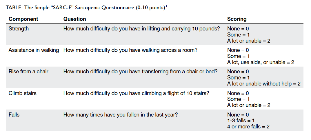
To the Editor—I thank Ho et al1 for their very interesting article “Prevalence of pre-sarcopenia and sarcopenia in Hong Kong Chinese geriatric patients with hip fracture and its correlation with different factors” in the February 2016 issue of the Hong Kong Medical Journal. I would like to mention another recent sarcopenia study in elderly Chinese men and women with a mean age of 81 years. The research group led by Hong et al2 showed that 42% of female patients and 84% of male patients with hip fracture were sarcopenic. In this study, the prevalence of sarcopenia with vertebral fracture was 34% in women and 40% in men. I agree with the authors that screening measures should be implemented more.1 In a general practice setting, measurement of SARC-F sarcopenia scale is feasible, simple, quick and inexpensive, and does not expose the patient to any particular strain (Table).3 The scale has also been evaluated in elderly patients in Hong Kong with direct measurement of muscle mass, strength, and physical performance.4 5 If a SARC-F score of ≥4 has been measured in an older patient, diagnosis of sarcopenia can be substantiated rather quickly.3 In my opinion, the SARC-F screen for sarcopenia should be routinely carried out among the Chinese elderly population every time they consult their doctor.
 

Table. The Simple “SARC-F” Sarcopenia Questionnaire (0-10 points)3
 
References
1. Ho AW, Lee MM, Chan EW, et al. Prevalence of pre-sarcopenia and sarcopenia in Hong Kong Chinese geriatric patients with hip fracture and its correlation with different factors. Hong Kong Med J 2016;22:23-9. Crossref
2. Hong W, Cheng Q, Zhu X, et al. Prevalence of sarcopenia and its relationship with sites of fragility fractures in elderly Chinese men and women. PLoS One 2015;10:e0138102. Crossref
3. Malmstrom TK, Morley JE. SARC-F: a simple questionnaire to rapidly diagnose sarcopenia. J Am Med Dir Assoc 2013;14:531-2. Crossref
4. Woo J, Leung J, Morley JE. Validating the SARC-F: a suitable community screening tool for sarcopenia? J Am Med Dir Assoc 2014;15:630-4. Crossref
5. Woo J, Leung J, Morley JE. Defining sarcopenia in terms of incident adverse outcomes. J Am Med Dir Assoc 2015;16:247-52. Crossref

Re: Alternatives to colonoscopy for population-wide colorectal cancer screening

DOI: 10.12809/hkmj164847
© Hong Kong Academy of Medicine. CC BY-NC-ND 4.0
 
LETTER TO THE EDITOR
Re: Alternatives to colonoscopy for population-wide colorectal cancer screening
John SM Leung, FCSHK, FHKAM (Surgery)
St Paul’s Hospital, Causeway Bay, Hong Kong
 
Corresponding author: Dr John SM Leung (leungsiumanjohn@yahoo.com.hk)
 
 Full paper in PDF
 
To the Editor—I much appreciate the timely publication of the review paper, “Alternatives to colonoscopy for population-wide colorectal cancer screening,” by Leung et al.1 We are facing an ageing population and a large proportion of our elderly patients have coronary and other arterial disorders for which they are prescribed aspirin. If stents have been deployed they are prescribed dual antiplatelet therapy. Direct interventional procedures such as colonoscopy might not be the ideal first-line screening procedure due to the risk of bleeding. Computed tomographic (CT) virtual colonoscopy might be an alternative. The CT imaging may not be comparable with the colonoscope, especially for the sub-centimetre polyp, but it has the advantage of picking up extraluminal lesions like epiploic appendagitis. More importantly the CT imaging also serves to screen the whole abdomen beyond the large intestine. The following case report illustrates this issue.
 
A 78-year-old retired professor complained of ill-defined pain in the chest. She was diagnosed to have coronary artery disease and two drug-eluting stents were deployed. The discomfort persisted and more medications were given. She became constipated and her haemorrhoids started to bleed. At this stage colonoscopy screening for colorectal cancer was considered but was reckoned inadvisable because of her dual antiplatelet therapy. A CT colonoscopy was done and revealed a few polyps and also a discreet tumour in the wall of the fundus of the stomach. Gastroscopy had likewise been vetoed because of the antiplatelet therapy. Nonetheless, the CT image helped to define the tumour outline to the extent that it was unlike the usual gastric carcinoma. Blood test for a panel of tumour markers showed that chromogranin A was strongly positive, thus narrowing the diagnosis to a gastric neuroendocrine tumour.
 
The standard treatment of a neuroendocrine tumour is surgical excision and so we are regrettably still faced with the problem of dual antiplatelet therapy and the risk of perioperative haemorrhage.
 
Reference
1. Leung WC, Foo DC, Chan TT, et al. Alternatives to colonoscopy for population-wide colorectal cancer screening. Hong Kong Med J 2016;22:70-7.

Pages