Antimicrobial resistance of clinical isolates in Hong Kong

ABSTRACT

Hong Kong Med J 1996;2:112–4 | Number 1, March 1996
LETTER TO THE EDITOR
Antimicrobial resistance of clinical isolates in Hong Kong
JM Ling
Department of Microbiology, Prince of Wales Hospital, Shatin, Hong Kong
 
 
No abstract available.
 
View this abstract indexed in MEDLINE:
 

A survey of pregnancies that ended in haemoglobin Bart's hydrops foetalis and Cooley's anaemia

ABSTRACT

Hong Kong Med J 1995;1:273 | Number 3, September 1995
LETTERS TO THE EDITOR
A survey of pregnancies that ended in haemoglobin Bart's hydrops foetalis and Cooley's anaemia
TN Leung, CY Li, A Chang
Department of Obstetrics and Gynaecology, Prince of Wales Hospital, Shatin, Hong Kong
 
Authors' reply
CS Feng, WC Tsoi
Haematology Laboratory and Blood Bank, Prince of Wales Hospital, Shatin, Hong Kong
 
 
No abstract available.
 

An addendum to “Doctor for Society” — Professor Lee Shiu Hung: “Pass on benevolence, pass on the legend”

Hong Kong Med J 2014;20:169 | Number 2, April 2014
DOI: 10.12809/hkmj144239
© Hong Kong Academy of Medicine. CC BY-NC-ND 4.0
 
LETTER TO THE EDITOR
An addendum to “Doctor for Society” — Professor Lee Shiu Hung: “Pass on benevolence, pass on the legend”
Jacob WT Ng, FCSHK, FHKAM (Surgery)1; John YH Ho, MEng (Oxon)2
1 Private practice
2 Love U All Charitable Foundation
 
Corresponding author: Dr Jacob WT Ng (ng_wtj@yahoo.com)
 
 
To the Editor—The section “Doctor for Society” written by medical students, featuring inspiring—at times, rather touching—stories, offered enjoyable reading at the end of a heavy diet of scientific and didactic papers. Not only did the remarkable feats of generations of compassionate doctors strike a chord with consummate fellows of the Academy, but they also stimulated and inspired many of our students. If we were to pass on one treasure to the latter, it should be the heart of benevolence.1
 
One of the most well-known and respected community medicine legends who deserves to be included in the series would be Prof Lee Shiu-hung. Regrettably, medical students no longer have the opportunity to interview him.
 
On 6 January this year, the senior author invited him (aged 81 years) to be the Guest of Honour at the Annual Winter Charity Dinner sponsored by his “Love U All Charitable Foundation”. As usual, he came on time with a warm smile.
 
Halfway through the dinner, he offered an impromptu entertainment for old folks and sang a Xianjiang folk song “That place from afar”. We were so taken by his enthusiasm that we took a snapshot of him while he was enjoying the lyrics, as did the ageing audience. Most interestingly, we found later right beside his head in the photograph the Chinese character for “Benevolence” from the backdrop, a word that aptly described his whole life to the very last minute (Fig). After singing the top notes at the end, he went back to his seat. Five minutes or so later, he fell to the carpeted floor and rapidly lapsed into deepening coma and passed away peacefully. It is most befitting for the premier medical journal of Hong Kong to be the first to publish this photo—the last glimpse of Prof Lee’s extraordinary long career and lifetime of devotion culminating in the realisation of true “Doctor for Society”.
 

Figure. The last photo of Prof Lee taken a few minutes before he fainted and passed away. Note the Chinese characters by the sides of his head
 
Reference
1. Wong I, Wong K. Pass on passion, pass on “loving heart”. Dr Jacob Wai-Tat Ng interviewed by medical students. Hong Kong Med J 2013;19:366-7.

Co-infection of influenza B and Streptococcus

Hong Kong Med J 2014;20:83 | Number 1, February 2014
DOI: 10.12809/hkmj134192
© Hong Kong Academy of Medicine. CC BY-NC-ND 4.0
 
LETTERS TO THE EDITOR
Co-infection of influenza B and Streptococcus
Beuy Joob, PhD1; Viroj Wiwanitkit, MD2
1 Sanitation, Medical Academic Center, Bangkok, Thailand
2 Hainan Medical University, China; Faculty of Medicine, University of Nis, Serbia
 
Corresponding author: Dr B Joob (beuyjoob@hotmail.com)
 
To the Editor—The recent report on 'co-infection of influenza B and Streptococcus' by Lam et al1 is very interesting. They reported "four cases infected with influenza B and streptococci that gave rise to severe pneumonia" and mentioned that "this is the second case report of severe invasive pneumococcal pneumonia secondary to influenza B infection."1 Indeed, both influenza B and Streptococcus infections are important infectious diseases that are encountered worldwide. In fact, there are more than two previous publications reporting the concurrent infection of influenza B and Streptococcus.2 3 In the report by Aebi et al,2 three cases were document and additional four cases were presented in the report by Scaber et al.3 Hence, the claim by Lam et al1 should not be correct. Nevertheless, in all reports, the clinical features of the pneumonia were serious and sometimes fatal. As Lam et al1 suggested, physicians should increase awareness of possible concurrent infection in the present era of emerging influenza.
 
References
1. Lam KW, Sin KC, Au SY, Yung SK. Uncommon cause of severe pneumonia: co-infection of influenza B and Streptococcus. Hong Kong Med J 2013;19:545-8. Crossref
2. Aebi T, Weisser M, Bucher E, Hirsch HH, Marsch S, Siegemund M. Co-infection of influenza B and streptococci causing severe pneumonia and septic shock in healthy women. BMC Infect Dis 2010;10:308. Crossref
3. Scaber J, Saeed S, Ihekweazu C, Efstratiou A, McCarthy N, O'Moore E. Group A streptococcal infections during the seasonal influenza outbreak 2010/11 in South East England. Euro Surveill 2011;16 pii:19780.
 
Authors' Reply
KW Lam, MB, BS, FHKAM (Medicine); KC Sin, MB, BS; SY Au, MB, BS; SK Yung, MB, BS
Intensive Care Unit, Queen Elizabeth Hospital, Jordan, Hong Kong
 
Corresponding author: Dr KW Lam (lamkw1@ha.org.hk)
To the Editor—Our report concerned severe pneumonia due to co-infection of influenza B and streptococcus. Scaber et al1 described a series of 19 invasive streptococcal infection cases affecting a variety of organs. Scaber's objective and emphasis were different from that of Aebi et al2 and our report.3 Moreover, Scaber et al1 did not provide much clinical details of the patients, such as laboratory investigation findings and complications. Therefore, their article1 was not included in our reference list. We would nevertheless like to thank Drs Joob and Wiwanitkit for their comments. The mentioned studies certainly enhance awareness of this serious co-infection of influenza B and streptococcus and alert physicians and other health care professionals.
 
References
1. Scaber J, Saeed S, Ihekweazu C, Efstratiou A, McCarthy N, O'Moore E. Group A streptococcal infections during the seasonal influenza outbreak 2010/11 in South East England. Euro Surveill 2011;16 pii:19780.
2. Aebi T, Weisser M, Bucher E, Hirsch HH, Marsch S, Siegemund M. Co-infection of influenza B and streptococci causing severe pneumonia and septic shock in healthy women. BMC Infect Dis 2010;10:308. Crossref
3. Lam KW, Sin KC, Au SY, Yung SK. Uncommon cause of severe pneumonia: co-infection of influenza B and Streptococcus. Hong Kong Med J 2013;19:545-8. Crossref

Laparoscopic removal of an eroding Mirena coil through the sigmoid colon

Hong Kong Med J 2014;20:82 | Number 1, February 2014
DOI: 10.12809/hkmj134203
© Hong Kong Academy of Medicine. CC BY-NC-ND 4.0
 
LETTERS TO THE EDITOR
Laparoscopic removal of an eroding Mirena coil through the sigmoid colon
Vincent YT Cheung, FRCOG, FRCSC
Department of Obstetrics and Gynaecology, Queen Mary Hospital, The University of Hong Kong, Pokfulam, Hong Kong
 
Corresponding author: Dr VYT Cheung (vytc@hku.hk)
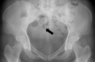
To the Editor—I read with interest the recently published article by Hussain et al,1 which reported a case of translocated intrauterine device (IUD) removed laparoscopically. I am most curious to know the meaning of "inconclusive X-rays" as stated by the authors. As Mirena is radiopaque, it can either be seen or not seen on X-rays, rather than inconclusive. For the purpose of illustration, I have included an X-ray of a 33-year-old woman who had a partially embedded Mirena in the uterine fundus (Fig). Interestingly, on reading an X-ray for a lost IUD, one really has to look for it to identify its presence. Further, in this reported case, if the X-rays were indeed inconclusive, on what basis did the authors decide that the IUD was expelled, without considering other imagings such as computed tomography (CT)? If the IUD was translocated close to the bowel, CT could also help to determine the presence and the degree of bowel penetration,2 3 4 so that preoperative counselling and preparation could be provided before proceeding with laparoscopy. The inference of "inconclusive X-rays" should have been pursued further, to avoid the surprise in finding at the first laparoscopy and the need for a second laparoscopy to remove the IUD.
 

Figure. A pelvic radiograph showing a Mirena intrauterine system (arrow)
 
References
1. Hussain A, Omar K, El-Hasani S. Laparoscopic removal of an eroding Mirena coil through the sigmoid colon. Hong Kong Med J 2013;19:560.e3-4. Crossref
2. Taras AR, Kaufman JA. Laparoscopic retrieval of intrauterine device perforating the sigmoid colon. JSLS 2010;14:453-5. Crossref
3. Zeino MY, Wietfeldt ED, Advani V, Ahad S, Younkin C, Hassan I. Laparoscopic removal of a copper intrauterine device from the sigmoid colon. JSLS 2011;15:568-70. Crossref
4. Boortz HE, Margolis DJ, Ragavendra N, Patel MK, Kadell BM. Migration of intrauterine devices: radiologic findings and implications for patient care. Radiographics 2012;32:335-52. Crossref
 
Authors’ Reply
A Hussain, FRCS (Eng), FRCS1; K Omar, FRCOG2; S El-Hasani, FRCS (Eng), FRCS1
1 General Surgery Department, Princess Royal University Hospital, Farnborough Common, Orpington, Kent, BR6 8ND, United Kingdom
2 Obstetric and Gynaecology Department, Princess Royal University Hospital, Farnborough Common, Orpington, Kent, BR6 8ND, United Kingdom
 
Corresponding author: Dr A Hussain (azahrahussain@yahoo.com)
 
To the Editor—Many thanks for Dr Cheung's interest in our article.1 First, all intrauterine contraceptive devices are radio-opaque as you correctly point out. The plain abdominal X-ray and ultrasound did not show evidence of the coil according to the reporting radiologists. The reasons were that the coil was unexpectedly located on the pelvic bones (left side) outside the uterine area and the colon was loaded with faeces. Retrospective analysis (after laparoscopy) and re-checking of the plain film actually showed the coil on the left side of the lower abdomen (outside the uterine area). Second, computed tomography (CT) had not been requested for two reasons. According to our hospital protocol, CT is to be avoided in young patients to reduce radiation risks, unless it is highly warranted for an acute abdomen or a lifesaving procedure. The other reason for laparoscopy and not CT was that this patient had lower abdominal pain and vaginal bleeding, of which gynaecological features are commonly investigated laparoscopically due to superior diagnostic yields than any form of imaging (including CT). Hence this woman was in need of a laparoscopy anyway and therefore there was no point of exposing her to the unnecessary risk of radiation via a CT.
 
Reference
1. Hussain A, Omar K, El-Hasani S. Laparoscopic removal of an eroding Mirena coil through the sigmoid colon. Hong Kong Med J 2013;19:560.e3-4. Crossref

Pages