Hong Kong Med J 2014;20:82 | Number 1, February 2014
DOI: 10.12809/hkmj134203
© Hong Kong Academy of Medicine. CC BY-NC-ND 4.0
LETTERS TO THE EDITOR
Laparoscopic removal of an eroding Mirena coil through the sigmoid colon
Vincent YT Cheung, FRCOG, FRCSC
Department of Obstetrics and Gynaecology, Queen Mary Hospital, The
University of Hong Kong, Pokfulam, Hong Kong
Corresponding author: Dr VYT Cheung (vytc@hku.hk)

To the Editor—I read with interest the recently
published article by Hussain et al,1 which reported
a case of translocated intrauterine device (IUD)
removed laparoscopically. I am most curious to
know the meaning of "inconclusive X-rays" as
stated by the authors. As Mirena is radiopaque,
it can either be seen or not seen on X-rays, rather
than inconclusive. For the purpose of illustration,
I have included an X-ray of a 33-year-old woman
who had a partially embedded Mirena in the uterine
fundus (Fig). Interestingly, on reading an X-ray for a
lost IUD, one really has to look for it to identify its
presence. Further, in this reported case, if the X-rays
were indeed inconclusive, on what basis did the
authors decide that the IUD was expelled, without
considering other imagings such as computed
tomography (CT)? If the IUD was translocated close
to the bowel, CT could also help to determine the
presence and the degree of bowel penetration,2 3 4 so
that preoperative counselling and preparation could
be provided before proceeding with laparoscopy.
The inference of "inconclusive X-rays" should have
been pursued further, to avoid the surprise in finding
at the first laparoscopy and the need for a second
laparoscopy to remove the IUD.
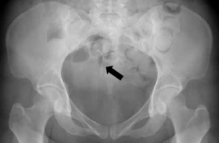
Figure. A pelvic radiograph showing a Mirena intrauterine system (arrow)
References
1. Hussain A, Omar K, El-Hasani S. Laparoscopic removal of an eroding Mirena coil through the sigmoid colon. Hong Kong Med J 2013;19:560.e3-4. Crossref
2. Taras AR, Kaufman JA. Laparoscopic retrieval of intrauterine device perforating the sigmoid colon. JSLS 2010;14:453-5. Crossref
3. Zeino MY, Wietfeldt ED, Advani V, Ahad S, Younkin C, Hassan I. Laparoscopic removal of a copper intrauterine device from the sigmoid colon. JSLS 2011;15:568-70. Crossref
4. Boortz HE, Margolis DJ, Ragavendra N, Patel MK, Kadell BM. Migration of intrauterine devices: radiologic findings and implications for patient care. Radiographics 2012;32:335-52. Crossref
Authors’ Reply
A Hussain, FRCS (Eng), FRCS1; K Omar, FRCOG2; S El-Hasani, FRCS (Eng),
FRCS1
1 General Surgery Department, Princess Royal University Hospital, Farnborough Common, Orpington,
Kent, BR6 8ND, United Kingdom
2 Obstetric and Gynaecology Department, Princess Royal University Hospital, Farnborough Common, Orpington,
Kent, BR6 8ND, United Kingdom
Corresponding author: Dr A Hussain (azahrahussain@yahoo.com)
To the Editor—Many thanks for Dr Cheung's interest in
our article.1 First, all intrauterine contraceptive
devices are radio-opaque as you correctly point out.
The plain abdominal X-ray and ultrasound did not
show evidence of the coil according to the reporting
radiologists. The reasons were that the coil was
unexpectedly located on the pelvic bones (left side)
outside the uterine area and the colon was loaded with
faeces. Retrospective analysis (after laparoscopy) and
re-checking of the plain film actually showed the coil
on the left side of the lower abdomen (outside the
uterine area). Second, computed tomography (CT)
had not been requested for two reasons. According
to our hospital protocol, CT is to be avoided in young
patients to reduce radiation risks, unless it is highly
warranted for an acute abdomen or a lifesaving
procedure. The other reason for laparoscopy and
not CT was that this patient had lower abdominal
pain and vaginal bleeding, of which gynaecological
features are commonly investigated laparoscopically
due to superior diagnostic yields than any form of
imaging (including CT). Hence this woman was in
need of a laparoscopy anyway and therefore there
was no point of exposing her to the unnecessary risk
of radiation via a CT.
Reference
1. Hussain A, Omar K, El-Hasani S. Laparoscopic removal of
an eroding Mirena coil through the sigmoid colon. Hong
Kong Med J 2013;19:560.e3-4. Crossref