Prevention of ventilator-associated pneumonia

Hong Kong Med J 2015 Feb;21(1):61–8 | Epub 16 Jan 2015
DOI: 10.12809/hkmj144367
© Hong Kong Academy of Medicine. CC BY-NC-ND 4.0
 
MEDICAL PRACTICE
Prevention of ventilator-associated pneumonia
Arthur CW Lau, FHKAM (Medicine)1; HM So, FHKAN (Critical Care)1; SL Tang, BSc (Nursing)2; Alwin Yeung, FHKAM (Medicine)2; SM Lam, FHKAM (Medicine)1; WW Yan, FHKAM (Medicine)1; for the Hong Kong East Cluster Task Force on Prevention of Ventilator-associated Pneumonia in Critical Care Areas
1Department of Intensive Care, Pamela Youde Nethersole Eastern Hospital, Chai Wan, Hong Kong
2Cardiac and Intensive Care Unit, Ruttonjee Hospital, Wanchai, Hong Kong
Corresponding author: Dr Arthur CW Lau (laucw3@ha.org.hk)
 Full paper in PDF
Abstract
Ventilator-associated pneumonia is the commonest, yet mostly preventable, infection in mechanically ventilated patients. Successful control of ventilator-associated pneumonia can save hospitalisation cost, and is possible by using a multidisciplinary clinical and administrative approach. The ventilator-associated pneumonia rate should be expressed as the number of ventilator-associated pneumonia days per 1000 ventilator days to take into account the device-utilisation duration for meaningful comparison. Various strategies address the issue, including general infection control measures, body positioning, intubation and mechanical ventilation, oral and gastro-intestinal tract, endotracheal tube, airway pressure, cuff pressure, selective digestive and/or oropharyngeal decontamination, and probiotic or early antibiotic treatment, as well as overall administration at a policy level. The rationale and controversy of these approaches are discussed in this article. The authors suggest that all units treating mechanically ventilated patients should have a ventilator-associated pneumonia prevention protocol in place, and ventilator-associated pneumonia should be seriously considered as a key performance indicator in local intensive care units.


Introduction
Ventilator-associated pneumonia (VAP) is the commonest, yet mostly preventable, infection in mechanically ventilated patients. It contributes to increased morbidity, mortality, and total hospitalisation cost. The estimated overall attributable mortality of VAP was 13%,1 and 52% of VAP was preventable.2 The VAP rate should be expressed as the number of VAP days per 1000 ventilator days to take into account the device-utilisation duration for meaningful comparison. From the reports of the National Health and Safety Network (NHSN) of the US Centers for Disease Control and Prevention (CDC),3 a decreasing trend for VAP has been noted in recent years. For example, from 2002 to 2012, for a mixed medical-surgical intensive care unit (ICU), the rate decreased from 5.1 to 0.9 days per 1000 ventilator days. The highest rates were reported for trauma (from 15.2 to 3.6 days) and burns (from 12.0 to 4.4 days) centres, while neurological and neurosurgical units reported intermediate rates (around 2 to 4 days). European rates are mostly higher, even if the same prevention strategies are used, and some possible reasons are explained below.4
 
Unlike many other conditions for which definitive diagnoses can be made pre-mortem, the gold standard of VAP diagnosis can only be made post-mortem. Clinicians diagnose VAP by radiology, signs and symptoms, with various methods of non-invasive and invasive microbiology sampling (tracheal aspirate, directed or blind broncho-alveolar lavage, protected specimen brush for qualitative and/or quantitative culture), and histology. However, when clinical criteria with microbiology were compared with autopsy findings, both the sensitivity and specificity were approximately 70%.5 Moreover, there has been no convincing evidence that qualitative culture of non-invasive samples, when compared with quantitative culture of invasive samples, produced any significant differences in clinical outcomes.6 In a survey of 27 ICUs of nine European countries, bronchoscopy was performed only in 23.3% of episodes of nosocomial pneumonia.7 Bronchoscopy might be considered more for immunocompromised hosts, both for detection of atypical organisms (if any) and to exclude infection.
 
The definition of VAP is therefore inherently subjective and, hence, contentious. The criteria used, interobserver variability, and subjectivity in chest X-ray interpretation affect the VAP rates by almost two-fold.8 9 All these factors influence findings in preventive trials and hamper comparisons. For surveillance purposes, most institutions use the surveillance definitions of radiology/signs/symptoms to monitor the rates. Commonly used surveillance definitions are those of the American Thoracic Society and Infectious Diseases Society of America (ATS/IDSA)10 and the CDC NHSN. The two sets of definitions are similar; the notable difference being that the ATS/IDSA guidelines exclude pneumonias occurring in the first 48 hours after intubation, whereas the CDC NHSN definition includes pneumonias in the first 48 hours; therefore, using the latter definition will result in a higher VAP incidence.
 
In an attempt to overcome the VAP definition’s inherent subjectivity, in 2014, the CDC NHSN started surveillance using the Ventilator-Associated Event protocol,11 under which a ventilator-associated condition (VAC) is defined if there are at least 2 days of stable or decreasing ventilator settings (positive end-expiratory pressure [PEEP] and fraction of inspired oxygen) followed by consistently higher settings for at least 2 days. The sensitivity of the VAC criteria for the detection of VAP was only 25.9%.12 When further microbiological and invasive techniques are employed, a VAC can be further classified into an infection-related VAC, and possible VAP. This modification enables greater objectivity and facilitates automated electronic capture, but captures changes due not only to VAP.
 
Pathophysiology and its implications for prevention strategies
In terms of pathophysiology, VAP is a misnomer because its occurrence is not related to the ventilator per se, but to the presence of the endotracheal tube (ETT). The ETT allows direct access to the lower respiratory tract, impairing the cough reflex and mucociliary clearance, but provides incomplete sealing to secretions above the cuff. Microaspiration (of materials from oropharyngeal cavities, sinuses, gastro-intestinal tract) and biofilm formation are the two most important mechanisms in VAP development, while inhalation, bacteraemia and haematogenous spread play smaller roles. Molecular analysis showed that most VAP patients had the same bacteria with the same sequencing in their oral cavities as in their lungs, and there were even pathogens found in the lungs that could not be detected by conventional culture-based methods.13 Types of bacteria vary with the time of onset; in early-onset VAP, they are commonly Enterobacteriaceae, Candida albicans and Staphylococcus aureus,14 while in late-onset VAP, they are Pseudomonas aeruginosa, Klebsiella pneumoniae, and Escherichia coli.
 
Evolution of prevention strategies
No single strategy is sufficient to prevent VAP, and several approaches are necessary. The most widely practised and well-known set of strategies is the Ventilator Bundle developed by the Institute for Healthcare Improvement (IHI) in 2001.15 The Ventilator Bundle is often mislabelled as the VAP Bundle, and its original aim was to improve better ventilator care overall, and not VAP alone, although its practice does reduce the VAP rate by 45%.15 The components of the Ventilator Bundle have now become the core of VAP prevention: (1) elevation of the head of bed to between 30° and 45°; (2) daily ‘sedative interruption’ and daily assessment of readiness to be extubated; (3) peptic ulcer disease prophylaxis; (4) deep venous thrombosis prophylaxis (unless contra-indicated); and (5) daily oral care with chlorhexidine. It is practice of the whole ‘bundle’ that decreases VAP, not just the individual components. Some components are not directly related to VAP, namely, deep venous thrombosis and peptic ulcer disease prophylaxis. Although specific data on VAP were not available, in general, the risk of hospital-acquired pneumonia increased when acid-suppressant medications were used. There was a slight trend for reduced VAP with sucralfate compared with a histamine 2 receptor antagonist, and proton pump inhibitors may be related to a higher rate of VAP.16 Therefore, acid-suppressant medications should not be overused. In Hong Kong, the Centre for Health Protection of the Department of Health promulgated a set of recommendations of VAP prevention in 2010.17 In this article, we review the latest literature regarding VAP prevention, and the strategies are summarised in the Table. The rationale and controversy are elaborated in the following discussion.
 

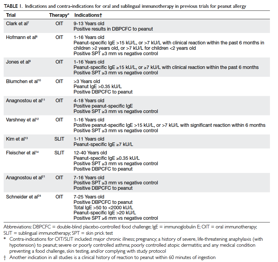
Table. Ventilator-associated pneumonia prevention strategies
Rationale and controversy behind some of the strategies are elaborated in the text. Strategies that have been suggested in the literature, but are not yet widely practised and/or require further research are listed in italics
 
Prevention strategies
Clinical strategies
General
General principles of infection control should be followed. Ventilator tubing should not be changed more frequently than every 1 week unless it is visibly soiled. In an early prospective randomised trial of 447 patients, decreasing the frequency of ventilator circuit changes from 3 times to once per week had no adverse effect on the overall rate of VAP.18 Subsequent trials also showed that circuit change intervals of 7 and 30 days had even lower risks for VAP than 2-day intervals.19 There was evidence that chlorhexidine bathing and hand hygiene compliance reduced global and specific infection rates, including VAP.20 A protocol for microbiological surveillance could be considered to aid in selection of empirical therapy.
 
Body positioning
The semi-recumbent position (30°-45° to the horizontal) is widely practised as one of the components of the IHI Ventilator Bundle, but which was based on only one randomised study with a non–intention-to-treat protocol of 86 mechanically ventilated patients, comparing the supine and semi-recumbent positions, in which the VAP rates were 34% and 8%, respectively.21 Subsequent studies were not able to reproduce these results, and found that a 45° position was difficult to maintain, and the mean angle achievable was only 28°. It has been suggested that avoidance of the supine position was beneficial as, while the semi-recumbent position might reduce gastro-oropharyngeal aspiration, the effect of gravity increases hydrostatic pressure of secretions pooled above the cuff. Recent animal studies found that mucus flow reversed towards the lungs in the semi-recumbent position, but drained out in the horizontal position. In human beings, the trachea/ETT axis is below the horizontal in the lateral Trendelenburg position (ie lying lateral, at 5°-10° below the horizontal). The Gravity VAP-Trial,22 an international randomised controlled trial (RCT) aiming at enrolment of 800 patients, is ongoing to compare the efficacy and safety of the two body positions, namely, the lateral Trendelenburg versus the semi-recumbent positions, in reducing the incidence of VAP. The estimated study completion date is December 2016. Continuous lateral rotation therapy, or kinetic bed therapy, ie lateral rotation of patient to ≥40° on one side (and 80° total arc) using specially designed beds, has been reported to reduce VAP, but was associated with other side-effects such as intolerance to rotation, unplanned extubation, loss of vascular access, and arrhythmias.23
 
Intubation and mechanical ventilation
Non-invasive ventilation (NIV) should be used to avoid the need for invasive ventilation and also shorten its duration. Non-invasive ventilation is particularly beneficial in chronic obstructive pulmonary disease (COPD), immunocompromised patients, and acute pulmonary oedema, and it allows early weaning from invasive ventilation in COPD. Summary estimates from 16 trials of moderate-to-good quality that included predominantly participants with COPD suggested that a weaning strategy that included NIV might reduce mortality and VAP rates without increasing the risk of weaning failure or reintubation.24
 
In a RCT of 128 adult mechanically ventilated patients, daily interruption of sedation resulted in a highly significant reduction in time spent on mechanical ventilation.25 The same authors also showed in the Awakening and Breathing Controlled trial that paired daily spontaneous awakening tests (ie interruption of sedatives) with daily spontaneous breathing tests resulted in better outcomes for mechanically ventilated patients.26 Although there is persistent concern of increased complications with this practice such as self-extubation, subsequent study found that this was not the case. A nurse-implemented sedation protocol has been shown to decrease VAP.27 Our ICU is also performing a nurse-led early weaning trial to aim at shortening the intubation period.
 
Oral and gastro-intestinal tract
Using chlorhexidine mouth rinse or gel as part of oral hygiene care, compared with placebo or usual care, was associated with a reduction in VAP, with a number needed to treat of 15 (95% confidence interval, 10-34).28 There is no consensus for the best practice for oral hygiene care. The optimal concentration of chlorhexidine solution to use is not known. Chlorhexidine has been studied in two strengths: 0.12% and 0.2%, and the US Food and Drug Administration recommends 0.12% oral chlorhexidine for use as mouth rinse. There is no evidence that the following is necessarily better than using chlorhexidine alone: addition of manual or powered tooth brushing, using Listerine (Johnson & Johnson Healthcare Products, New Brunswick [NJ], US), sodium bicarbonate oral rinses, or use of povidone-iodine solution. Povidone-iodine seemed to increase the rate of acute respiratory distress syndrome.29 A local study showed that continuous clearance of oral secretion by the saliva ejector might reduce the rate of VAP, decreasing the duration of mechanical ventilation, and shortening the duration of stay of patients in the ICU.30 Although gastroesophageal aspiration is implicated in VAP development, in an intention-to-treat study of 449 patients on mechanical ventilation, the absence of gastric volume monitoring was not inferior to routine residual gastric volume monitoring in terms of development of VAP (16.7% in the intervention group and 15.8% in the control group), nor were there any significant between-group differences in other ICU-acquired infections, mechanical ventilation duration, ICU stay duration, or mortality.31 There were more VAPs with early enteral feeding, and less with post-pyloric feeding. Further studies are required to confirm this.
 
Endotracheal tube
Novel designs of ETTs target two major mechanisms of VAP, namely, microaspiration and biofilm formation.32 33 Microaspiration is controlled by incorporating changes in cuff shape, material, and addition of a subglottic suction port. Prevention of biofilm formation is managed by addition of coating, surface modification, and removal of biofilm by mechanical means. The conventional ETT cuff (eg Portex; Smiths Medical International Ltd, Ashford, UK) is made of polyvinyl chloride (PVC), of which, when inflated, redundant parts fold on themselves and, because of the thickness of the material (>50 µ), microchannels form through which leakage is possible. By modification of the shape from conical to tapered (eg TaperGuard; Covidien, Irvine [CA], US), better contact of the cuff with the trachea can be achieved, and microchannel formation is reduced. By modification of the material from PVC to the thin 7-µ thick polyurethane (eg Microcuff; Kimberly-Clark Health Care, Roswell [GA], US), microchannel formation can almost be prevented. Our benchtop study showed that the Microcuff consistently outperformed the Portex and the TaperGuard in terms of leakage, especially during zero PEEP mechanical ventilation, laboured breathing, disconnection, and airway suction.34 One disadvantage of the Microcuff is that the material is so thin that even moisture diffuses across, leading to fluid accumulation inside the cuff with resultant blockage of the pilot balloon tube. This can be solved by purging the tube with small amount of air. Other materials being studied are Lycra fibre (Invista, Wichita [KS], US), silicone, and latex. Addition of a subglottic suction drainage (SSD) port removes secretions collected above the cuff (eg TaperGuard Evac, Covidien). In a meta-analysis of 10 RCTs with 2213 patients, SSD significantly reduced the incidence of VAP and early-onset VAP, shortened ventilation duration by 1.55 days, and prolonged time to VAP by 3.90 days.35 Ports of SSD are now available in tracheostomy tubes. One study of such tracheostomy tubes showed that the prevalences of VAP were 56% in the control group and 11% in the suction tracheostomy group.36 The SSD port can be put into intermittent or continuous suction, with similar efficacy of VAP prevention. However, the port could become blocked secondary to suctioned tracheal mucosa in one third of cases, with the possibility of tracheal injury.
 
Of the various coatings of ETT studied, so far, only silver-based coating (eg Agento IC; Bard Medical, Covington [GA], US) has been tested in clinical trials. A meta-analysis identified two high-quality RCTs with a total of 1630 participants, and showed that compared with non-coated ETTs, silver-coated ETTs resulted in a lower incidence of VAP, device-related adverse events, and microbiological burden, with no significant difference in total mortality.37 Recently, an ETT with a newly engineered micro-pattern surface was found to reduce colonisation and biofilm formation of key VAP-associated pathogens in vitro by 99.9% compared with unpatterned control ETTs, including methicillin-resistant S aureus and P aeruginosa biofilm formation.38 Another modification of the ETT is by addition of mucus slurping holes near the distal end of the ETT to keep the inner walls of the ETT free of mucus deposits. Attempts are also being made to develop mechanical means of removing adherent ETT secretions (eg endOclear catheter; endOclear LLC, Petoskey [MI], US), which could even relieve life-threatening ETT obstruction. Another example of mechanical removal is the Mucus Shaver.39
 
Airway pressure
Maintenance of a positive pressure gradient across the ETT cuff is an important factor to prevent secretions collecting above the cuff to trickle down across the cuff-trachea interface due to gravity. This pressure gradient is positive during positive pressure ventilation, but becomes zero during airway disconnection, and periodically negative during laboured inspiration through a narrow ETT or during airway suction. Without positive pressure, leakage around the cuff is significant, especially if the conventional PVC cuff is used and the cuff pressure is less than 30 cm H2O.34 During routine care, disconnections of the airway circuit are frequent, for instance, use of another ventilator for patient transport, change of the heat-and-moisture exchanger, spontaneous breathing trials, and change of ventilator tubing. Such disconnections should be avoided as far as possible. Spontaneous breathing trials should not be unduly prolonged without the intention for resumption of positive pressure ventilation or direct extubation. There is no evidence that the two methods of humidification, namely, using either a heated humidifier or a heat-and-moisture exchanger, affect the VAP rate.
 
Cuff pressure
A polyurethane cuff allows for a much lower sealing pressure than the conventional PVC cuff.34 Periodical manual checking of cuff pressure maintains adequate cuff pressure, but the benefit could be offset if the cuff pressure is accidentally released during checking. To overcome this issue, a continuous cuff pressure maintenance device can be used (Fig). A disposable device called the TRACOE Smart Cuff Manager (TRACOE Medical GmbH, Nieder-Olm, Germany; Fig) can also provide the similar functions of being both a pressure maintenance device (at 30 cm H2O) and a visual indicator of the adequacy of cuff pressure. In the largest study to date on the incidence of VAP comparing a continuous and an intermittent cuff pressure control system on 284 patients, a lower incidence of VAP was found (22.0% vs 11.2%).40
 

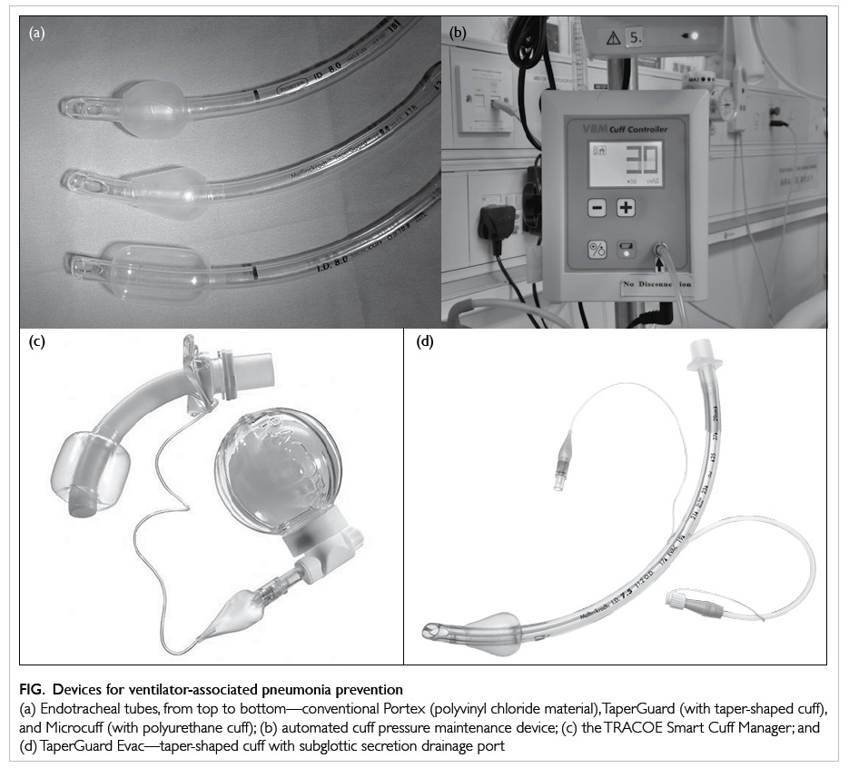
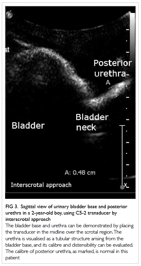
Figure. Devices for ventilator-associated pneumonia prevention
(a) Endotracheal tubes, from top to bottom—conventional Portex (polyvinyl chloride material), TaperGuard (with taper-shaped cuff), and Microcuff (with polyurethane cuff); (b) automated cuff pressure maintenance device; (c) the TRACOE Smart Cuff Manager; and (d) TaperGuard Evac—taper-shaped cuff with subglottic secretion drainage port
 
Selective digestive decontamination, selective oropharyngeal decontamination, and probiotic or early antibiotic treatment
Most meta-analyses have shown reductions in VAP with the use of selective digestive decontamination or selective oropharyngeal decontamination, but these interventions are still not being widely implemented because of concerns of emergence of antimicrobial resistance in pathogens. A recent systematic review of 64 studies suggested that such perceived risk of long-term harm could not be justified by the available data, but admitted that the effect on ICU-level antimicrobial resistance rates was understudied.41 Further studies are also required for the use of probiotics regarding efficacy and the risk of colonisation or infection with probiotics. Around 10% to 30% of ventilator-associated tracheobronchitis may progress to VAP, and appropriate antibiotic treatment was independently associated with reduced risk for transition.42 For antibiotic prophylaxis, a prospective cohort study of 129 comatose patients (Glasgow Coma Score ≤8) showed that a single dose of antibiotic within 4 hours of intubation might lower the incidence of early-onset VAP, with no difference in late-onset VAP.43 Further RCTs are needed.
 
Administrative strategies and coordination
Good administrative strategies and coordination are especially important for the implementation of this bundle of strategies all the way from patient care, equipment, infection control, and antibiotic stewardship. Considering the situation in Hong Kong that mechanically ventilated patients are being looked after not only in the ICU, but also in various critical care areas such as high dependency units, respiratory wards, and even general wards, it is imperative that a VAP protocol is in place and diligently adhered to in every such unit, with regular audits to ensure compliance. The existence of written standards for management of mechanically ventilated patients and the availability of VAP surveillance systems were positively associated with compliance with VAP-prevention measures and should be fostered at a policy level.45 Cost-effectiveness data for the whole Ventilator Bundle are scarce and such analysis should be performed in future research. In one Danish cost-effectiveness analysis, implementation of the Ventilator Bundle was found to be potentially cost-effective from the perspective of the hospital, using outcomes of prevention of VAP and prevention of death.46 In view of the necessity for multidisciplinary involvement, VAP surveillance is better coordinated at a higher administrative level. The Hong Kong East Cluster of Hospitals has established a Task Force on Prevention of VAP in Critical Care Areas since 2013. Ventilator-associated pneumonia should also be seriously considered as a key performance indicator in Hong Kong ICUs, as in other advanced countries.
 
Conclusion
Ventilator-associated pneumonia is the commonest, yet mostly preventable, cause of morbidity and mortality in mechanically ventilated patients. Successful control of VAP can save total hospitalisation cost, and is possible by using a multidisciplinary clinical and administrative approach. All units taking care of mechanically ventilated patients should have a VAP-prevention protocol in place, and VAP should be seriously considered to become a key performance indicator in local ICUs.
 
References
1. Melsen WG, Rovers MM, Groenwold RH, et al. Attributable mortality of ventilator-associated pneumonia: a meta-analysis of individual patient data from randomised prevention studies. Lancet Infect Dis 2013;13:665-71. CrossRef
2. Lambert ML, Silversmit G, Savey A, et al. Preventable proportion of severe infections acquired in intensive care units: case-mix adjusted estimations from patient-based surveillance data. Infect Control Hosp Epidemiol 2014;35:494-501. CrossRef
3. Ventilator-associated pneumonia (VAP). Available from: http://www.cdc.gov/hai/vap/vap.html. Accessed 21 Jul 2014.
4. Kalanuria AA, Zai W, Mirski M. Ventilator-associated pneumonia in the ICU. Crit Care 2014;18:208. CrossRef
5. Fàbregas N, Ewig S, Torres A, et al. Clinical diagnosis of ventilator associated pneumonia revisited: comparative validation using immediate post-mortem lung biopsies. Thorax 1999;54:867-73. CrossRef
6. Berton DC, Kalil AC, Teixeira PJ. Quantitative versus qualitative cultures of respiratory secretions for clinical outcomes in patients with ventilator-associated pneumonia. Cochrane Database Syst Rev 2012;(10):CD006482.
7. Koulenti D, Lisboa T, Brun-Buisson C, et al. Spectrum of practice in the diagnosis of nosocomial pneumonia in patients requiring mechanical ventilation in European intensive care units. Crit Care Med 2009;37:2360-8. CrossRef
8. Novosel TJ, Hodge LA, Weireter LJ, et al. Ventilator-associated pneumonia: depends on your definition. Am Surg 2012;78:851-4.
9. Klompas M. Interobserver variability in ventilator-associated pneumonia surveillance. Am J Infect Control 2010;38:237-9. CrossRef
10. American Thoracic Society; Infectious Diseases Society of America. Guidelines for the management of adults with hospital-acquired, ventilator-associated, and healthcare-associated pneumonia. Am J Respir Crit Care Med 2005;171:388-416. CrossRef
11. Ventilator-Associated Event (VAE). Available from: http://www.cdc.gov/nhsn/PDFs/pscManual/10-VAE_FINAL.pdf. Accessed 1 Jan 2015.
12. Muscedere J, Sinuff T, Heyland DK, et al. The clinical impact and preventability of ventilator-associated conditions in critically ill patients who are mechanically ventilated. Chest 2013;144:1453-60. CrossRef
13. Bahrani-Mougeot FK, Paster BJ, Coleman S, et al. Molecular analysis of oral and respiratory bacterial species associated with ventilator-associated pneumonia. J Clin Microbiol 2007;45:1588-93. CrossRef
14. Charles MP, Easow JM, Joseph NM, Ravishankar M, Kumar S, Sivaraman U. Aetiological agents of ventilator-associated pneumonia and its resistance pattern—a threat for treatment. Australas Med J 2013;6:430-4. CrossRef
15. How-to guide: Prevent ventilator-associated pneumonia. Available from: http://www.ihi.org/resources/Pages/Tools/HowtoGuidePreventVAP.aspx. Accessed 20 Jul 2014.
16. Khorvash F, Abbasi S, Meidani M, Dehdashti F, Ataei B. The comparison between proton pump inhibitors and sucralfate in incidence of ventilator associated pneumonia in critically ill patients. Adv Biomed Res 2014;3:52. CrossRef
17. Recommendations on prevention of ventilator-associated pneumonia. Scientific Committee on Infection Control, and Infection Control Branch, Centre for Health Protection, Department of Health; 2010.
18. Long MN, Wickstrom G, Grimes A, Benton CF, Belcher B, Stamm AM. Prospective, randomized study of ventilator-associated pneumonia in patients with one versus three ventilator circuit changes per week. Infect Control Hosp Epidemiol 1996;17:14-9. CrossRef
19. Fink JB, Krause SA, Barrett L, Schaaff D, Alex CG. Extending ventilator circuit change interval beyond 2 days reduces the likelihood of ventilator-associated pneumonia. Chest 1998;113:405-11. CrossRef
20. Martínez-Reséndez MF, Garza-González E, Mendoza-Olazaran S, et al. Impact of daily chlorhexidine baths and hand hygiene compliance on nosocomial infection rates in critically ill patients. Am J Infect Control 2014;42:713-7. CrossRef
21. Drakulovic MB, Torres A, Bauer TT, Nicolas JM, Nogué S, Ferrer M. Supine body position as a risk factor for nosocomial pneumonia in mechanically ventilated patients: a randomised trial. Lancet 1999;354:1851-8. CrossRef
22. The Gravity VAP network. Gravity VAP-Trial. Available from: http://compartint.net/gravityvaptrial/joomla/. Accessed 21 Jul 2014.
23. Staudinger T, Bojic A, Holzinger U, et al. Continuous lateral rotation therapy to prevent ventilator-associated pneumonia. Crit Care Med 2010;38:486-90. CrossRef
24. Burns KE, Meade MO, Premji A, Adhikari NK. Noninvasive positive-pressure ventilation as a weaning strategy for intubated adults with respiratory failure. Cochrane Database Syst Rev 2013;(12):CD004127.
25. Kress JP, Pohlman AS, O’Connor MF, Hall JB. Daily interruption of sedative infusions in critically ill patients undergoing mechanical ventilation. N Engl J Med 2000;342:1471-7. CrossRef
26. Girard TD, Kress JP, Fuchs BD, et al. Efficacy and safety of a paired sedation and ventilator weaning protocol for mechanically ventilated patients in intensive care (Awakening and Breathing Controlled trial): a randomised controlled trial. Lancet 2008;371:126-34. CrossRef
27. Quenot JP, Ladoire S, Devoucoux F, et al. Effect of a nurse-implemented sedation protocol on the incidence of ventilator-associated pneumonia. Crit Care Med 2007;35:2031-6. CrossRef
28. Shi Z, Xie H, Wang P, et al. Oral hygiene care for critically ill patients to prevent ventilator-associated pneumonia. Cochrane Database Syst Rev 2013;(8):CD008367.
29. Seguin P, Laviolle B, Dahyot-Fizelier C, et al. Effect of oropharyngeal povidone-iodine preventive oral care on ventilator-associated pneumonia in severely brain-injured or cerebral hemorrhage patients: a multicenter, randomized controlled trial. Crit Care Med 2014;42:1-8. CrossRef
30. Chow MC, Kwok SM, Luk HW, Law JW, Leung BP. Effect of continuous oral suctioning on the development of ventilator-associated pneumonia: a pilot randomized controlled trial. Int J Nurs Stud 2012;49:1333-41. CrossRef
31. Reignier J, Mercier E, Le Gouge A, et al. Effect of not monitoring residual gastric volume on risk of ventilator-associated pneumonia in adults receiving mechanical ventilation and early enteral feeding: a randomized controlled trial. JAMA 2013;309:249-56. CrossRef
32. Fernandez JF, Levine SM, Restrepo MI. Technologic advances in endotracheal tubes for prevention of ventilator-associated pneumonia. Chest 2012;142:231-8. CrossRef
33. Lam SM, Lau AC. Prevention of ventilator-associated pneumonia (VAP) by novel endotracheal tube designs. Hong Kong Lung Foundation, Hong Kong Thoracic Society & ACCP (HK & Macau Chapter) Newsletter 2011: 32-5.
34. Lau AC, Lam SM, Yan WW. Benchtop study of leakages across the Portex, TaperGuard, and Microcuff endotracheal tubes under simulated clinical conditions. Hong Kong Med J 2014;20:7-15. CrossRef
35. Wang F, Bo L, Tang L, et al. Subglottic secretion drainage for preventing ventilator-associated pneumonia: an updated meta-analysis of randomized controlled trials. J Trauma Acute Care Surg 2012;72:1276-85.
36. Ledgerwood LG, Salgado MD, Black H, Yoneda K, Sievers A, Belafsky PC. Tracheotomy tubes with suction above the cuff reduce the rate of ventilator-associated pneumonia in intensive care unit patients. Ann Otol Rhinol Laryngol 2013;122:3-8. CrossRef
37. Li X, Yuan Q, Wang L, Du L, Deng L. Silver-coated endotracheal tube versus non-coated endotracheal tube for preventing ventilator-associated pneumonia among adults: a systematic review of randomized controlled trials. J Evid Based Med 2012;5:25-30. CrossRef
38. May RM, Hoffman MG, Sogo MJ, et al. Micro-patterned surfaces reduce bacterial colonization and biofilm formation in vitro: potential for enhancing endotracheal tube designs. Clin Transl Med 2014;3:8. CrossRef
39. Berra L, Coppadoro A, Bittner EA, et al. A clinical assessment of the Mucus Shaver: a device to keep the endotracheal tube free from secretions. Crit Care Med 2012;40:119-24. CrossRef
40. Lorente L, Lecuona M, Jiménez A, et al. Continuous endotracheal tube cuff pressure control system protects against ventilator-associated pneumonia. Crit Care 2014;18:R77. CrossRef
41. Daneman N, Sarwar S, Fowler RA, Cuthbertson BH; SuDDICU Canadian Study Group. Effect of selective decontamination on antimicrobial resistance in intensive care units: a systematic review and meta-analysis. Lancet Infect Dis 2013;13:328-41. CrossRef
42. Nseir S, Martin-Loeches I, Makris D, et al. Impact of appropriate antimicrobial treatment on transition from ventilator-associated tracheobronchitis to ventilator-associated pneumonia. Crit Care 2014;18:R129. CrossRef
43. Vallés J, Peredo R, Burgueño MJ, et al. Efficacy of single-dose antibiotic against early-onset pneumonia in comatose patients who are ventilated. Chest 2013;143:1219-25. CrossRef
44. Kaier K, Lambert ML, Frank UK, et al. Impact of availability of guidelines and active surveillance in reducing the incidence of ventilator-associated pneumonia in Europe and worldwide. BMC Infect Dis 2014;14:199. CrossRef
45. Møller AH, Hansen L, Jensen MS, Ehlers LH. A cost-effectiveness analysis of reducing ventilator-associated pneumonia at a Danish ICU with ventilator bundle. J Med Econ 2012;15:285-92. CrossRef
 
Find HKMJ in MEDLINE:
 

Non-surgical treatment of lung cancer: personalised stereotactic ablative radiotherapy

Hong Kong Med J 2014;20(6):529–36 | Epub 26 Sep 2014
DOI: 10.12809/hkmj144269
© Hong Kong Academy of Medicine. CC BY-NC-ND 4.0
 
MEDICAL PRACTICE
Non-surgical treatment of lung cancer: personalised stereotactic ablative radiotherapy
Maverick WK Tsang, FRCR, FHKAM (Radiology)1; Michael KM Kam, FRCR, FHKAM (Radiology)1; SF Leung, MD, FHKAM (Radiology)1; Anthony TC Chan, MD, FRCP2
1 Department of Clinical Oncology, Prince of Wales Hospital, Shatin, Hong Kong
2 State Key Laboratory in Oncology in South China, The Chinese University of Hong Kong, Shatin, Hong Kong
 
Corresponding author: Dr Maverick WK Tsang (wk_tsang@clo.cuhk.edu.hk)
 Full paper in PDF
Abstract
Stereotactic ablative radiotherapy has emerged as a standard treatment for medically inoperable stage I non–small-cell lung cancer and selected cases of lung metastasis. Techniques to freeze or limit tumour movement during treatment and image-guided radiation delivery are integral to a successful stereotactic ablative treatment without overdose of surrounding normal structures. In this article, the practice in a local oncology institution will be used to illustrate the concept of personalised stereotactic ablative radiotherapy.


Introduction
Lung cancer is the second most common cancer and number one killer among all cancers in Hong Kong.1 Non–small-cell lung cancer (NSCLC) accounts for about 80% of all lung cancer cases. Surgery remains the mainstay of treatment for early-stage NSCLC. For patients who refuse or are medically unfit for surgery, stereotactic ablative radiotherapy (SABR) has emerged as a standard treatment. In the era of personalised medicine, SABR should be executed with techniques which are most suitable for the patient. In this article, the concepts of SABR, tumour motion control, and image-guided radiation delivery will be introduced. Then, using the Prince of Wales Hospital as an example, the approach to selecting the appropriate techniques for execution of personalised SABR will be explained.
 
Stereotactic ablative radiotherapy
Stereotactic ablative radiotherapy, also named as stereotactic body radiotherapy, is defined as “an external beam radiotherapy method used to very precisely deliver a high dose of radiation to an extracranial target within the body, using either a single dose or a small number of fractions”.2 The goal of SABR is to give a very high (ablative) radiation dose to kill all cancer cells within the target while avoiding radiation damage to the surrounding normal tissues. For lung SABR, a radiation dose of 10 to 20 Gray (Gy, a radiation dose unit) per treatment fraction is delivered for a total of 48 to 60 Gy in five or less fractions within 2 weeks.
 
Indications for stereotactic ablative radiotherapy in lung cancer
Stage I non–small-cell lung cancer: medically inoperable disease or patient refuses surgery
Nowadays, SABR is the gold-standard treatment for patients with stage I NSCLC who refuse surgical intervention or are medically unfit to undergo surgery (mostly due to poor pulmonary function). Prospective phase I/II studies document local control rate of over 90% with SABR, with overall survival approaching that of surgery (Table 13 4 5 6 7 8 9 10). In Hong Kong, the reported 2-year local control and 2-year overall survival rates were 89% to 95% and 53% to 87%, respectively.11 12 13 Generally, SABR is well tolerated, even by patients with poor pulmonary function. Guckenberger et al14 demonstrated that SABR had only very limited acute and chronic pulmonary toxicity even in patients with poor pulmonary function and there was no requirement of minimal pulmonary function for safe practice of SABR.
 

Table 1. Results of prospective studies of stereotactic ablative radiotherapy for early-stage non–small-cell lung cancer3 4 5 6 7 8 9 10
 
In view of the encouraging data on local control and mild toxicity of SABR for medically inoperable cases, the gold-standard role of surgery for operable stage I NSCLC is now being challenged. At least two retrospective studies have shown that survival of patients with operable stage I NSCLC treated with SABR paralleled that of lobectomy.15 16 Another two prospective phase 2 trials reported 76% to 84% 2-to-3-year overall survival rates for operable stage I disease after SABR, which compares favourably to surgical outcomes.3 7 Unfortunately, all phase 3 trials comparing SABR with surgery in operable stage I NSCLC were terminated prematurely due to poor accrual. Thus, the race between SABR and surgery for the title of standard treatment for operable stage I NSCLC continues without a foreseeable end.
 
Oligometastasis involving the lungs with controlled primary tumour
Generally, oligometastasis is defined as one to five metastatic lesions besides the primary tumour. Evidence has emerged that patients with limited metastases can be cured by removal of all metastases. The reported 10- and 15-year survival rates of patients undergoing complete lung metastasectomy were 26% and 22%, respectively. Patients with fewer metastases and longer disease-free interval fared even better.17 Prospective studies of limited lung metastases treated with SABR reported 2-year local control and 2-year survival rates of 89% to 96% and 39% to 84%, respectively, which are not inferior to the surgical results.18 19 20 21
 
Stereotactic ablative radiotherapy versus conventional radiotherapy
The local control rate of stage I NSCLC after SABR is ≥90%, in contrast to 50% rate with conventional radiotherapy.22 23 24 A clinical dose-response curve of human malignant lung tumours has been established.25 Thus, a high local tumour control can be achieved by delivering a high radiation dose. In lung cancer, however, the intrathoracic normal structures (normal lung tissues, spinal cord, brachial plexus, oesophagus, trachea and main bronchi, heart, great vessels, ribs and skin) close to the tumour may also receive a high radiation dose which may cause severe or even fatal treatment complications. It is this radiation damage of normal structures that limits the possible radiation dose to the lung tumour in conventional radiotherapy.
 
Stereotactic ablative radiotherapy is able to deliver a very high radiation dose to the target while sparing the surrounding normal tissues, thanks to its intrinsic physical advantage. On the other hand, many normal structures can tolerate a small volume of high-dose radiation without complications. Thus, we can deliver high-dose tumour radiation and yet limit volumes of the surrounding normal tissues exposed to high-dose radiation by reducing the size of the radiation field. In SABR, radiation field size reduction can be achieved through incorporation of techniques to limit tumour movement during radiotherapy (tumour motion management) and image-guided radiation delivery.
 
Physical property
Compared with conventional radiotherapy, SABR is able to create a very rapid dose fall-off at the target normal tissue interface so that radiation can be precisely delivered to the target without damaging the surrounding normal tissues. This can be achieved by aligning the treatment field borders or multi-leaf collimators close to the planning target volume (PTV) borders (refer to the “Individualised radiation target volume” section below for definition of PTV) and by prescribing dose at the part of the radiation dose depth curve with a steep slope, ie 60% to 90% isodose line. Such rapid dose fall-off property of SABR can be further enhanced by adoption of intensity-modulated radiotherapy or volumetric-modulated arc therapy techniques which, in addition, can create a concave radiation dose distribution to further improve radiation dose conformity to the target.
 
Tumour movement restriction during radiotherapy
A lung tumour will move with respiration. In conventional radiotherapy, the radiation field is enlarged to encompass the tumour in all respiratory phases. In SABR, however, tumour movement during radiotherapy is restricted by various tumour motion management techniques. As a result, the radiation field can be smaller, thereby, limiting the amount of normal tissues exposed to a high radiation dose.
 
Image-guided radiotherapy
Even with proper tumour motion management, there will be residual tumour movement both during the same treatment fraction (intra-fraction) and between different fractions on different days (inter-fractions). Therefore, daily pretreatment verification of tumour position by various image-guided radiotherapy (IGRT) techniques is essential to avoid geographical tumour miss. Radiation field size reduction can only be realised with IGRT.
 
Techniques to freeze or restrict movement of a lung tumour during stereotactic ablative radiotherapy
Active breathing control/voluntary inspiratory breath-hold
Breath-holding SABR can be achieved actively by active breathing control (ABC) or voluntarily by self-initiated breath-hold. The ABC apparatus is a modified spirometer consisting of two pairs of flow monitors and scissor valves to control inspiration and expiration, respectively. The operator activates ABC at a predefined lung volume by closing both valves to immobilise the breathing motion for 15 to 20 seconds. At the same time, radiation beam is switched on. Then, the patient is allowed to breathe freely until the next ABC activation. The cycle is repeated until complete delivery of a treatment fraction, which typically takes 30 minutes. Mostly, ABC will be activated in inspiration when lungs expand, resulting in less normal lung irradiation (Fig 1a). A study showed that ABC reduced normal lung V20 (volume of normal lung tissues receiving a dose ≥20 Gy) by 34% compared with free breathing.26 There is a good reproducibility of lung tumour position under ABC both inter-fractionally and intra-fractionally, with mean tumour displacement in supero-inferior direction of 1.1 mm and 0.3 mm, respectively.27 28 Voluntary inspiratory breath-hold technique is used in case a patient can hold his/her breath for at least 15 seconds but is unable to hold the mouthpiece of an ABC apparatus without air leakage.
 

Figure 1. Tumour motion management techniques
(a) Breathing waveform shows that active breathing control is activated at deep inspiratory level, and (b) Real-time Patient Management system respiratory gating, where radiation beam is on only at end of expiration
 
Respiratory gating
Respiratory gating involves delivery of radiation only in certain phases of respiration. The gating window (respiratory phases at which radiation beam will be turned on) is usually selected at late expiratory phases as a lung tumour stays for a longer period in the expiratory phase than in the inspiratory phase, resulting in a shorter treatment time. In addition, the tumour position will be more consistent and reproducible at end of expiration. A four-dimensional computed tomography (4D-CT; 4D means 3D + time) for radiotherapy planning purpose is done with the Real-time Patient Management (RPM) system (Varian Medical Systems, US), which consists of an infrared reflective block and an infrared tracking camera. The reflective block is placed on the anterior abdominal skin surface midway between the xiphisternum and umbilicus. The infrared camera tracks motion of the reflective block. The up-and-down breathing movement of the abdominal wall and thus position of the reflective block now reflects the respiratory phase during which a particular set of CT images are captured. As a result, positions of the tumour in various respiratory phases can be displayed on the 4D-CT images. A radiation field is designed according to the tumour positions at selected gating windows. Respiratory gating can be executed with either the RPM or the ExacTrac Adaptive Gating systems (Brainlab AG, Germany).
 
Real-time Patient Management system
During SABR, the infrared reflective block is placed on the patient’s abdominal wall and serves as an external indicator to predict the tumour location. The infrared camera will track movements of the reflective block to turn the radiation beam on (at gating window) and off (Fig 1b).
 
ExacTrac Adaptive Gating system
Similar to the RPM system, ExacTrac has an optical infrared tracking system comprising several infrared reflective body markers (usually five to eight) and an infrared tracking camera mounted on the ceiling of the radiation treatment room (Fig 2a). The radiation beam will be turned on only at a predefined gating window. The advantages and disadvantages of different tumour motion management techniques are tabulated in Table 2.
 

Figure 2. Image-guided radiotherapy facilities
(a) ExacTrac Adaptive Gating system. The two X-ray tubes are embedded on the room floor. The two amorphous silicon flat panel X-ray detectors, and the infrared tracking camera are mounted on the ceiling. (b) Onboard Imager. The X-ray tube and X-ray detector are mounted on the linear accelerator so that the X-ray tube-detector pair is always at 90º to the treatment head of the linear accelerator
 

Table 2. Comparison of different techniques
 
Radiation delivery under image guidance
Multiple studies have concluded that neither external indicators (infrared reflective block in the RPM system29 30) nor internal indicators (diaphragm,31 bony anatomy such as vertebral bodies32 33) have a consistent correlation with tumour position over time. Therefore, direct visualisation of the lung tumour itself is required for accurate and precise radiation delivery. Image-guided radiotherapy is a procedure that uses various imaging techniques (eg X-ray and CT) to identify a tumour to guide the radiation beam during SABR treatment. It makes radiotherapy more accurate and causes less damage to healthy tissues.
 
Pretreatment verification of tumour position by a CT or X-ray imaging device mounted on a linear accelerator (Linac; a machine for generation and delivery of radiation beam) should be done daily for detection of inter-fractional tumour displacement, with necessary correction if there is significant tumour displacement from its planned position (Fig 3). Moreover, interval treatment verification with X-ray imaging during one treatment fraction is required for identification of intra-fractional tumour displacement.
 

Figure 3. Pretreatment verification of tumour position. The small circles indicate the area where the fiducial marker is expected to stay during radiotherapy
(a) No displacement of the tumour. (b) Tumour displacement from its expected position, as indicated by displacement of the fiducial markers outside the small circles. The patient has to be repositioned for correction of tumour displacement before start of radiotherapy
 
Onboard Imager
Onboard Imager (OBI; Varian Medical Systems, US) is a high-resolution X-ray device mounted on the treatment head of a Linac to display real-time tumour location (Fig 2b). Radiographic images can be taken at the gating window for online (ie when a patient lies on treatment table of the Linac) verification of tumour position in RPM respiratory-gated radiotherapy. In ABC treatment, the tumour position under ABC can also be verified online. It should be noted that X-ray images can be taken only when the radiation beam is turned off.
 
Cone-beam computed tomography
Cone-beam CT (CBCT) is a 3D mode of OBI, which is able to acquire and reconstruct 3D volumetric data in one rotation of treatment head of the Linac in 1 minute. Because of the long image acquisition time, CBCT is unsuitable for treatment verification if breath-holding or respiratory gating techniques are used. Rather, it is a useful and accurate tool for daily treatment verification of the Tumour Encompassing Targeting radiotherapy.
 
ExacTrac Adaptive Gating
The ExacTrac Adaptive Gating system has two components: the optical infrared tracking system for respiratory gating (mentioned above) and the stereoscopic X-ray imaging system for online detection and correction of tumour position shift. The stereoscopic X-ray imaging system consists of two X-ray tubes embedded in the Linac room floor and two amorphous silicon flat panel detectors mounted on the ceiling; the angle between the two X-ray tube-detector pairs is approximately 90° (Fig 2a). Stereoscopic X-ray can be taken at the gating window for verification of tumour position. Its advantage over OBI is that periodic X-ray acquisition at gating window is possible during both beam-on and beam-off periods. That means tumour displacement can be detected anytime during treatment, including during the beam-on period. Radiation beam can be turned off automatically if the tumour is displaced outside its allowed region. Table 2 compares the pros and cons of various IGRT techniques.
 
Personalised stereotactic ablative radiotherapy adapted to patient’s needs and limitations
Individualised tumour motion management
The criteria for selection of an appropriate tumour motion management technique include: (1) ability of the patient to tolerate inspiratory breath-hold for ≥15 seconds, (2) extent of tumour movement at tidal breathing, and (3) selected IGRT technique (refer to the ‘Individualised image-guided radiotherapy’ section below for details).
 
Ideally, all patients should be treated under breath-holding condition as the lung tumour will be frozen and a minimal safety margin is required for creation of a radiation field. In practice, many lung cancer patients are elderly and smokers with compromised pulmonary function. These patients cannot hold the breath long enough for SABR treatment. For patients unsuitable for breath-holding techniques, a 4D-CT is done under tidal breathing to assess the magnitude of tumour movement. If it is ≤1 cm, the Tumour Encompassing Targeting technique (‘3.5D’ radiotherapy) is used in which the radiation field will cover all possible tumour positions at any respiratory phase as shown on 4D-CT. For tumours with excessive (>1 cm) movement under tidal breathing, the respiratory gating technique should be utilised.
 
A flowchart showing the approach to selecting appropriate tumour motion management technique for lung SABR is shown in Figure 4a.
 

Figure 4. Flowcharts showing the approach to selecting appropriate tumour motion management and image-guided radiotherapy techniques for lung stereotactic ablative radiotherapy: (a) tumour motion management and (b) image-guided radiotherapy
 
Individualised image-guided radiotherapy
An IGRT technique should be selected to match requirements of the selected tumour motion management technique and characteristics of the tumour (Fig 4b). A fiducial marker (a marker made of pure gold that can be visualised clearly on X-ray) can be implanted into or close to the tumour as an indicator of tumour location under OBI. There are various commercially available fiducial markers of different shapes and sizes, such as a cylindrical marker measuring 0.75 mm in diameter and 10 mm in length. Fiducial markers are implanted under CT guidance. In theory, all SABR treatments should be executed either under fiducial markers guidance or with CBCT as these are the most accurate methods for pretreatment tumour position verification. In fact, CBCT is the IGRT technique of choice for ‘3.5D’ radiotherapy. Nearly all patients cannot hold the breath long enough for performing CBCT, which typically takes a minute for 360° acquisition of a full set of CT images. Furthermore, 4D-CBCT is unavailable in most oncology centres in Hong Kong. As a result, CBCT pretreatment verification is impractical for both breath-holding and respiratory gating techniques. On the other hand, fiducial marker implantation under CT guidance will result in significant pneumothorax necessitating insertion of a chest drain. Thus, it is not recommended in old and/or frail patients. Provided that the tumour can be visualised on X-ray, OBI alone can be used for pretreatment verification in such patients. Unfortunately, a lung tumour may not be visible on X-ray because of its small size or close proximity to the ribs, mediastinum, or heart. If a fiducial marker is not implanted and the tumour is invisible on OBI, neither breath-holding nor respiratory-gated SABR treatment is realistic. Instead, the ‘3.5D’ radiotherapy technique should be utilised with CBCT pretreatment verification.
 
Individualised radiation target volume
The International Commission on Radiation Units and Measurements Report 62 clearly defines various target volumes for radiation.34 Gross tumour volume (GTV) is the tumour mass shown on clinical examination or by imaging. Clinical target volume (CTV) encompasses the subclinical microscopic disease around GTV. In SABR, the tissue immediately around GTV will receive a dose high enough to eradicate all possible microscopic disease. Therefore, CTV margin is not required in most of the cases, ie GTV = CTV. To compensate for possible inter-fractional and intra-fractional tumour movement, a further margin (internal margin [IM]) is added to CTV to create the internal target volume (ITV). As 4D-CT scan for radiotherapy planning only delineates the snapshot tumour movement at the time of scanning, an IM is still required to allow for residual tumour movement. A final setup margin (SM) for all uncertainties in patient-radiation beam positioning during radiotherapy treatment is added to ITV to become the final PTV. The required IM and SM depend on the selected tumour motion management and IGRT techniques. As the breathing pattern of patients may change significantly both within one treatment and between different treatment sessions, a larger IM is indicated for respiratory gating techniques. Compared with fiducial markers and CBCT, OBI is less accurate for determination of tumour position during treatment, thereby, requiring a larger SM (Table 3).
 

Table 3. Determination of target volumes in accordance with the selected tumour motion management and image-guided radiotherapy techniques
 
Conclusion
Stereotactic ablative radiotherapy is the standard treatment for medically inoperable stage I NSCLC. Phase 3 trials are eagerly awaited to settle the debate on superiority of SABR or surgery in the treatment of operable stage I disease. Various tumour motion management and IGRT techniques are available for effective execution of SABR. Personalised SABR should be offered to suit each patient’s needs and limitations.
 
References
1. Hong Kong Cancer Registry. Available from: http://www3.ha.org.hk/cancereg/. Accessed Jun 2014.
2. Practice guideline for the performance of stereotactic body radiation therapy. Reston VA: American College of Radiology (ACR); 2009: 8.
3. Timmerman RD, Paulus R, Pass HI, et al. RTOG 0618: Stereotactic body radiation therapy (SBRT) to treat operable early-stage lung cancer patients [abstract]. J Clin Oncol 2013;31(Suppl):abstract 7523.
4. Nagata Y, Hiraoka M, Shibata T, et al. Stereotactic body radiation therapy for T1N0M0 non–small cell lung cancer: first report for inoperable population of a phase II trial by Japan Clinical Oncology Group (JCOG 0403). Int J Radiat Oncol Biol Phys 2012;84(Suppl):S46. CrossRef
5. Ricardi U, Filippi AR, Guarneri A, et al. Stereotactic body radiation therapy for early stage non–small cell lung cancer: results of a prospective trial. Lung Cancer 2010;68:72-7. CrossRef
6. Timmerman R, Paulus R, Galvin J, et al. Stereotactic body radiation therapy for inoperable early stage lung cancer. JAMA 2010;303:1070-6. CrossRef
7. Nagata Y, Hiraoka M, Shibata T, et al. A phase II trial of stereotactic body radiation therapy for operable T1N0M0 non–small cell lung cancer: Japan Clinical Oncology Group (JCOG0403). Int J Radiat Oncol Biol Phys 2010;78(Suppl):S27-8. CrossRef
8. Fakiris AJ, McGarry RC, Yiannoutsos CT, et al. Stereotactic body radiation therapy for early-stage non–small-cell lung carcinoma: four-year results of a prospective phase II study. Int J Radiat Oncol Biol Phys 2009;75:677-82. CrossRef
9. Baumann P, Nyman J, Hoyer M, et al. Outcome in a prospective phase II trial of medically inoperable stage I non–small-cell lung cancer patients treated with stereotactic body radiotherapy. J Clin Oncol 2009;27:3290-6. CrossRef
10. Nagata Y, Takayama K, Matsuo Y, et al. Clinical outcomes of a phase I/II study of 48 Gy of stereotactic body radiotherapy in 4 fractions for primary lung cancer using a stereotactic body frame. Int J Radiat Oncol Biol Phys 2005;63:1427-31. CrossRef
11. Chan OS, Yeung RM, Hung AW, et al. Stereotactic ablative radiotherapy for medically inoperable early stage lung cancer: early outcomes. Hong Kong Med J 2012;8:412-8.
12. Tsang WK. Stereotactic and respiratory-gated radiotherapy: local experience. Proceedings of the 4th Asia Pacific Perspectives in Lung Cancer 2010; 2010 Sep 3-4. Hong Kong.
13. Ng AW, Tung SY, Wong VY. Hypofractionated stereotactic radiotherapy for medically inoperable stage I non–small cell lung cancer—report on clinical outcome and dose to critical organs. Radiother Oncol 2008;87:24-8. CrossRef
14. Guckenberger M, Kestin LL, Hope AJ, et al. Is there a lower limit of pretreatment pulmonary function for safe and effective stereotactic body radiotherapy for early-stage non–small cell lung cancer? J Thorac Oncol 2012;7:542-51. CrossRef
15. Shirvani SM, Jiang J, Chang JY, et al. Comparative effectiveness of 5 treatment strategies for early-stage non–small cell lung cancer in the elderly. Int J Radiat Oncol Biol Phys 2012;84:1060-70. CrossRef
16. Onishi H, Shirato H, Nagata Y, et al. Stereotactic body radiotherapy (SBRT) for operable stage I non–small-cell lung cancer: can SBRT be comparable to surgery? Int J Radiat Oncol Biol Phys 2011;81:1352-8. CrossRef
17. Long-term results of lung metastasectomy: prognostic analyses based on 5206 cases. The International Registry of Lung Metastases. J Thorac Cardiovasc Surg 1997;113:37-49. CrossRef
18. Ricardi U, Filippi AR, Guarneri A, et al. Stereotactic body radiation therapy for lung metastases. Lung Cancer 2012;75:77-81. CrossRef
19. Rusthoven KE, Kavanagh BD, Burri SH, et al. Multi-institutional phase I/II trial of stereotactic body radiation therapy for lung metastases. J Clin Oncol 2009;27:1579-84. CrossRef
20. Norihisa Y, Nagata Y, Takayama K, et al. Stereotactic body radiotherapy for oligometastatic lung tumors. Int J Radiat Oncol Biol Phys 2008;72:398-403. CrossRef
21. Yoon SM, Choi EK, Lee SW, et al. Clinical results of stereotactic body frame based fractionated radiation therapy for primary or metastatic thoracic tumors. Acta Oncol 2006;45:1108-14. CrossRef
22. Haffty BG, Goldberg NB, Gerstley J, Fischer DB, Peschel RE. Results of radical radiation therapy in clinical stage I, technically operable non–small cell lung cancer. Int J Radiat Oncol Biol Phys 1988;15:69-73. CrossRef
23. Kaskowitz L, Graham MV, Emami B, Halverson KJ, Rush C. Radiation therapy alone for stage I non–small cell lung cancer. Int J Radiat Oncol Biol Phys 1993;27:517-23. CrossRef
24. Krol AD, Aussems P, Noordijk EM, Hermans J, Leer JW. Local irradiation alone for peripheral stage I lung cancer: could we omit the elective regional nodal irradiation? Int J Radiat Oncol Biol Phys 1996;34:297-302. CrossRef
25. Fletcher GH. Clinical dose-response curves of human malignant epithelial tumours. Br J Radiol 1973;46:1-12. CrossRef
26. Barnes EA, Murray BR, Robinson DM, Underwood LJ, Hanson J, Roa WH. Dosimetric evaluation of lung tumor immobilization using breath hold at deep inspiration. Int J Radiat Oncol Biol Phys 2001;50:1091-8. CrossRef
27. Kashani R, Balter JM, Hayman JA, Henning GT, van Herk M. Short-term and long-term reproducibility of lung tumor position using active breathing control (ABC). Int J Radiat Oncol Biol Phys 2006;65:1553-9. CrossRef
28. Cheung PC, Sixel KE, Tirona R, Ung YC. Reproducibility of lung tumor position and reduction of lung mass within the planning target volume using active breathing control (ABC). Int J Radiat Oncol Biol Phys 2003;57:1437-42. CrossRef
29. Koch N, Liu HH, Starkschall G, et al. Evaluation of internal lung motion for respiratory-gated radiotherapy using MRI: Part I—correlating internal lung motion with skin fiducial motion. Int J Radiat Oncol Biol Phys 2004;60:1459-72. CrossRef
30. Berbeco RI, Nishioka S, Shirato H, Chen GT, Jiang SB. Residual motion of lung tumours in gated radiotherapy with external respiratory surrogates. Phys Med Biol 2005;50:3655-67. CrossRef
31. Wang J, Liang J, Hugo G, et al. Distance between thoracic tumor position and diaphragm position during the course of radiotherapy: does it remain constant? Int J Radiat Oncol Biol Phys 2005;63:S524. CrossRef
32. Guckenberger M, Meyer J, Wilbert J, et al. Cone-beam CT based image-guidance for extracranial stereotactic radiotherapy of intrapulmonary tumors. Acta Oncol 2006;45:897-906. CrossRef
33. Purdie TG, Bissonnette JP, Franks K, et al. Cone-beam computed tomography for on-line image guidance of lung stereotactic radiotherapy: localization, verification, and interfraction tumor position. Int J Radiat Oncol Biol Phys 2007;68:243-52. CrossRef
34. Prescribing, recording and reporting photon beam therapy (Report 62 by International Commission on Radiation Units and Measurements). Available from: http://www.icru.org/home/reports/prescribing-recording-and-reporting-photon-beam-therapy-report-62. Accessed Sep 2014.

Paediatric vesicoureteric reflux imaging: where are we? Novel ultrasound-based voiding urosonography

Hong Kong Med J 2014 Oct;20(5):437–43 | Epub 18 Jul 2014
DOI: 10.12809/hkmj144215
© Hong Kong Academy of Medicine. CC BY-NC-ND 4.0
 
MEDICAL PRACTICE     CME 
Paediatric vesicoureteric reflux imaging: where are we? Novel ultrasound-based voiding urosonography
KS Tse, FHKCR, FHKAM (Radiology); LS Wong, PDDR, MAS (Sonography); HY Lau, FHKCR, FHKAM (Radiology); WS Fok, MB, BS, FRCR; YH Chan, MB, ChB, FRCR; KW Tang, FHKCR, FHKAM (Radiology); Susan CH Chan, FHKCR, FHKAM (Radiology)
Department of Radiology and Imaging, Queen Elizabeth Hospital, Jordan, Hong Kong
 
Corresponding author: Dr KS Tse (sunnytse@ha.org.hk)
 Full paper in PDF
Abstract
Vesicoureteric reflux is an important association of paediatric urinary tract infection. Fluoroscopic micturating cystourethrography and radionuclide cystography have been employed for detecting and grading vesicoureteric reflux. However, both modalities involve ionising radiation, which can pose significant radiation risk to growing children. They also have a lower detection rate due to intermittent fluoroscopic technique in micturating cystourethrography, and lower spatial resolution in radionuclide cystography. Therefore, newer radiation-free ultrasound-based contrast-enhanced voiding urosonography has been developed in Europe for 15 years. This article aimed to summarise the current literature and discuss the first local pilot study in our institution on detection of vesicoureteric reflux by contrast-enhanced voiding urosonography. Contrast-enhanced voiding urosonography is a valid alternative to micturating cystourethrography in assessing vesicoureteric reflux, based on its superior diagnostic performance, reliability, safety, feasibility, and radiation safety for children. Therefore, it should be incorporated in the guideline for investigating paediatric urinary tract infection.
 
 
Introduction
Urinary tract infection (UTI) is a common emerging paediatric condition. It has a multifactorial aetiology, with multiple host factors implicated in its pathogenesis. Vesicoureteric reflux is considered one of the most important associations of paediatric UTI. Vesicoureteric reflux refers to the abnormal retrograde flow of urine from urinary bladder back into the ureter or, even, to the kidney. It accounts for about 25% to 40% of UTIs in children, with no significant difference in the prevalence among boys and girls presenting with UTI, except in infancy.1 A local cross-sectional study2 demonstrated that vesicoureteric reflux was prevalent in 30% of boys and 43% of girls presenting with symptomatic UTI in infancy. It is not only a developmental anomaly related to inadequate length of intravesical submucosal ureter, but also a dysfunctional problem in which many patients have associated bladder emptying and bowel dysfunction.3 For decades, it has been thought to be associated with reflux nephropathy and renal scarring.4 Nevertheless, there are disputes about the role of vesicoureteric reflux in the development of UTI,5 as well as the effectiveness of antibiotic prophylaxis in preventing pyelonephritis and scarring.6 Nonetheless, it is recommended to exclude vesicoureteric reflux in high-risk patients, including those with hydronephrosis, renal scarring, or other findings that suggest high-grade vesicoureteric reflux or obstructive uropathy on renal ultrasound, and in those suffering from atypical UTI or complex clinical circumstances.7 Conventional reflux imaging modalities for diagnosing the condition include micturating cystourethrography (MCU)/voiding cystourethrography and radionuclide cystography (RNC).
 
Micturating cystourethrography
Micturating cystourethrography has been the gold standard of imaging for diagnosing and grading vesicoureteric reflux. It is a fluoroscopic examination utilising radiographic contrast medium and fluoroscopic (X-ray) screening. The procedure involves bladder catheterization and intravesical administration of radiographic contrast via the urinary catheter, followed by fluoroscopic examination of the lower abdomen and pelvis. The presence of opacification of the upper urinary tract with radiographic contrast during bladder filling and voiding phases is diagnostic of vesicoureteric reflux (Fig 1). A standardised international system is used for grading the reflux as shown in Table 1.8 Occurrence of reflux during filling and voiding phases, which represents low-pressure low-volume and high-pressure high-volume conditions, respectively, has different prognostic implications.9
 

Figure 1. Anteroposterior projection of micturating cystourethrography of a 3-year-old boy during voiding phase showing opacification of left renal pelvis and ureter by radiographic contrast (arrowhead), suggesting grade 4 vesicoureteric reflux on the left
 

Table 1. International grading of voiding cystourethrography8
 
Micturating cystourethrography involves fluoroscopy and, thus, exposure to ionising radiation. The standard mean effective dose of MCU is approximately 0.4 to 0.9 mSv.10 To reduce radiation exposure in both patients and operators, intermittent fluoroscopic screening and last image hold on pulsed digital fluoroscopy are employed. Nevertheless, children are more susceptible than adults to the long-term hazards of radiation, because growing tissues in children are more sensitive to radiation effects than the fully mature tissues of adults. Furthermore, children have longer life expectancy during which potential oncogenic effects of radiation may be manifested.10 Recent literature shows a dramatic increase in medical radiation burden to children arising from radiological examinations with the expansion of medical imaging. In the United States, the number of computed tomographic examinations doubled for children younger than 5 years of age, and tripled for those aged 5 to 14 years between 1996 and 2005.11 It is postulated that medical radiation can contribute to radiation-induced cancers.10 Hence, radiation exposure is a major drawback of MCU. Of note, as vesicoureteric reflux is an intermittent phenomenon,12 it can sometimes be missed by intermittent fluoroscopic screening techniques. The dilution of small amount of radiographic contrast in the already-dilated collecting system, and obscuration by overlying bowel shadow, also contribute to the lower sensitivity of MCU.12
 
Radionuclide cystography
Direct RNC also involves bladder catheterization and intravesical administration of radiopharmaceuticals. It carries the advantages of continuous examination of kidneys and bladder during filling phase, and lower gonadal radiation dose.13 The estimated dose to the ovary is 0.005 to 0.01 mGy, and even smaller dose to the testis.14 In general, it has comparable diagnostic performance with MCU, with no significant difference in the detection rate.13 The mean direct cost of RNC, including the cost of labour, as well as materials and consumables, is also lower than that of MCU.15 However, owing to its lower spatial resolution and impaired anatomical delineation, RNC is generally used for follow-up of patients with known vesicoureteric reflux.14 It is not recommended as the first diagnostic test for vesicoureteric reflux, particularly in boys due to its limited efficacy in examining the urethral abnormality in detail. Besides, RNC also involves ionising radiation to both children and parents.
 
Novel technique: contrast-enhanced voiding urosonography
Ultrasound-based reflux imaging has been investigated in Europe for about 20 years.16 This modality obviates exposure of children to ionising radiation and allows prolonged, continuous scanning.17 It is now called ‘contrast-enhanced voiding urosonography’ (ceVUS), previously known as reflux sonography, echocystography, cystosonography, and echo-enhanced cystography.18 19 20 The ceVUS is technically analogous to conventional MCU, in that an ultrasound contrast agent is administered intravesically via the urinary catheter, followed by continuous, alternate examination of the kidneys, urinary bladder, and retrovesical region during filling and voiding phases, as well as the urethra via transperineal or interscrotal approach during voiding phase. The diagnosis of vesicoureteric reflux is determined by the presence of moving echogenic (bright) microbubbles from ultrasound contrast in the upper urinary tract (Fig 2). Its five-tier grading system by Darge and Troeger21 is similar to the international reflux system, based on the presence of reflux and dilatation of the collecting system. It allows analogous correlation by the clinicians with the well-established radiographic grading system. The diagnostic performance of ceVUS was only improved since the introduction of stabilised ultrasound contrast agent on intravesical application,20 as well as the advances in the ultrasound techniques, namely, harmonic imaging.22 Levovist (Levovist Schering, Berlin, Germany) was the first-generation stabilised ultrasound contrast composed of palmitic-acid stabilised microbubbles employed in ceVUS.23 It was first introduced for intravenous use in assessing cardiac shunts and defects in mid-1990s, and, later, approved for intravesical application. Currently, second-generation ultrasound contrast SonoVue (SonoVue, Bracco, Italy) has several intrinsic advantages over Levovist.24 SonoVue is a stabilised aqueous suspension of sulphur hexafluoride microbubbles with a phospholipid shell, which resonate by asymmetric contraction and expansion, and strongly increase the ultrasound backscatter allowing visualisation. It is not readily soluble in water, and, hence, remains stable for up to 6 hours.25 In addition to the improved intrinsic property of ultrasound contrast, tissue harmonic imaging technique is now employed in ultrasound imaging. Tissue harmonic imaging is based on the phenomenon of non-linear distortion of an acoustic signal as the ultrasound wave insonates and travels through the body tissues. It improves contrast and spatial resolution, and reduces artefacts compared with conventional grayscale ultrasound.26 Together with subtraction technique, contrast-specific harmonic imaging mode further increases the conspicuity of the microbubbles.
 

Figure 2. Longitudinal view of left kidney in voiding urosonography of a 16-month-old boy, using C5-2 transducer by transabdominal approach
 
With the application of newer-generation ultrasound contrast agent and ultrasound techniques, ceVUS is currently regarded as a valid, radiation-free imaging modality for examining vesicoureteric reflux in Europe.27 In addition to the previous literature, the first local pilot comparative study in Hong Kong by the authors also supports ceVUS as a valid alternative to MCU in most clinical indications, based on its high efficacy, reliability, high safety profile and feasibility, and radiation safety for children.28
 
High diagnostic efficacy
The utilisation of stabilised ultrasound contrast agent has revolutionised ultrasound-based reflux imaging, by enabling prolonged sonographic examination of the upper urinary tract. Darge29 has confirmed the diagnostic performance of ceVUS by the first-generation stabilised ultrasound contrast Levovist. Using MCU as the reference method, the sensitivity of ceVUS ranged from 57% to 100%, and specificity from 85% to 100%. The diagnostic accuracy, measuring the concordance of both positive and negative cases, ranged from 78% to 96%. Approximately 10% of all reflux units were diagnosed by MCU alone, and 9% were diagnosed by ceVUS alone. However, the majority of reflux units missed on ceVUS were of low grade, while most missed refluxes on MCU were of medium-to-high grade.29 The intermittent nature of vesicoureteric reflux, together with intermittent fluoroscopy, and dilution of radiographic contrast were postulated to result in lower detection rate of high-grade reflux on MCU. On the other hand, the lower detection rate of low-grade reflux on ceVUS is attributed to the difficulty in visualising retrovesical regions and non-dilated ureter related to the acoustic shadow casted by the intravesical contrast.
 
Currently, the second-generation contrast SonoVue-enhanced VUS has superior sensitivity ranging from 80% to 100%, and a specificity of 77% to 97% (Table 2).27 28 30 31 32 33 34 Diagnostic accuracy is similar to that of Levovist, at about 80% to 98%.27 28 30 31 32 33 34 Moreover, SonoVue-enhanced VUS has consistently higher reflux detection rate than MCU. Data show that MCU misses 6% to 62% of all reflux units. In the study by Ključevšek et al,31 26 (62%) out of 42 reflux units were additionally identified by ceVUS alone, but none by MCU alone. On the other hand, ceVUS misses only 0% to 12% of all reflux units.27 28 30 31 32 33 34 Diagnostic accuracy is similar to that of Levovist, at about 80% to 98%.27 28 30 31 32 33 34 Similarly, our pilot study showed that ceVUS achieved 100% sensitivity and 85% specificity, as well as 85% accuracy, in 31 patients (ie 62 kidney-ureter units). Higher detection rate was, once again, achieved by ceVUS, where MCU had missed 64% of all reflux units (9 out of 14 reflux units), half of which were of high grade.28 Therefore, ceVUS is not only highly concordant with MCU on reflux detection, but also more sensitive than MCU.
 

Table 2. Diagnostic performance of voiding urosonography with intravesical second-generation ultrasound contrast agent, SonoVue, in primary diagnostic comparative studies using micturating cystourethrography as reference method27 28 30 31 32 33 34
 
Reliability
Sonographic techniques entail specialised scanning and interpretation skills, and are considered to be operator-dependent. According to a recent review by Prasad and Cheng,35 the techniques of ceVUS remained operator-dependent and required highly skilled sonographers. Hence, our pilot study had specifically examined the reliability of ceVUS by independent review of the saved images and cine video clips of all the ceVUS examinations by two operators after study completion. Perfect inter-observer agreement was achieved, with Cohen’s Kappa statistics of 1.0 (P<0.001). Therefore, with harmonic imaging and modified ultrasound techniques, ceVUS has good reliability in diagnosing vesicoureteric reflux in children.
 
Safety profile and feasibility
Voiding urosonography involves intravesical application of ultrasound contrast and continuous sonographic examination. The ultrasound contrast is not administered intravenously and, hence, systemic complications are extremely rare. In a recent European territory-wide questionnaire-based survey,36 there were no allergic reactions or systemic complications related to SonoVue in 5079 paediatric ceVUS examinations performed in 45 European centres. Only few minor complications related to catheterization were encountered. Our pilot study also confirmed the high safety profile of SonoVue-enhanced VUS. No complications related to the contrast agent, catheterization, or infection were noticed.28 Apart from high safety profile, technical feasibility is another advantage of ceVUS. As mentioned earlier, ceVUS is technically analogous with MCU, except that it involves sonographic examination of the urinary tract instead of fluoroscopy. In terms of manpower, a ceVUS examination requires a radiologist and two sonographers, which is similar to that for MCU. Therefore, the examination duration and manpower involved in ceVUS are similar to that for MCU.28 Finally, the dosage of SonoVue in each ceVUS examination is 0.8 mL to 1 mL, which is adequate for at least three cycles of filling and voiding phases. Therefore, a vial of SonoVue can be shared among several patients in each session, thus, allowing effective usage of the contrast agent.28
 
Radiation protection
With the use of ultrasound examination in ceVUS, many clinical indications of MCU can be performed by ceVUS. The ceVUS had been incorporated in the joint guideline for urological examination by the European Society of Urogenital Radiology (ESUR) and European Society of Paediatric Radiology (ESPR) in 2007.37 The indications of ceVUS include follow-up examination of known vesicoureteric reflux, investigation of UTI in girls, as well as screening for familial history of vesicoureteric reflux and fetal hydronephrosis. With the application of urethral imaging in ceVUS, examination of the urethra is technically feasible (Fig 3). Duran et al38 revealed that diagnosis of urethral pathologies, such as posterior urethral valve, diverticulum of prostatic utricle, and anterior urethral stricture could be achieved by using interscrotal and transperineal approaches in boys. The application of ceVUS has extended to investigation of UTI in boys and urethral imaging in genitogram in the ESUR and ESPR guideline 2012.39
 

Figure 3. Sagittal view of urinary bladder base and posterior urethra in a 2-year-old boy, using C5-2 transducer by interscrotal approach
 
Micturating cystourethrography is by far the most common fluoroscopic examination performed in children, accounting for 40% of the examinations.40 In a recent study on radiation dose of paediatric MCU by Sulieman et al,41 the mean entrance surface dose for MCU with positive reflux was 1.45 mGy, and negative reflux was 1.05 mGy. As gonads were inside the radiation field during the examination, there was a higher organ equivalent dose to ovaries (0.44 mSv) and testes (0.33 mSv) than to thyroid (0.006 mSv). The estimated risks of malignancy of ovaries and testes were 4.4 x 10⁻⁷ and 3.3 x 10⁻⁷, respectively. Although the risks are small, cumulative radiation exposure and radiation to developing gonads are inevitable in patients with positive reflux who require repeat examinations for follow-up. Taking 20% as the positive rate of MCU, a large proportion of patients and parents are exposed to ionising radiation for ruling out vesicoureteric reflux. As ceVUS can provide most of the diagnostic information offered by MCU, it can be a valid radiation-free alternative to MCU. According to Giordano et al,42 radiation dose has significantly reduced since the application of ceVUS in routine clinical practice.
 
Limitations of contrast-enhanced voiding urosonography
As discussed in the previous section, the acoustic shadowing produced by the high concentration of ultrasound contrast can obscure the retrovesical region and, thus, decrease the sensitivity of ceVUS in detecting grade I reflux.29 This is remedied by dilution of ultrasound contrast by continuous saline infusion, and is best assessed during the second cyclical examination.28 Besides, ceVUS has limitation in those examinations that require detailed anatomical assessment, such as in evaluation of recto-urethral fistula in distal loopogram in neonates with anorectal malformation.28 However, the majority of indications of MCU, as mentioned in previous sections, can also be performed by ceVUS.
 
Conclusion
In the era of heightened radiation awareness and protection, radiation doses to infants and children should be kept as low as reasonably achievable. Contrast-enhanced voiding urosonography using intravesical ultrasound contrast agent should be introduced as a valid alternative diagnostic modality for detecting vesicoureteric reflux, based on its radiation-free, highly efficacious, reliable, and safe characteristics43; MCU can be reserved for patients requiring detailed anatomical assessment.
 
Declaration
No conflicts of interest were declared by the authors.
 
References
1. Sargent MA. What is the normal prevalence of vesicoureteric reflux? Pediatr Radiol 2000;30:587-93. CrossRef
2. Fong KW, Wong SN. Symptomatic urinary tract infection in children: experience in a regional hospital in Hong Kong. Hong Kong J Paediatr 2004;9:30-6.
3. Koff SA, Wagner TT, Jayanthi VR. The relationship among dysfunctional elimination syndromes, primary vesicoureteral reflux and urinary tract infections in children. J Urol 1998;160:1019-22. CrossRef
4. Bailey RR. The relationship of vesico-ureteric reflux to urinary tract infection and chronic pyelonephritis—reflux nephropathy. Clin Nephrol 1973;1:132-41.
5. Ditchfield MR, De Campo JF, Cook DJ, et al. Vesicoureteral reflux: an accurate predictor of acute pyelonephritis in childhood urinary tract infection? Radiology 1994;190:413-5.
6. Pennesi M, Travan L, Peratoner L, et al. North East Italy Prophylaxis in VUR study group. Is antibiotics prophylaxis in children with vesicoureteral reflux effective in preventing pyelonephritis and renal scars? A randomized, controlled trial. Pediatrics 2008;121:e1489-94. CrossRef
7. Subcommittee on Urinary Tract Infection, Steering Committee on Quality Improvement and Management, Roberts KB. Urinary tract infection: clinical practice guideline for the diagnosis and management of the initial UTI in febrile infants and children 2 to 24 months. Pediatrics 2011;128:595-610. CrossRef
8. Lebowitz RL, Olbing H, Parkkulainen KV, Smellie JM, Tamminen-Mobius TE. International system of radiographic grading of vesicoureteric reflux. International Reflux Study in Children. Pediatr Radiol 1985;15:105-9. CrossRef
9. Arsanjani A, Alagiri M. Identification of filling versus voiding reflux as predictor of clinical outcome. Urology 2007;70:351-4. CrossRef
10. Perisinakis K, Raissaki M, Damilakis J, Stratakis J, Neratzoulakis J, Gourtsoyiannis N. Fluoroscopy-controlled voiding cystourethrography in infants and children: are the radiation risks trivial? Eur Radiol 2006;16:846-51. CrossRef
11. Miglioretti DL, Johnson E, Williams A, et al. The use of computed tomography in pediatrics and the associated radiation exposure and estimated cancer risk. JAMA Pediatr 2013;167:700-7. CrossRef
12. Sukan A, Bayazit AK, Kibar M, et al. Comparison of direct radionuclide cystography and voiding direct cystography in the detection of vesicoureteral reflux. Ann Nucl Med 2003;17:549-53. CrossRef
13. Unver T, Alpay H, Biyikli NK, Ones T. Comparison of direct radionuclide cystography and voiding cystourethrography in detecting vesicoureteral reflux. Pediatr Int 2006;48:287-91. CrossRef
14. Fettich J, Colarinha P, Fischer S, et al. Guidelines for direct radionuclide cystography in children. Eur J Nucl Med Mol Imaging 2003;30:39-44. CrossRef
15. Medina LS, Aquirre E, Altman NR. Vesicoureteral reflux imaging in children: comparative cost analysis. Acad Radiol 2003;10:139-44. CrossRef
16. Atala A, Wible JH, Share JC, Carr MC, Retik AB, Mandell J. Sonography with sonicated albumin in the detection of vesicoureteral reflux. J Urol 1993;150:756-8.
17. Valentini AL, De Gaetano AM, Destito C, Marino V, Minordi LM, Marano P. The accuracy of voiding urosonography in detecting vesico-ureteral reflux: a summary of existing data. Eur J Pediatr 2002;161:380-4. CrossRef
18. Radmayr C, Klauser A, Pallwein L, Zurnedden D, Bartsch G, Frauscher F. Contrast enhanced reflux sonography in children: a comparison to standard radiological imaging. J Urol 2002;167:1428-30. CrossRef
19. Escape I, Martinez J, Bastart F, Solduga C, Sala P. Usefulness of echocystography in the study of vesicoureteral reflux. J Ultrasound Med 2001;20:145-9.
20. Bosio M. Cystosonography with echocontrast: a new imaging modality to detect vesicoureteric reflux in children. Pediatr Radiol 1998;28:250-5. CrossRef
21. Darge K, Troeger J. Vesicoureteral reflux grading in contrast-enhanced voiding urosonography. Eur J Radiol 2002;43:122-8. CrossRef
22. Tranquart F, Grenier N, Eder V, Pourcelot L. Clinical use of ultrasound tissue harmonic imaging. Ultrasound Med Biol 1999;25:889-94. CrossRef
23. Fritzsch T, Schlief R. Levovist. Drugs Fut 1995;20:1224-7.
24. Schneider M. SonoVue, a new ultrasound contrast agent. Eur Radiol 1999;9 Suppl 3:347S-348S. CrossRef
25. Rossling G. Physico-chemical properties of Levovist. Proceedings of the 2nd European Meeting on Sonographic Diagnosis of Vesicoureteral Reflux; 2000 Mar; Heidelberg, Germany.
26. Shapiro RS, Wagreich J, Parsons RB, Stancato-Pasik A, Yeh HC, Lao R. Tissue harmonic imaging sonography: evaluation of image quality compared with conventional sonography. Am J Roentgenol 1998;171:1203-6. CrossRef
27. Papadopoulou F, Anthopoulou A, Siomou E, Efremidis S, Tsamboulas C, Darge K. Harmonic voiding urosonography with a second-generation contrast agent for the diagnosis of vesicoureteral reflux. Pediatr Radiol 2009;39:239-44. CrossRef
28. Tse KS, Wong LS, Fan TW, et al. New radiation-free era in reflux imaging for paediatric urinary tract infection (UTI): voiding urosonography with intravesical ultrasound contrast—first local pilot study. Paper presented at 2013 Hospital Authority Convention; 2013 May 15-16; Hong Kong.
29. Darge K. Voiding urosonography with US contrast agents for the diagnosis of vesicoureteric reflux in children. II. Comparison with radiological examinations. Pediatr Radiol 2008;38:54-63; quiz 126-7. CrossRef
30. Papadopoulou F, Katzioti F, Arkoumani E, et al. Voiding urosonography harmonic imaging with 2nd generation contrast agent for the diagnosis of reflux [abstract]. Pediatr Radiol 2005;35:130S.
31. Ključevšek D, Battelino N, Tomažič M, Kersnik Levart T. A comparison of echo-enhanced voiding urosonography with X-ray voiding cystourethrography in the first year of life. Acta Paediatr 2012;101:e235-9. CrossRef
32. Ascenti G, Zimbaro G, Mazziotti S, et al. Harmonic US imaging of vesicoureteric reflux in children: usefulness of a second generation US contrast agent. Pediatr Radiol 2004;34:481-7. CrossRef
33. Kis E, Nyitrai A, Varkonyi I, et al. Voiding urosonography with second-generation contrast agent versus voiding cystourethrography. Pediatr Nephrol 2010;25:2289-93. CrossRef
34. Darge K, Beer M, Gordjani N, Riedmiller H. Contrastenhanced voiding urosonography with the use of a 2nd generation US contrast medium: preliminary results. Pediatr Radiol 2004;34:97S.
35. Prasad MM, Cheng EY. Radiographic evaluation of children with febrile urinary tract infection: bottom-up, top-down, or none of the above? Adv Urol 2012;2012:716739.
36. Riccabona M. Application of a second-generation US contrast agent in infants and children—a European questionnaire-based survey. Pediatr Radiol 2012;42:1471- 80. CrossRef
37. Riccabona M, Avni FE, Blickman JG, et al. Imaging recommendations in paediatric uroradiology: minutes of the ESPR workgroup session on urinary tract infection, fetal hydronephrosis, urinary tract ultrasonography and voiding cystourethrography, Barcelona, Spain, June 2007. Pediatr Radiol 2008;38:138-45. CrossRef
38. Duran C, Valera A, Alguersuari A, et al. Voiding urosonography: the study of the urethra is no longer a limitation of the technique. Pediatr Radiol 2009;39:124-31. CrossRef
39. Riccabona M, Avni FE, Damasio MB, et al. ESPR Uroradiology Task Force and ESUR Paediatric Working Group—Imaging recommendations in paediatric uroradiology, part V: childhood cystic kidney disease, childhood renal transplantation and contrast-enhanced ultrasonography in children. Pediatr Radiol 2012;42:1275-83. CrossRef
40. Schneider K, Kruger-Stollfuss I, Ernst G, Kohn NM. Paediatric fluoroscopy—a survey of children’s hospitals in Europe. I. Staffing, frequency of fluoroscopic procedures and investigation technique. Pediatr Radiol 2001;31:238-46. CrossRef
41. Sulieman A, Theodorou K, Vlychou M, et al. Radiation dose measurement and risk estimation for paediatric patients undergoing micturating cystourethrography. Br J Radiol 2007;80:731-7. CrossRef
42. Giordano M, Marzolla R, Puteo F, Scianaro L, Caringella DA, Depalo T. Voiding urosonography as first step in the diagnosis of vesicoureteral reflux in children: a clinical experience. Pediatr Radiol 2007;37:674-7. CrossRef
43. Wong LS, Tse KS, Fan TW, et al. Voiding urosonography with second-generation ultrasound contrast versus micturating cystourethrography in the diagnosis of vesicoureteric reflux. Eur J Pediatr 2014 Mar 23. Epub ahead of print. CrossRef

Immunotherapy for peanut allergy

Hong Kong Med J 2014 Aug;20(4):325–30 | Epub 20 June 2014
DOI: 10.12809/hkmj144243
© Hong Kong Academy of Medicine. CC BY-NC-ND 4.0
 
MEDICAL PRACTICE     CME 
Immunotherapy for peanut allergy
TH Lee, ScD, FRCP1; June Chan, BSc, MSc1; Vivian WY Lau, BSc, MSc1; WL Lee, BNurs, MNurs1; PC Lau, BNurs1; MH Lo, BSc, MSc2
1 Allergy Centre, Hong Kong Sanatorium and Hospital, 2 Village Road, Happy Valley, Hong Kong
2 Department of Pathology, Hong Kong Sanatorium and Hospital, 2 Village Road, Happy Valley, Hong Kong
 
Corresponding author: Dr TH Lee (thlee@hksh.com)
 Full paper in PDF
Abstract
Peanut allergy is one of the commonest food hypersensitivities causing fatal or near-fatal reactions. There is, currently, no preventive treatment and the incidence of severe allergic reactions during peanut desensitisation has limited its clinical use. Anti–immunoglobulin E therapy has been shown to be effective in preventing peanut-induced reactions but it does not result in long-term tolerance. Two important advances have recently been reported. One involves gradual oral introduction of peanut protein to desensitise, whereas the other approach uses a combination of anti–immunoglobulin E and oral peanut immunotherapy. Both approaches could offer a way to desensitise with a far greater margin of safety than has, hitherto, been reported. This article provides an overview of the literature on peanut immunotherapy and describes the experience in a small group of children in Hong Kong who were treated successfully using anti–immunoglobulin E combined with oral peanut desensitisation.
 
 
Introduction
Peanut allergy is the commonest food hypersensitivity causing fatal or near-fatal reactions in the western world.1 There has been a longstanding but erroneous belief that peanut allergy is less prevalent in Hong Kong compared with other countries. Two studies have estimated the prevalence of allergic reactions after eating peanuts in children living in Hong Kong to be 0.6% and 0.3%, respectively,2 3 which is similar to pooled international data. Strikingly, 700/100 000 of the population in Hong Kong aged 14 years or younger is estimated to have a risk of anaphylaxis3 and peanut is a leading causative food allergen alongside shellfish, egg, milk, beef, and tree nuts.2 3
 
The current medical management of peanut allergy is to encourage strict avoidance of peanuts and to use self-administered adrenaline for anaphylaxis due to inadvertent ingestion. Dietary restrictions are not only difficult but also stressful for the patient and families. Reactions from accidental exposure are common and annual incidence rates range from 3% to 50%.4 Furthermore, adrenaline is not always accessible for emergency use. It is, therefore, essential to discover ways to prevent allergic reactions caused by peanut exposure. While herbal remedies may show some promise,5 6 most of the previous studies have tested the efficacy and safety of desensitisation.
 
Food desensitisation means an increase in threshold of food antigen causing allergic symptoms and depends on the regular (usually daily) consumption of the food. When dosing is interrupted, any protective effect may be lost or attenuated. Mechanisms for desensitisation include decreased allergen-specific immunoglobulin E (IgE), increased allergen-specific IgG4, and reduced responsiveness of mast cells and basophils. In established oral tolerance, the food can be eaten without allergic problems even when regular dosing ceases. Mechanisms responsible for oral tolerance likely involve recruitment of regulatory T cells with a shift away from the pro-allergic T helper cell subtype 2 (TH2) phenotype. There is scant information on long-term outcomes and tolerance following oral immunotherapy (OIT) in food allergy.
 
Previous immunotherapy trials
There are no immunotherapy regimens in routine use for peanut allergy. Most (but not all) peanut immunotherapy protocols involve an initial escalation phase (range, 0-7 days) of orally administered peanut, or a pre-immunotherapy oral peanut challenge, to determine the starting dose for OIT. This is followed by administration of further build-up doses (range, 0-22 months) and then maintenance doses (range, 1-36 months).
 
The maximum maintenance doses are between 300 mg and 4000 mg peanut protein. While some studies have shown encouraging results,7 8 9 10 11 12 the risk of severe reactions during peanut OIT is of concern.
 
Clark et al7 reported that four children underwent successful peanut OIT starting from 5 mg peanut protein to reach a maintenance dose of 800 mg peanut protein after 12 biweekly increments. During the final open challenge, all four subjects could ingest between 2380 mg and 2760 mg peanut protein reflecting an increase in dose threshold of at least 48-478 fold. Hofmann et al8 showed that 20 of 28 subjects were able to complete peanut OIT to reach a daily maintenance dose of 300 mg. Jones et al9 showed that 27 of 29 subjects with peanut allergy could be desensitised. Before OIT, they were developing reactions to eating less than 50 mg peanut protein but after 4 to 22 months of daily maintenance dosing with 300 mg, they were able to ingest 3900 mg. Similarly, Blumchen et al10 reported successfully desensitising 14 of 23 subjects with OIT to reach a maintenance dose of 500 mg peanut. Anagnostou et al11 reported successful desensitisation in 19 of 22 patients. Thirty weeks into the maintenance phase of OIT and ingesting 800 mg peanut protein daily, the subjects could eat a mean dose of peanut that was 1000-fold greater than baseline. Varshney et al12 published the first double-blind placebo-controlled study of peanut OIT and showed that 16 of 19 subjects were able to consume 4000 mg after 12 months of OIT.
 
In these reports, while allergic symptoms were uncommon during maintenance dosing (2.1%-3.7% of doses), they were very common during the initial escalation phase (47%-100% of patients) and the build-up phase (1.2%-46% of doses).7 8 9 10 11 12 Up to 10.5% of the subjects required adrenaline treatment on the initial escalation day. The dropout rate was high (4.5%-10.7%) due to the severity of allergic complications. These problems have greatly restricted the use of oral peanut desensitisation.
 
Use of sublingual immunotherapy (SLIT) may hold promise but there is limited experience with this form of desensitisation in peanut allergy. Kim et al13 successfully desensitised 18 children with peanut allergy using SLIT over 12 months. As assessed by double-blind placebo-controlled food challenges, the treatment group was able to ingest 20 times more peanut protein compared with the placebo group (median, 1710 vs 85 mg peanut protein). In 2013, Fleischer et al14 showed that after 44 weeks of SLIT, 14 out of 20 peanut-allergic subjects showed increased ability to ingest peanut protein from 3.5 mg to 496 mg; and after 68 weeks of SLIT, the increase was twice as high at 996 mg. Allergic symptoms developing during SLIT were reported with 11.5% of peanut doses and 8.6% of placebo doses. Of the 4182 active peanut doses, only 0.26% of the doses taken at home required antihistamine treatment and 0.02% required use of salbutamol. Thus, with the limited data available, SLIT appeared to have fewer allergic side-effects than OIT.
 
Anti-IgE administration has the potential to prevent peanut allergy,15 16 as it reduces free-circulating IgE levels and inhibits expression of the high-affinity IgE receptor on mast cells and other immune cells.17 18 19 20 Leung et al15 showed that 450 mg of a humanised IgG1 monoclonal antibody against IgE significantly increased the threshold of sensitivity to peanut on oral food challenge from approximately half a peanut to almost nine peanuts. Similarly, Sampson et al16 have suggested that the anti-IgE monoclonal antibody omalizumab (Xolair; Novartis, Basel, Switzerland), which is approved in Hong Kong and in many other countries for treating severe asthma, could increase the tolerability to peanut. Unfortunately, this latter study was terminated early because of two severe anaphylactic reactions after oral peanut challenge during the recruitment phase.
 
These results are encouraging but Xolair has to be administered by subcutaneous injection. As the dose and frequency of administration are determined by total serum IgE and body weight, it is suited optimally for only those within 20% of the ideal body weight. Furthermore, the drug is expensive and peanut allergy relapses soon after anti-IgE is discontinued; thus, it cannot induce long-term tolerance, which may likely require specific allergen immunotherapy.
 
Recent developments
There have been some recent advances in peanut OIT that look promising. Anagnostou et al21 conducted a randomised controlled cross-over trial comparing OIT using peanut flour with peanut avoidance. They reported successful OIT in 62% of a group of children aged 7 to 16 years with peanut allergy. There was an initial updosing schedule of biweekly increments up to a maximum oral intake of 800 mg peanut protein/day. This was followed by a maintenance period when the highest dose that could be safely eaten was taken daily for 26 weeks. By this time, 91% could ingest 800 mg peanut protein daily versus none in the control group, and 54% had no reactions to a 1400 mg peanut challenge. Side-effects were reported in 20% of subjects but they were mostly mild consisting mainly of gastro-intestinal symptoms and oral pruritus. The median peanut threshold dose had increased by 25.5-fold.
 
In light of the biological activities of Xolair, it was logical to combine it with peanut OIT to test whether the drug can facilitate allergen-specific desensitisation by reducing incidence of side-effects. A period of pretreatment with anti-IgE has already been reported to decrease acute allergic reactions developing during rush immunotherapy for ragweed-induced seasonal rhinitis and milk allergy.22 23
 
Schneider et al24 treated 13 children with a brief course of Xolair over 20 weeks. At 12 weeks of Xolair administration, OIT was started. On the first day of OIT, 11 desensitising doses of peanut flour were given over 6 hours (rush OIT). This was followed by a slower escalation phase of peanut allergen doses at weekly intervals for 7 to 12 weeks until the subjects were receiving 4000 mg of peanut flour (equivalent to about 9-10 peanuts) daily at which time Xolair was discontinued. The children then continued to ingest 4000 mg peanut flour daily during maintenance phase. On this regimen, the subjects were able to ingest 160 to 400 times the dose that could be eaten before OIT. The rapidity with which the patients reached 4000 mg was notable and this was achieved with only about 2% of the peanut doses associated with mild allergic reactions. The initial rush desensitisation allowed the patients to ingest a cumulative dose of 992 mg peanut flour (about 2 peanuts) after only 24 hours of OIT. This would have removed the patient very rapidly from risk of anaphylaxis caused by accidental exposure.
 
Schneider et al’s report24 is very similar to our experience in Hong Kong. We have completed the first phase of a small pilot desensitisation study in four children with mild-to-moderately severe peanut allergy in which Xolair and peanut OIT were combined. The inclusion criteria for the study were volunteers aged 8 years or older with a history of peanut allergy manifested by any of the following: urticaria, angioedema, asthma, gastro-intestinal symptoms, or anaphylaxis within 60 minutes of ingestion; a serum total IgE between 30 and 1500 IU/mL; a positive double-blind placebo-controlled oral peanut challenge; good general health; within 20% of ideal body weight; a positive skin prick test (at least 3 x 3 mm wheal greater than diluent control); a positive serum-specific IgE to peanut as measured by radioallergosorbent test (RAST); and no prior exposure to monoclonal antibodies. Asthma must have been stable with a forced expiratory volume in 1 second of at least 80% predicted value. Systemic glucocorticoids, beta blockers, and angiotensin-converting enzyme inhibitors were prohibited before screening and throughout the study. Aspirin, antihistamines, and antidepressants were not permitted for 3 days, 1 week, and 2 weeks, respectively, before skin testing or oral food challenge. If patients had poorly controlled asthma and/or atopic dermatitis, or inability to discontinue antihistamines or other medications for skin testing and oral challenges, they were excluded. They were also deemed ineligible if it seemed unlikely that they would be able to comply with the study protocol for any reason. The subjects were recruited from patients attending the Allergy Centre at the Hong Kong Sanatorium and Hospital. The study was approved by the Hospital Research Ethics Committee; both written informed consent from the children’s parents and the children’s informed verbal assent were obtained. The inclusion and exclusion criteria for the previous trials cited in this review are included in Table 1 7 8 9 10 11 12 13 14 21 24 for comparison.
 

Table 1. Indications and contra-indications for oral and sublingual immunotherapy in previous trials for peanut allergy
 
The children in our study had a history of peanut allergy manifested by urticaria, angioedema, asthma, sore mouth, and anaphylaxis within minutes of ingestion (Table 2). Their serum total IgE levels were raised and they had a positive skin prick test and RAST to peanut. They were also positive for specific IgE to Ara h 2, a molecular component of peanut protein which, at high levels, is reported to identify a subgroup of subjects allergic to peanut with more severe symptoms, although this issue is considered debatable.25 Each child had a positive, double-blinded oral peanut challenge at recruitment confirming their clinical allergy.
 

Table 2. Characteristics, IgE, IgG4, FEV1, and peanut sensitivity (skin testing and oral challenge) before and following omalizumab (Xolair, Novartis) combined with oral peanut immunotherapy in four subjects
 
The study protocol had three stages. In stage 1, each subject received Xolair for 16 to 18 weeks. At 12 weeks of Xolair treatment, each subject had a graded oral peanut challenge to ensure that Xolair had increased the amount of peanut protein that could be ingested. If the challenge showed at least a two-step increase in the threshold dose of peanut provoking a reaction compared with baseline, OIT was started. If the increase in threshold was less than two-dose steps, the peanut challenge was repeated 4 weeks later to ensure that the threshold target had been met before OIT was initiated; if not, the subject was withdrawn. In stage 2, OIT had an escalation phase of peanut oral administration with updosing at biweekly intervals. In the most sensitive subjects, the doses could be: 0.5, 1, 2, 5, 12, 25, 50, 100, 200, 400, 800, 1200, 1600, and 2000 mg of peanut protein, given as defatted peanut flour with 50% peanut protein by weight. However, if subjects became less sensitised to peanut during Xolair treatment, as was the case in all our four subjects, the escalation phase might start in the mid-range of the dose range indicated above, thus, shortening the escalation phase considerably. The escalation phase was followed by maintenance phase when subjects continued to ingest the top dose of peanut (4000 mg peanut flour) for 36 months. Stage 3 was started when OIT ceased after 36 months and subsequent progress was monitored to assess whether long-term tolerance had been induced over the next 36 months (end of stage 3). Our study subjects are in stage 2 of the pilot study.
 
The Hong Kong protocol differed from Schneider et al’s24 in some respects. We treated the children with Xolair for 16 to 18 weeks and not 20 weeks. The Xolair treatment only overlapped the initial few weeks of OIT in the Hong Kong subjects whereas in Schneider’s protocol, Xolair was administered during the entire build-up phase of OIT. The serum elimination half-life of Xolair averaged about 26 days, so even when the injections were stopped, the drug effect would likely have persisted significantly longer. We did not have a rush OIT phase, preferring to updose more slowly at biweekly intervals to give a wider margin of safety. As a consequence, the duration of our escalation phase was slightly longer (14 weeks) compared with 7 to 12 weeks in the Schneider et al’s study.24 Despite these differences in protocol design, the results were very similar between the two studies.
 
One subject (subject 1) experienced mild abdominal cramps and mild oral itching when eating 4000 mg peanut flour (2000 mg peanut protein; equivalent to about 9 peanuts as each peanut contains about 240 mg peanut protein) as a single daily dose at home, but was able to ingest the dose when administered in two 2000 mg doses separated by at least 30 minutes. Compared with baseline, when subjects could only eat 2 to 12 mg peanut flour, at the end of the escalation phase on formal challenge under supervision, three subjects could eat a cumulative maximum dose of 9600 mg peanut flour (about 20 peanuts) [Table 2]. Subject 1 could eat a cumulative dose of 5600 mg (about 11 peanuts) but reacted at 9600 mg with mild abdominal cramps which resolved spontaneously. On the combination regimen, the children were, therefore, able to eat between 466- and 4800-fold more peanut protein than before they were desensitised. Subjects’ threshold skin prick test reactions to peanut extract had also increased (10-100 fold) [Table 2]. Thus, at the end of the escalation phase, all the children could ingest many more peanuts than would have been eaten inadvertently, and were protected from severe allergic reactions after accidental ingestion.
 
The clinical improvement was accompanied by an increase in each subject’s peanut-specific IgG4, suggesting mechanistic recruitment of the interleukin-10/Treg pathway and a shift away from the pro-allergic TH2 phenotype. It was noted that serum peanut-specific IgE increased in three out of the four children following Xolair and updosing of allergen, when concentrations might have been expected to decrease, as in other forms of allergen-specific desensitisation (Table 2). Interpretation of IgE measurements following Xolair administration is difficult because the drug complexes with free-circulating IgE resulting in an apparent increase in total IgE levels that may last for many weeks after treatment.26 Measurement of free-serum IgE would circumvent this problem but this is technically difficult to assay and was not performed in our laboratory. Instead, we used extinction skin prick tests as a surrogate marker of mast cell-bound peanut-specific IgE.
 
The incidence of side-effects during desensitisation in our limited experience was 0.2% of total number of peanut doses, which is much less than the incidence reported previously in the absence of Xolair cover7 8 9 10 11 12 and even less than the 2% reported recently.24
 
Conclusion
The results of recent studies taken together are encouraging and strongly suggest that there are several new strategies, including the use of anti-IgE with OIT, that could now allow desensitisation to peanut to be undertaken safely and, in one study, very rapidly. These approaches may have merit in the future for treating severe peanut allergy once protocols have been refined and results validated. However, these treatment regimens should always be used by experienced and appropriately trained clinicians, in an environment where facilities are available for emergency resuscitation in case a serious adverse event occurs. Whether the regimens can induce long-term tolerance will have to await review of progress when OIT ceases after 3 years.
 
Acknowledgements
The authors thank The Hong Kong Sanatorium and Hospital for support and to the study steering group (Dr YC Tsao, Dr Walton Li, Prof Raymond Liang, Prof Kar-nang Lai, Dr Edmond Ma, and Dr Stephen Till) for advice. We also thank Ms Melissa Tung for secretarial assistance.
 
Declaration
No conflicts of interests were declared by authors.
 
References
1. Wang J, Sampson HA. Food anaphylaxis. Clin Exp Allergy 2007;37:651-60. CrossRef
2. Leung TF, Yung E, Wong YS, Lam CW, Wong GW. Parent-reported adverse food reactions in Hong Kong Chinese pre-schoolers: epidemiology, clinical spectrum and risk factors. Pediatr Allergy Immunol 2009;20:339-46. CrossRef
3. Ho MH, Lee SL, Wong WH, Ip P, Lau YL. Prevalence of self-reported food allergy in Hong Kong children and teens—a population survey. Asian Pac J Allergy Immunol 2012;30:275-84.
4. Sicherer SH, Noone SA, Muñoz-Furlong A. The impact of childhood food allergy on quality of life. Ann Allergy Asthma Immunol 2001;87:461-4. CrossRef
5. Li XM, Zhang TF, Huang CK, et al. Food Allergy Herbal Formula-1 (FAHF-1) blocks peanut-induced anaphylaxis in a murine model. J Allergy Clin Immunol 2001;108:639-46. CrossRef
6. Srivastava KD, Kattan JD, Zou ZM, et al. The Chinese herbal medicine formula FAHF-2 completely blocks anaphylactic reactions in a murine model of peanut allergy. J Allergy Clin Immunol 2005;115:171-8. CrossRef
7. Clark AT, Islam S, King Y, Deighton J, Anagnostou K, Ewan PW. Successful oral tolerance induction in severe peanut allergy. Allergy 2009;64:1218-20. CrossRef
8. Hofmann AM, Scurlock AM, Jones SM, et al. Safety of a peanut oral immunotherapy protocol in children with peanut allergy. J Allergy Clin Immunol 2009;124:286-91. CrossRef
9. Jones SM, Pons L, Roberts JL, et al. Clinical efficacy and immune regulation with peanut oral immunotherapy. J Allergy Clin Immunol 2009;124:292-300, 300.e1-97.
10. Blumchen K, Ulbricht H, Staden U, et al. Oral peanut immunotherapy in children with peanut anaphylaxis. J Allergy Clin Immunol 2010;126:83-91. CrossRef
11. Anagnostou K, Clark A, King Y, Islam S, Deighton J, Ewan P. Efficacy and safety of high-dose peanut oral immunotherapy with factors predicting outcome. Clin Exp Allergy 2011;41:1273-81. CrossRef
12. Varshney P, Jones SM, Scurlock AM, et al. A randomized controlled study of peanut oral immunotherapy: clinical desensitization and modulation of the allergic response. J Allergy Clin Immunol 2011;127:654-60. CrossRef
13. Kim EH, Bird JA, Kulis M, et al. Sublingual immunotherapy for peanut allergy: clinical and immunologic evidence of desensitization. J Allergy Clin Immunol 2011;127:640-6. CrossRef
14. Fleischer DM, Burks AW, Vickery BP, et al. Sublingual immunotherapy for peanut allergy: a randomized, double-blind, placebo-controlled multicenter trial. J Allergy Clin Immunol 2013;131:119-27.e1-7.
15. Leung DY, Sampson HA, Yunginger JW, et al. Effect of anti-IgE therapy in patients with peanut allergy. N Engl J Med 2003;348:986-93. CrossRef
16. Sampson HA, Leung DY, Burks AW, et al. A phase II, randomized, double-blind, parallel-group, placebo-controlled oral food challenge trial of Xolair (omalizumab) in peanut allergy. J Allergy Clin Immunol 2011;127:1309-10. CrossRef
17. Djukanovi&cacute; R, Wilson SJ, Kraft M, et al. Effects of treatment with anti-immunoglobulin E antibody omalizumab on airway inflammation in allergic asthma. Am J Respir Crit Care Med 2004;170:583-93. CrossRef
18. Milgrom H, Fick RB Jr, Su JQ, et al. Treatment of allergic asthma with monoclonal anti-IgE antibody. rhuMAb-E25 Study Group. N Engl J Med 1999;341:1966-73. CrossRef
19. MacGlashan DW Jr, Bochner BS, Adelman DC, et al. Down-regulation of Fc(epsilon)RI expression on human basophils during in vivo treatment of atopic patients with anti-IgE antibody. J Immunol 1997;158:1438-45.
20. Prussin C, Griffith DT, Boesel KM, Lin H, Foster B, Casale TB. Omalizumab treatment downregulates dendritic cell FcepsilonRI expression. J Allergy Clin Immunol 2003;112:1147-54. CrossRef
21. Anagnostou K, Islam S, King Y, et al. Assessing the efficacy of oral immunotherapy for the desensitisation of peanut allergy in children (STOP II): a phase 2 randomised controlled trial. Lancet 2014;383:1297-304. CrossRef
22. Klunker S, Saggar LR, Seyfert-Margolis V, et al. Combination treatment with omalizumab and rush immunotherapy for ragweed-induced allergic rhinitis: inhibition of IgE-facilitated allergen binding. J Allergy Clin Immunol 2007;120:688-95. CrossRef
23. Nadeau KC, Schneider LC, Hoyte L, Borras I, Umetsu DT. Rapid oral desensitization in combination with omalizumab therapy in patients with cow’s milk allergy. J Allergy Clin Immunol 2011;127:1622-4. CrossRef
24. Schneider LC, Rachid R, LeBovidge J, Blood E, Mittal M, Umetsu DT. A pilot study of omalizumab to facilitate rapid oral desensitization in high risk peanut-allergic patients. J Allergy Clin Immunol 2013;132:1368-74. CrossRef
25. Lopes de Oliveira LC, Aderhold M, Brill M, et al. The value of specific IgE to peanut and its component Ara h 2 in the diagnosis of peanut allergy. J Allergy Clin Immunol Pract 2013;1:394-8. CrossRef
26. Hamilton RG, Marcotte GV, Saini SS. Immunological methods for quantifying free and total serum IgE levels in allergy patients receiving omalizumab (Xolair) therapy. J Immunol Methods 2005;303:81-91. CrossRef

Cytogenetic biodosimetry: what it is and how we do it

ABSTRACT

Hong Kong Med J 2013;19:168–73 | Number 2, April 2013
MEDICAL PRACTICE
Cytogenetic biodosimetry: what it is and how we do it
KF Wong, Lisa LP Siu, E Ainsbury, J Moquet
Department of Pathology, Queen Elizabeth Hospital, Jordan, Hong Kong
 
 
Dicentric assay is the international gold standard for cytogenetic biodosimetry after radiation exposure, despite being very labour-intensive, time-consuming, and highly expertise-dependent. It involves the identification of centromeres and structure of solid-stained chromosomes and the enumeration of dicentric chromosomes in a large number of first-division metaphases of cultured T lymphocytes. The dicentric yield is used to estimate the radiation exposure dosage according to a statistically derived and predetermined dose-response curve. It can be used for population triage after large-scale accidental over-exposure to ionising radiation or with a view to making clinical decisions for individual patients receiving substantial radiation. In this report, we describe our experience in the establishment of a cytogenetic biodosimetry laboratory in Queen Elizabeth Hospital, Hong Kong. This was part of the contingency plan for emergency measures against radiation accidents at nuclear power stations.
 
Key words: Acute radiation syndrome; Cytogenetic analysis; Nuclear power plants; Radiation dosage; Radiometry
 
View this abstract indexed in MEDLINE:
 

Angioplasty and stenting for intracranial atherosclerotic stenosis: position statement of the Hong Kong Society of Interventional and Therapeutic Neuroradiology

ABSTRACT

Hong Kong Med J 2013;19:69–73 | Number 1, February 2013
MEDICAL PRACTICE
Angioplasty and stenting for intracranial atherosclerotic stenosis: position statement of the Hong Kong Society of Interventional and Therapeutic Neuroradiology
Simon CH Yu, Harold KM Cheng, PW Cheng, WM Lui, KM Leung, CB Tan, KY Pang, George KC Wong, YL Cheung, Raymand Lee, YC Wong, CK Wong, John CK Kwok; Hong Kong Society of Interventional and Therapeutic Neuroradiology
Department of Imaging and Interventional Radiology, The Chinese University of Hong Kong, Prince of Wales Hospital, Shatin, Hong Kong
 
 
As a means of preventing secondary ischaemic stroke, angioplasty and stenting are considered potentially beneficial for patients with severe intracranial atherosclerotic stenosis. However, the role of stenting has been challenged since the publication of the first randomised controlled trial on Stenting versus Aggressive Medical Management for Preventing Recurrent stroke in Intracranial arterial Stenosis (SAMMPRIS). This indicated that aggressive medical management was superior to stenting using Wingspan to prevent recurrent stroke, because stenting has a high peri-procedural stroke and death rate. In this paper, we review the management of intracranial atherosclerosis, revisit the skepticism on stenting, and state our position on the topic in the form of recommendations. These are based on the prevalence of the disease in Hong Kong, the high risk of recurrent stroke despite medical therapy in the presence of haemodynamic intracranial stenosis without sufficient collaterals, an analysis of the weak points of SAMMPRIS, and results of clinical studies in Hong Kong.
 
Key words: Cerebral angiography; Intracranial arteriosclerosis; Risk factors; Stents
 
View this abstract indexed in MEDLINE:
 

Use of biologics for inflammatory bowel disease in Hong Kong: consensus statement

ABSTRACT

Hong Kong Med J 2013;19:61–8 | Number 1, February 2013
MEDICAL PRACTICE
Use of biologics for inflammatory bowel disease in Hong Kong: consensus statement
WK Leung, Siew C Ng, Dorothy KL Chow, WC Lao, Vincent KS Leung, Michael KK Li, YT Hui, Simon SM Ng, Aric J Hui, ST Lai, Jodis TW Lam, Jensen TC Poon, Annie OO Chan, H Yuen, Justin CY Wu; Hong Kong IBD Society
Department of Medicine, Queen Mary Hospital, University of Hong Kong, Hong Kong
 
 
OBJECTIVE. With the increasing use of biologics in patients with inflammatory bowel disease, the Hong Kong IBD Society developed a set of consensus statements intended to serve as local recommendations for clinicians about the appropriate use of biologics for treating inflammatory bowel disease.
 
PARTICIPANTS. The consensus meeting was held on 9 July 2011 in Hong Kong. Draft consensus statements were developed by core members of the Hong Kong IBD Society, including local gastroenterologists and colorectal surgeons experienced in managing patients with inflammatory bowel disease.
 
EVIDENCE. Published literature and conference proceedings on the use of biologics in management of inflammatory bowel disease, and guidelines and consensus issued by different international and regional societies on recommendations for biologics in inflammatory bowel disease patients were reviewed.
 
CONSENSUS PROCESS. Four core members of the consensus group drafted 19 consensus statements through the modified Delphi process. The statements were first circulated among a clinical expert panel of 15 members for review and comments, and were finalised at the consensus meeting through a voting session. A consensus statement was accepted if at least 80% of the participants voted “accepted completely” or “accepted with some reservation”.
 
CONCLUSIONS. Nineteen consensus statements about inflammatory bowel disease were generated by the clinical expert panel meeting. The statements were divided into four parts which covered: (1) epidemiology of the disease in Hong Kong; (2) treatment of the disease with biologics; (3) screening and contra-indications pertaining to biologics; and (4) patient monitoring after use of biologics. The current statements are the first to describe the appropriate use of biologics in the management of inflammatory bowel disease in Hong Kong, with an aim to provide guidance for local clinical practice.
 
Key words: Biological agents; Hong Kong; Gastrointestinal agents; Inflammatory bowel diseases; Practice guidelines as topic
 
View this abstract indexed in MEDLINE:
 

Haemophagocytic lymphohistiocytosis: an uncommon clinical presentation of tuberculosis

ABSTRACT

Hong Kong Med J 2012;18:517–25 | Number 6, December 2012
MEDICAL PRACTICE
Haemophagocytic lymphohistiocytosis: an uncommon clinical presentation of tuberculosis
YF Shea, Jasper FW Chan, WC Kwok, YY Hwang, TC Chan, Michael YX Ni, Iris WS Li, Patrick KC Chiu, James KH Luk, LW Chu
Department of Medicine, Queen Mary Hospital, The University of Hong Kong, Pokfulam, Hong Kong
 
 
Secondary haemophagocytic lymphohistiocytosis is a rare but fatal complication of tuberculosis. We describe two cases, and review the local and international experience on the management of this clinical entity. Prompt treatment with anti-tuberculous drugs forms the cornerstone of therapeutic success.
 
Key words: Lymphohistiocytosis, hemophagocytic; Tuberculosis
 
View this abstract indexed in MEDLINE:
 

Developing primary care in Hong Kong: evidence into practice and the development of reference frameworks

ABSTRACT

Hong Kong Med J 2012;18:429–34 | Number 5, October 2012
MEDICAL PRACTICE
Developing primary care in Hong Kong: evidence into practice and the development of reference frameworks
Sian M Griffiths, Jeff PM Lee
School of Public Health and Primary Care, The Chinese University of Hong Kong, Shatin, Hong Kong
 
 
Enhancing primary care is one of the proposals put forward in the Healthcare Reform Consultation Document "Your Health, Your Life" issued in March 2008. In 2009, the Working Group on Primary Care, chaired by the Secretary for Food and Health, recommended the development of age-group and disease-specific primary care conceptual models and reference frameworks. Drawing on international experience and best evidence, the Task Force on Conceptual Model and Preventive Protocols of the Working Group on Primary Care has developed two reference frameworks for the management of two common chronic diseases in Hong Kong, namely diabetes and hypertension, in primary care settings. Adopting a population approach for the prevention and control of diabetes and hypertension across the life course, the reference frameworks aim to provide evidence-based and appropriate recommendations for the provision of continuing and comprehensive care for patients with chronic diseases in the community.
 
Key words: Delivery of health care; Health services research; Primary health care
 
View this abstract indexed in MEDLINE:
 

Ultrasound-guided thrombin injection for pseudoaneurysms: a case series at a local hospital

ABSTRACT

Hong Kong Med J 2012;18:333–7 | Number 4, August 2012
MEDICAL PRACTICE
Ultrasound-guided thrombin injection for pseudoaneurysms: a case series at a local hospital
Adrian XN Lo, Timothy YW Hon, WH Luk, Tony KL Loke, SS Lo, James CS Chan
Department of Radiology and Organ Imaging, United Christian Hospital, Kwun Tong, Kowloon, Hong Kong
 
 
Post-catheterization pseudoaneurysms are increasingly prevalent due to widespread use of endovascular procedures. Ultrasound-guided thrombin injection has emerged as a treatment of choice for these pseudoaneurysms. We review our experience performing this procedure for a series of cases from 2007 to 2010 with different clinical manifestations at a single hospital in Hong Kong. We achieved a high technical success rate with no complications.
 
Key words: Aneurysm, false; Catheterization; Thrombin; Ultrasonography
 
View this abstract indexed in MEDLINE:
 

Pages