© Hong Kong Academy of Medicine. CC BY-NC-ND 4.0
REMINISCENCE: ARTEFACTS FROM THE HKMMS
The mobile X-ray machine
Shiobhon Y Luk, FHKAM (Radiology)
Member, Education and Research Committee,
Hong Kong Museum of Medical Sciences Society
 Full
paper in PDF
Full
paper in PDF
The specialty of radiology and radiotherapy began
in 1895 when Wilhelm Conrad Roentgen, Professor
and Head of the Department of Physics at the Julius
Maximilian University at Wurzburg in Germany,
discovered the X-ray.1 The medical community
throughout the world soon realised the importance
of this brilliant discovery and was eager to learn
more about this ‘shadowgraph’ or ‘roentgenogram’
that was named after Prof Roentgen.2
In the first two decades following its discovery,
X-ray was mainly used to localise bullets in wounded
soldiers and to diagnose fractures. The first X-ray
machine in Hong Kong was installed at the Alice
Memorial Hospital in 1910.2 In the early days, this
‘Roentgen Ray Apparatus’ was mainly used for
diagnosing tuberculosis and fractures.3 Acquisition
of radiology equipment and the field of application
was rapid in Hong Kong after 1910. A government
radiology service in Hong Kong was first provided in
1929 in the Government Civil Hospital, and replaced
in 1937 by the radiology department of Queen Mary
Hospital in Pokfulam.2
As the X-ray machine became more generally
used, its design was also steadily improved. The
Metalix X-ray machine was first introduced in 1925
and was the first X-ray tube that was shielded to
prevent unwanted X-ray exposure.4 The main body
of the X-ray tube was a chromium iron cylinder
shielded by lead and sealed directly by glass at its end.
This design ensured that X-rays could only leave the
tube via the glass window.1 5 Later models provided
insulation against high voltages, significantly
decreasing the number of electrical accidents that
were not uncommon during the 1920s to 1940s.4 5
Another significant technological advancement was
the development of a rotating anode. The anode,
being the target bombarded by an accelerated stream
of electrons to generate the X-ray, becomes heated
in the process. Its rotation exposes a different part
to the electrons and allows for rapid and repeated
X-ray generation without exceeding the maximum
tolerated temperature, thus making it possible to
image fast-moving organs.1 4 In the 1960s and 1970s,
screens for radiography were improved with better
film speed so that less radiation was required for
each exposure. There was also improvement in the
design of image intensifiers producing better quality
images. In the 1980s, an innovative change occurred
in radiography with the development of digital
radiographs.1 Today, mobile wireless digital X-ray
systems are available, allowing X-ray images to be
acquired and processed within a short time.6
On the Philips Metalix machine featured in this
article (Fig), the X-ray tube and the cassette holder
are mounted at opposite ends of a rotating arm
connected to a mobile stand. The rotating arm can be
adjusted to various angles and heights, making this
machine suitable for numerous types of radiological
examination.7 8 Based on the available historical
information of X-ray machine development over
the decades, it is likely that this machine that was
acquired in the 1950s was a more advanced version,
shielded for unwanted X-rays, protective against
high voltages, possessing a rotating anode, and
mobile. The mobility of the X-ray machine enabled
bedside imaging and facilitated its use in ambulant
mass chest radiography for screening of tuberculosis
which was globally prevalent in the pre-1960s.3 9
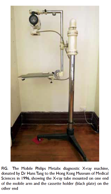
Figure. The Mobile Philips Metalix diagnostic X-ray machine, donated by Dr Hans Tang to the Hong Kong Museum of Medical Sciences in 1996, showing the X-ray tube mounted on one end of the mobile arm and the cassette holder (black plate) on the other end
The late Dr Hans Tang (湯于翰 1913-2014),
who donated this mobile machine to the Hong
Kong Museum of Medical Sciences in 1996, was
a renowned philanthropist and the vice-patron
of the Hong Kong Museum of Medical Sciences
Society. He was born in Hong Kong but grew up
in Zhenhai in Ningbo City, Zhejiang Province. He
graduated from the Shanghai Medical University
in 1934 and underwent postgraduate training at
Louvain University in Belgium, where he served as
Research Fellow and Resident Physician. It was here
that he also completed his thesis on the ‘Principles of
radiotherapy’, for which he was awarded the degree
of Senior Doctorate of Medicine. His interest in
medical specialties included radiology, microbiology,
and oncology.10
He returned to China in 1938 during the
War of Resistance against Japanese Aggression and
became the President and Physician-in-Charge of
the Shanghai Sino-Belgian Radium Institute, at the
time one of the only two centres with radiotherapy
equipment. The late Dr Tang also became President
of the Shanghai Red Cross Hospital and leader of
several other medical organisations. He settled
in Hong Kong in 1945 shortly after the Second
World War and started his private practice in 1948
specialising in cardiology. He was elected Member
of the Royal College of Physicians of Edinburgh and
Royal College of Physicians of London in 1948. At
the time, he was one of the few physicians in private
specialist practice in Hong Kong. The X-ray machine
featured in this article was used in his clinic during
the 1950s to 1970s.10
The establishment of private practice in Hong
Kong with diagnostic radiology equipment as far back
as the 1930s has been documented in the literature.
The use of X-ray equipment by traditional Chinese
bonesetters and in medical and dental clinics in the
private sector in the 1950s has also been reported.
In the early days, there appears to have been a
lack of concern regarding the potential dangers of
radiation. Nonetheless, the medical community in
Hong Kong gradually realised the importance of
enforcing radiation safety and a Radiation Board was
established in 1952 and the Radiation Ordinance and
Regulations were legally gazetted in 1965.3 Dr Tang’s
clinic was one of the few private clinics from the
1950s to 1970s to have installed an X-ray equipment.
With his studies in radiotherapy in Belgium and
experience in the Shanghai Sino-Belgian Radium
Institute, his training would have stood him in good
stead regarding the installation and operation of the
X-ray machine, and personnel radiation protection.
In 1956, Dr Tang founded the Society of
Physicians of Hong Kong with his colleagues. In the
following decades he was elected fellow of several
colleges, including the International College of
Surgeons, American College of Chest Physicians,
American College of Cardiology, Royal College of
Physicians of Edinburgh, Royal College of Physicians
of London, Hong Kong College of Physicians, Hong
Kong College of Cardiology, Hong Kong Academy
of Medicine, and Patron of its Foundation. He also
served as a member of the Radiation Board of the
Hong Kong Government and the Medical Licensing
Committee of the Hong Kong Medical Council,
and was President of the International College of
Surgeons in Hong Kong, the Hong Kong Chinese
Medical Association, and the Society of Physicians of
Hong Kong. In 1997, he helped establish the Medical
School of Ningbo University and the Dr Hans Tang
Medical Centre in his hometown.10
References
1. Hofman JA. The art of medical imaging: Philips and the evolution of medical x-ray technology. Medicamundi
2010;54:5-21.
2. The first 100 years of radiology. Hong Kong: Hong Kong College of Radiologists; 1995.
3. Ram V. Emperor extraordinaire: Life and work of John H.C. Ho. Hong Kong: Scientific Communications (HK)
Limited; 2003.
4. Philips Historical Products. Philips X-ray. Available from: http://www.philips-historische-producten.nl/xray-uk.html. Accessed Aug 2015.
5. Bouwers A. Self-protecting tubes and their influence on the development of x-ray technique. Radiology 1929;13:95-110. Crossref
6. Howell WL. The rise of mobile X-ray technology. Diagnostic Imaging. Available from: http://www.diagnosticimaging.
com/articles/rise-mobile-x-ray-technology. Accessed Sep 2015.
7. Brochure on Philips ‘Unipractix’. Catalogue Sheet No. B753, 1953.
8. Brochure on Philips ‘Practix’ X-ray Apparatus. Catalogue Sheet No. A125, 1955.
9. Golub JE, Mohan CI, Comstock GW, Chaisson RE. Active case finding of tuberculosis: historical perspective and
future prospects. Int J Tuberc Lung Dis 2005;9:1183-203.
10. Lu YY. A Movie Queen Chan Yunshang. Xinhua Publishing House; 2001.