DOI: 10.12809/hkmj144210
© Hong Kong Academy of Medicine. CC BY-NC-ND 4.0
CASE REPORT
Synthetic fibre granuloma of the conjunctiva
ST Mak, FRCSEd (Ophth), FHKAM (Ophthalmology)1,2;
YH Lui, FRCPA, FHKCPath3 #;
Kenneth KW Li, FRCS (Ed), FHKAM (Ophthalmology)1,2
1 Department of Ophthalmology, United Christian Hospital, Kwun Tong, Hong Kong
2 Department of Ophthalmology, LKS Faculty of Medicine, The University of Hong Kong, Pokfulam, Hong Kong
3 Department of Pathology, United Christian Hospital, Kwun Tong, Hong Kong
# YH Lui is now with the Department of Clinical Pathology, Pamela Youde Nethersole Eastern Hospital, Chai Wan, Hong Kong
Corresponding author: Dr ST Mak (dr.makst@gmail.com)
 Full
paper in PDF
Full
paper in PDF
Abstract
Synthetic fibre granuloma of the conjunctiva,
sometimes known as ‘teddy bear granuloma’,
results from granulomatous foreign body reaction
of the conjunctiva to synthetic fibres. It is often
an incidental finding, most commonly found in
children, is unilateral, and occurs in the lower eyelid.
We present here, what we believe is the first reported
case of synthetic fibre conjunctival granuloma in
Hong Kong, together with a review of the condition.
An awareness of this clinical entity allows early and
accurate diagnosis and early treatment.
Introduction
Synthetic fibre granuloma of the conjunctiva,
sometimes known as ‘teddy bear granuloma’, was
first described by Weinberg et al in 1984.1 It is a
rare granulomatous foreign body reaction of the
conjunctiva to synthetic fibres. It occurs most
commonly in children, and usually presents as a
unilateral, inferior conjunctival mass of the lower
eyelid. The lesion is known as ‘teddy bear granuloma’
because some cases were caused by materials used in
stuffed toy animals.2
Seventeen cases of conjunctival synthetic fibre
‘teddy bear granuloma’ have been reported in the
literature. To the best of our knowledge, this is the
first reported case of this condition in Hong Kong.
Case report
A 7-year-old girl with good health presented to the
ophthalmology clinic of United Christian Hospital,
Hong Kong, in December 2012 with a left lower
eyelid conjunctival mass for 1 month. There was no
history of trauma. It was an incidental finding by the
girl’s mother and the girl did not complain of any
pain or discomfort. There was no change in visual
acuity.
Examination showed a 3.5 mm x 1.5 mm
conjunctival mass in the inferior fornix of her left
lower eyelid (Fig a). It was embedded with a bunch
of hair-like material. The lesion prolapsed easily with
gentle pressure over the lower eyelid but could not
be removed during slit-lamp examination. The rest
of her ophthalmological examination was normal.
The girl’s mother was very keen on removal of the
mass. Excisional biopsy of the mass was performed
under general anaesthesia. The mass was excised and
sent for histopathological analysis.
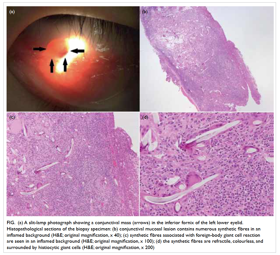
Figure. (a) A slit-lamp photograph showing a conjunctival mass (arrows) in the inferior fornix of the left lower eyelid. Histopathological sections of the biopsy specimen: (b) conjunctival mucosal lesion contains numerous synthetic fibres in an inflamed background (H&E; original magnification, x 40); (c) synthetic fibres associated with foreign-body giant cell reaction are seen in an inflamed background (H&E; original magnification, x 100); (d) the synthetic fibres are refractile, colourless, and surrounded by histiocytic giant cells (H&E; original magnification, x 200)
Microscopic examination revealed a piece
of conjunctival mucosa with stromal granulation
tissue showing heavy chronic inflammation, mild
activity, and aggregates of foreign body consistent
with synthetic fibres, associated with giant cell
reaction (Figs b to d). The fibres were refractile
and colourless. In another section, scanty hair was
seen in the stroma. The picture was compatible
with a diagnosis of synthetic fibre granuloma of the
conjunctiva.
Postoperatively, the wound healed well and
there was no recurrence of the lesion at 1.5 years
after excision.
Discussion
Protective mechanisms of the eye including blinking
and tearing normally remove any foreign body
that comes into contact with the ocular surface.
Occasionally, foreign body may be retained in the
eyelid fornix, encapsulated by mucous, embedded
in the underlying stroma, and, subsequently, induces
a local inflammatory response.2 Synthetic fibre
granuloma of the conjunctiva occurs when synthetic
fibres are inoculated in the conjunctiva of the eyelid
fornix leading to an inflammatory reaction. The lesion
is also commonly known as ‘teddy bear granuloma’
because some cases were caused by materials used
in stuffed toy animals.2 Various other objects have
been suggested as the source of the lesion, including
blankets, beddings, and pullover sweaters.2 3 4
The majority of patients were brought in
by parents or caretakers who identified a mass
in the child’s eyelid. The patients were usually
asymptomatic, without a history of trauma. Affected
children may rarely present with symptoms of ocular
irritation and foreign body sensation.5 Synthetic fibre
conjunctival granuloma is usually unilateral, and
mainly occurs in the inferior eyelid fornix, except in
one reported case where it presented superiorly.1
Differential diagnoses of synthetic fibre
conjunctival granuloma include chalazion,
pyogenic granuloma, papillary hyperplasia,
sarcoidosis, dermoid, or neoplasm including
rhabdomyosarcoma.2 6 7 It has been proposed that the
most reliable clinical sign to suggest this diagnosis
was the presence of a unilateral inferior conjunctival
mass in a child or adolescent.2 In addition, the
histological features of synthetic fibre conjunctival
granuloma are characteristic and diagnostic.
Microscopic examination reveals granulomatous
inflammatory cell response with lymphocytes,
plasma cells and eosinophils, and foreign-body giant
cells surrounding the exogenous synthetic fibres.4 8
Treatment of synthetic fibre conjunctival
granuloma involves surgical removal of the foreign
body and excision of the granuloma.2 Should
the granuloma present early and the patient be
compliant, it has been suggested to remove the
lesion during slit-lamp examination under topical
anaesthesia with minimal bleeding and discomfort.9
However, since the granuloma is usually present for a
long duration before being noticed, the lesion could
be deeply embedded. As a result, excision in the
operating theatre under general anaesthesia is often
needed, particularly when patients are very young
and anxious. Prognosis following surgical excision is
excellent.6
Although the entity of synthetic fibre
conjunctival granuloma was recognised more than two
decades ago, clinicians, including ophthalmologists
and pathologists, are unfamiliar with this condition.4
While the number of reports in the literature is
limited, accurate reporting may actually reveal a
higher incidence of this entity.9 An awareness of this
condition will allow early and accurate diagnosis and
treatment, which subsequently spare the risks and
expense associated with general anaesthesia.3
Declaration
No conflicts of interests were declared by authors.
References
1. Weinberg JC, Eagle RC Jr, Font RL, Streeten BW,
Hidayat A, Morris DA. Conjunctival synthetic fiber
granuloma. A lesion that resembles conjunctivitis nodosa.
Ophthalmology 1984;91:867-72. CrossRef
2. Schmack I, Kang SJ, Grossniklaus HE, Lambert SR.
Conjunctival granulomas caused by synthetic fibers: report
of two cases and review of literature. J AAPOS 2005;9:567-71. CrossRef
3. Enzenauer RW, Speers WC. Teddy bear granuloma of the
conjunctiva. J Pediatr Ophthalmol Strabismus 2002;39:46-8.
4. Ferry AP. Synthetic fiber granuloma. ‘Teddy bear’
granuloma of the conjunctiva. Arch Ophthalmol
1994;112:1339-41. CrossRef
5. Farooq MK, Prause JU, Heegaard S. Synthetic fiber from a
teddy bear causing keratitis and conjunctival granuloma: case report. BMC Ophthalmol 2011;11:17. CrossRef
6. Shields JA, Augsburger JJ, Stechschulte J, Repka M.
Synthetic fiber granuloma of the conjunctiva. Am J
Ophthalmol 1985;99:598-600. CrossRef
7. Lueder GT. Synthetic fiber granuloma. Arch Ophthalmol
1995;113:848-9. CrossRef
8. Batta B, Robin A, George JL, Angioi K. “Teddy bear
granuloma”, a rare condition: a case report of a 3-year-old
child [in French]. J Fr Ophtalmol 2012;35:117-20. CrossRef
9. Resnick SC, Schainker BA, Ortiz JM. Conjunctival
synthetic and nonsynthetic fiber granulomas. Cornea
1991;10:59-62. CrossRef
Find HKMJ in MEDLINE: