Hong Kong Med J 2014;20:168.e3 | Number 2, April 2014
DOI: 10.12809/hkmj133976
© Hong Kong Academy of Medicine. CC BY-NC-ND 4.0
PICTORIAL MEDICINE
Biliary papillomatosis
YQ Hsu, FRCP, FHKAM (Medicine)
Department of Medicine, St. Teresa’s Hospital, 327 Prince Edward Road West, Kowloon, Hong Kong
Corresponding author: Dr YQ Hsu (hsuyauque@yahoo.com)

A 65-year-old man presented with cholangitis which
was treated effectively with antibiotics in October
2012. Abdominal computed tomography revealed
gallstones inside the gallbladder; the intrahepatic
duct (IHD) and common bile duct (CBD) were
dilated but no stones were detected therein.
However, a mural lesion was suspected inside the
upper CBD. Subsequently, endoscopic retrograde
cholangiopancreatography (ERCP) was performed
and revealed dilated IHD and CBD, and multiple
filling defects throughout the entire dilated CBD (Fig a). The right and left hepatic ducts were dilated but no
filling defects were seen. After biliary sphincterotomy,
and sweeping of the CBD with a balloon catheter,
abundant gelatinous material mixed with tissue
and three small pigmented stones were extracted
through the papilla. Repeated sweeping of the CBD
and imaging showed an extensive frondy mass
attached to the CBD wall floating inside the lumen (Fig b). Some tissues were extracted for histological
examination which revealed dysplastic cells. The
ERCP findings were highly suggestive of the diagnosis
of intraductal papillary mucinous neoplasm. Because
of its malignant potential, resection of extrahepatic
bile duct was performed. Pathological examination
showed the CBD was extensively involved by high-grade
dysplastic glands forming papillomatosis; no
invasive malignancy was seen. The patient recovered
uneventfully after the operation.
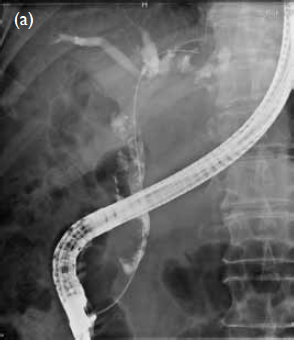
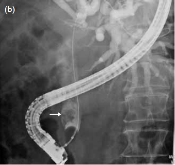
Figure. Endoscopic retrograde cholangiopancreatogram view showing (a) multiple irregular filling defects inside the common bile duct (CBD), and (b) an extensive frondy mass (arrow) attached to the CBD wall floating inside the lumen
Biliary papillomatosis1 is a rare disorder
characterised by multiple papillary adenomas in the
biliary tree. It affects mainly middle-aged, or elderly
persons and commonly presents with obstructive
jaundice and cholangitis. The papillomatosis varies
in extent and distribution within the intrahepatic
and/or extrahepatic biliary tree. The papillomas can
be classified into mucin or non-mucin secreting,
and are premalignant with definite malignant
potential. The pathogenesis of this condition is
unknown, although it has been suggested that the
malignant transformation follows the pathway of
adenoma to carcinoma sequence, similar to colonic
polyps adenoma. The definitive treatment is surgical
resection.
Reference
1. Lee SS, Kim MH, Lee SK, et al. Clinicopathologic review of 58 patients with biliary papillomatosis. Cancer
2004;100:783-93. CrossRef