© Hong Kong Academy of Medicine. CC BY-NC-ND 4.0
REMINISCENCE: ARTEFACTS FROM THE HONG KONG MUSEUM OF MEDICAL SCIENCES
Brown-Buerger cystoscope
CH Leong, FHKAM (Surgery)
Honorary Advisor, Hong Kong Museum of Medical Sciences Society
 Full paper in PDF
It is no exaggeration to say that urologists were
the pioneers in using the endoscope. In 1805,
Phillipp Bozzini made his first attempt to visualise
the living body through a hollow tube he created,
called 'Lichtleiter' (Light Conductor). In 1853,
Antoine Désormeaux developed an instrument to
examine the urethra and bladder, which he called an
endoscope.1
The most common cystoscope used today is
the Brown-Buerger cystoscope, invented by Frederic
Brown in 1899, which utilises two different lens
systems to visualise the whole bladder. Leo Buerger
expanded on Brown’s idea in 1907 by passing
different instruments through the same outer sheath
for endoscopic procedures (Fig 1).
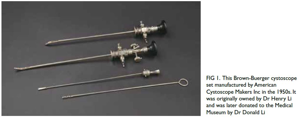
Figure 1. This Brown-Buerger cystoscope set manufactured by American Cystoscope Makers, Inc. in the 1950s. It was originally owned by Dr Henry Li and was later donated to the Medical Museum by Dr Donald Li
For the Brown-Buerger cystoscopy to be
effective, it requires the following components:
a metal sheath with a curved beak to manipulate
the curve of the bulbous and membranous
urethra in the male (Fig 2);
different lens systems to visualise the whole
bladder (Fig 3);
a channel to introduce fluid to distend the bladder
for visualisation;
channels for introducing instruments for
endoscopic procedures; and
light sources to illuminate the inside of the
bladder and the whole length of the urethra.
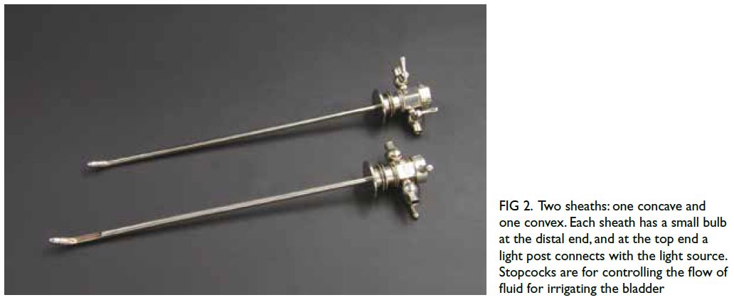
Figure 2. Two sheaths: one concave and one convex. Each sheath has a small bulb at the distal end, and at the top end a light post connects with the light source. Stopcocks are for controlling the flow of fluid for irrigating the bladder
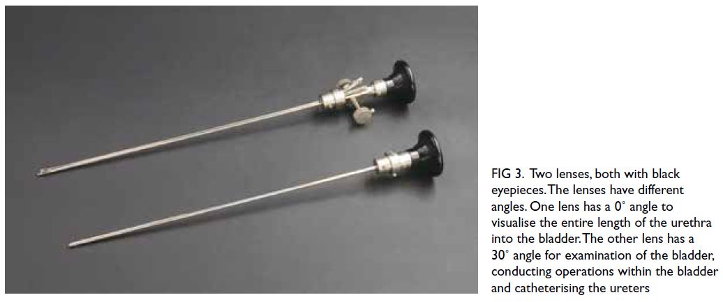
Figure 3. Two lenses, both with black eyepieces. The lenses have different angles. One lens has a 0˚ angle to visualise the entire length of the urethra into the bladder. The other lens has a 30˚ angle for examination of the bladder, conducting operations within the bladder and catheterising the ureters
It is of interest to trace the development of
the light source in cystoscopy. The light source of the first generation is a reflected candlelight that
my ancient urologist colleagues must have fantastic
eyesight. The second-generation light source was a
small electric bulb at the tip of the curved beak of
the sheath connected by a fine wire to a portable
battery pack. I was brought up with this secondgeneration
light source; regrettably the bulbs fused
easily, necessitating the replacement of many bulbs
during a single examination. The advent of fibre
optics marked the arrival of the third-generation
light source, which was a 'god send'. Urologists
now are blessed with a good illumination for as
long as it takes to do the examination and necessary procedures.
What are the drawbacks of the Brown-Buerger
cystoscopy? Being a rigid instrument, the procedure
could be uncomfortable particularly when performed
under local anaesthesia. The absence of a retrograde
lens in the cystoscope also limits the urologist’s
ability to examine the bladder neck opening. The
invention of a flexible cystoscopy has eliminated
all these drawbacks and made bladder and urethra
examination complete.
The Brown-Buerger cystoscopy, despite
its drawbacks, remains one of the most used
cystoscopes.
References
1. Samplaski MK, Jones JS. Two centuries of cystoscopy: the development of imaging, instrumentation and synergistic technologies. BJU Int 2009;103:154-8. Crossref