Hong Kong Med J 2023 Aug;29(4):355–7 | Epub 25 Jul 2023
© Hong Kong Academy of Medicine. CC BY-NC-ND 4.0
CASE REPORT
Metastatic adenocarcinoma of the stomach presenting as malignant acanthosis nigricans and tripe palms: a case report
Carla PM Lam, MB, BS, FHKAM (Medicine)1; Mandy WM Chan, MB, BS (UCL), MRCP (London)2
1 Division of Gastroenterology and Hepatology and Department of Medicine, Queen Mary Hospital, Hong Kong SAR, China
2 Division of Dermatology, Department of Medicine, Queen Mary Hospital, Hong Kong SAR, China
Corresponding author: Dr Carla PM Lam (lpm496@ha.org.hk)
 Full paper in PDF
Case report
A 46-year-old lady with good past health presented
to a tertiary hospital in August 2020 with a 6-month
history of chronic cough, epigastric discomfort, and
weight loss of 20 kg. She also reported progressive
darkening and thickening of skin over both hands,
neck, axilla, and groins since March 2020. She
had consulted general practitioners and Chinese
medicine practitioners and been given topicals and
herbal treatment with no improvement. Physical
examination revealed velvety hyperkeratosis and
hyperpigmentation over both palms (Fig a), nape (Fig b), bilateral axilla (Fig c), and inguinal regions.
The mucosal surfaces were not involved. Physical
examination revealed stony dullness to percussion
over the right mid-to-lower zone of the lungs and
absent breath sounds on auscultation. A non-tender
enlarged supraclavicular lymph node of 1 cm
was palpable over the left supraclavicular region.
Chest radiograph showed moderate right pleural
effusion. Therapeutic thoracocentesis drained 2 L
of clear straw-coloured fluid. Pleural fluid analysis
revealed an exudative pleural effusion with 47.3 g/L
fluid protein (70 g/L serum protein) and 224 U/L fluid lactate dehydrogenase (226 U/L serum lactate
dehydrogenase). Pleural fluid cytology revealed
suspicious cells with high nuclear-to-cytoplasmic
ratio, coarse chromatin and enlarged, irregular
hyperchromatic nuclei. Other laboratory findings
were unremarkable: haemoglobin level 14 g/dL,
white blood cell count 3.62 × 10⁹/L, platelet count
156 × 109/L, creatinine level 71 μmol/L, bilirubin
level 8 μmol/L, albumin level 28 g/L, alanine
aminotransferase level 13 U/mL, and aspartate
aminotransferase level 20 U/mL. Tumour markers
including carcinoembryonic antigen, cancer antigen
(CA) 15-3, CA 19-9, CA-125, and alpha-fetoprotein
were all within the normal limits.
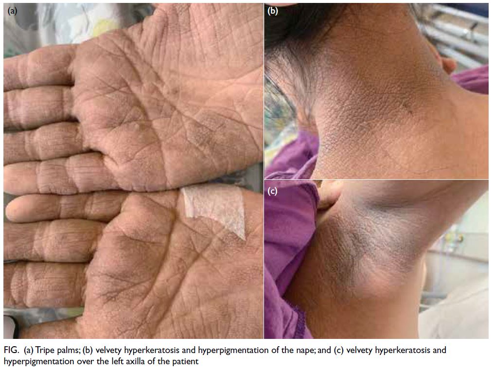
Figure. (a) Tripe palms; (b) velvety hyperkeratosis and hyperpigmentation of the nape; and (c) velvety hyperkeratosis and hyperpigmentation over the left axilla of the patient
A clinical diagnosis of malignant acanthosis
nigricans (AN) with tripe palms was made after
dermatology review. Upper endoscopy revealed
two Forrest class III gastric ulcers in the proximal
greater curvature surrounded by abnormal 3-cm
mucosal thickening with irregular mucosal surface
and microvascular pattern under narrow-band
imaging. Biopsy of the gastric ulcers was negative
for Helicobacter pylori but confirmed poorly
differentiated adenocarcinoma on histopathology.
Positron emission tomography–computed
tomography scan revealed a hypermetabolic focus in
the stomach, multiple intra-abdominal lymph nodes,
and a large hypermetabolic pelvic tumour. A clinical
diagnosis was made of Krukenberg tumour. The
patient was referred to medical oncology for palliative
chemotherapy. She subsequently developed massive
pulmonary embolism and succumbed 3 months
after the initial diagnosis.
Discussion
Acanthosis nigricans is a velvety hyperkeratotic,
hyperpigmentation of the skin that occurs most
commonly in intertriginous areas such as the back of
the neck, axilla, and groins. Eight types of AN have
been described and all share a common mechanism.
They stimulate receptor tyrosine kinase signalling
pathways; epidermal growth factor receptor (EGFR),
insulin-like growth factor (IGF-1), and fibroblast
growth factor receptors. Increased circulating insulin
stimulates keratinocyte IGF receptors, especially
IGF-1 and at high concentrations, displaces IGF-1
from IGF-1–binding protein. Increased serum-free
IGF-1 in turn stimulates the proliferation of
keratinocytes and dermal fibroblasts.1 2
Malignant AN is a paraneoplastic
phenomenon most commonly associated with
gastric adenocarcinoma with an incidence of 55% to
61%, followed by pancreatic cancer, gynaecological
malignancies, and lung carcinoma.2 Increased
transforming growth factor alpha (TGF-α) is
postulated to be the underlying mechanism. The
TGF-α acts on EGFR, stimulating soft tissue growth.1 Amelioration of malignant AN following tumour
resection, associated with a reduction in elevated
circulating TGF-α, supports the participation of
EGFR signalling in malignant AN. Malignant AN
can manifest preceding, together, or after diagnosis
of an underlying malignancy. Rapid evolution of the
velvety hyperpigmentation, tripe palms and signs
of Leser-Trélat, a rare finding of sudden eruption
of seborrhoeic keratoses, are strongly indicative
of malignant AN.3 Affected patients are typically
not obese and may be cachectic because of the
underlying malignancies. Histological features are
non-specific and commonly include hyperkeratosis,
papillomatosis, basal layer hyperpigmentation, and
some dermal papillae that project upwards in the
form of finger-like projections.4 Malignant AN may
resolve following tumour resection but can recur if
there is tumour recurrence.
Krukenberg tumours are defined by the
World Health Organization as ovarian carcinomas
characterised by the presence of stromal involvement,
mucin-producing neoplastic signet ring cells, and
ovarian stromal sarcomatoid proliferation. The most
common sites of primary malignancies are from the
gastrointestinal tract and the breasts. The mean age
at diagnosis of Krukenberg tumours is 49.3 ± 13.3
years. The prognosis is generally very poor, probably
because of the late stage of diagnosis. The median
survival time is 35.0 ± 3.5 months while the 5-year
overall survival is around 25%.5
This case illustrates the classic presentation of
malignant AN and tripe palms that are associated
with metastatic gastric adenocarcinoma. Physicians
need to be aware of these features since they may
be the only presenting symptoms of the underlying
malignancies. Full systemic evaluation for underlying
malignancies is warranted to enable early diagnosis
and timely management.
Author contributions
Concept or design: CPM Lam.
Acquisition of data: CPM Lam.
Analysis or interpretation of data: CPM Lam.
Drafting of the manuscript: CPM Lam.
Critical revision of the manuscript for important intellectual content: Both authors.
Acquisition of data: CPM Lam.
Analysis or interpretation of data: CPM Lam.
Drafting of the manuscript: CPM Lam.
Critical revision of the manuscript for important intellectual content: Both authors.
Both authors had full access to the data, contributed to the study, approved the final version for publication, and take responsibility for its accuracy and integrity.
Conflicts of interest
Both authors have disclosed no conflicts of interest.
Funding/support
This study received no specific grant from any funding agency in the public, commercial, or not-for-profit sectors.
Ethics approval
The patient was treated in accordance with the Declaration of Helsinki. Written informed consent for publication was
obtained from the patient’s next-of-kin.
References
1. Phiske MM. An approach to acanthosis nigricans. Indian Dermatol Online J 2014;5:239-49. Crossref
2. DermNetNZ. Acanthosis nigricans. December 2021. Available from: https://dermnetnz.org/topics/acanthosis-nigricans. Accessed 18 Jul 2023.
3. Kilickap S, Yalcin B. Images in clinical medicine. The sign of Leser-Trélat. N Engl J Med 2007;356:2184. Crossref
4. Yu Q, Li XL, Ji G, et al. Malignant acanthosis nigricans: an early diagnostic clue for gastric adenocarcinoma. World J Surg Oncol 2017;15:208. Crossref
5. Xu KY, Gao H, Lian ZJ, Ding L, Li M, Gu J. Clinical analysis of Krukenberg tumours in patients with colorectal cancer—a review of 57 cases. World J Surg Oncol 2017;15:25. Crossref