© Hong Kong Academy of Medicine. CC BY-NC-ND 4.0
CASE REPORT
Diagnostic dilemma between skull base
osteomyelitis and nasopharyngeal carcinoma: a case series
KY Wong, MB, ChB; KC Wong, FRCSEd (ORL), FHKCORL
Department of Ear, Nose and Throat, Pamela Youde Nethersole Eastern Hospital, Hong Kong
Corresponding author: Dr KY Wong (wky053@ha.org.hk)
 Full
paper in PDF
Full
paper in PDF
Case report
Skull base osteomyelitis (SBO) is a rare and life-threatening
complication of otorhinological
infection. The anatomical location of the disease and
clinical features allow it to mimic nasopharyngeal
carcinoma (NPC): a malignancy that is endemic in
Southeast Asia. Suspicious clinical or radiological
findings warrant prompt histological investigation
for confirmation. Misdiagnosis of either entity can
lead to devastating consequences of late treatment
and disease progression. Our centre identified
four cases of SBO between 2019 and 2020, all of
which mimicked NPC at presentation. The caveats
encountered during the diagnostic process are
highlighted here.
Case 1
An 81-year-old diabetic man presented to our
department with left middle ear effusion. Endoscopy
revealed vague bulging over the left nasopharynx
with obliteration of the left fossa of Rosenmuller.
Gadolinium-enhanced magnetic resonance imaging
(MRI) showed enhancing signal intensities in the
left nasopharynx involving multiple skull base
structures (Fig). Multiple biopsies yielded only
benign results. Tissue culture grew methicillin-sensitive
Staphylococcus aureus. Blood test results
revealed elevated inflammatory markers including
erythrocyte sedimentation rate (ESR) and C-reactive
protein (CRP). His plasma Epstein–Barr virus (EBV)
DNA level was 0 copies/mL. The final diagnosis was
SBO and the patient was treated accordingly with a
good response.
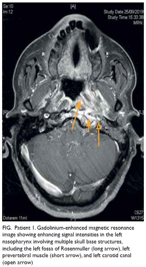
Figure. Patient 1. Gadolinium-enhanced magnetic resonance image showing enhancing signal intensities in the left nasopharynx involving multiple skull base structures, including the left fossa of Rosenmuller (long arrow), left prevertebral muscle (short arrow), and left carotid canal (open arrow)
Case 2
A 76-year-old man presented with left-sided
headache and left jaw pain with a recent history of
lower molar tooth extraction. Physical examination
revealed left otitis media with effusion, and tongue
and uvula deviation. Endoscopy showed a vague
swelling over the left nasopharynx. The MRI images
indicated locally invasive NPC, involving the clivus
and ipsilateral skull base foramina. The patient
underwent multiple biopsies including under general anaesthesia with computed tomography
(CT) navigation. Blood ESR and CRP levels were
elevated and tissue cultures grew Candida, Pichia,
and Lactobacillus. All results indicated inflammation
only. The patient was treated with a prolonged course
of meropenem and antifungal medication with
a favourable clinical response. His headache and
inflammatory markers improved, and interval MRI
showed reduced signal intensities at the skull base.
Case 3
A 71-year-old man was referred to our clinic for
persistent left otalgia, headache, and facial pain.
Initial endoscopy demonstrated bulging of the
left nasopharynx and left cord palsy. Computed
tomography and subsequent MRI revealed a
neoplastic process in the left nasopharynx with
invasion of the parapharyngeal space and skull
base. Prominent retropharyngeal lymph nodes
were suggestive of nodal spread. Multiple biopsies
were carried out, once with MRI navigation via
a transsphenoidal approach towards the clivus.
Results were all benign. His plasma EBV DNA
level was 0 copies/mL. Cultures grew methicillin-resistant
S aureus and Corynebacterium. White cell
count and blood ESR and CRP levels were elevated.
A prolonged course of meropenem was prescribed
with consequent improvement of symptoms and
reducing trend of inflammatory markers. The
3-month interval MRI demonstrated reduced
enhancement of skull base structures.
Case 4
A 59-year-old man with poorly controlled diabetes
underwent left mastoidectomy for malignant
otitis externa. After surgery, he complained of
persistent left temporal headache and dysphagia.
Pathology from the operation was suggestive of
inflammation only. Endoscopy revealed bulging of
the left nasopharynx and left cord palsy. Computed
tomography showed left otomastoiditis whilst MRI
demonstrated an aggressive process with extensive
involvement of skull base structures. Nasopharynx
biopsies failed to confirm malignancy. Cultures
were positive for methicillin-resistant S aureus,
Staphylococcus spp, and Klebsiella. His plasma EBV
DNA was 0 copies/mL. White cell count and blood
ESR and CRP levels were elevated. After 6 weeks
of treatment with meropenem and vancomycin,
his headache resolved, inflammatory markers
normalised, and tissue culture results from a new
biopsy were negative.
Despite the apparent clinical resolution,
interval MRI 8 weeks after cessation of treatment
showed disease progression. There was new right-sided
contrast enhancement in the nasopharynx
with extension to the right carotid space, hypoglossal
canal, and petrous apex. The patient was soon re-hospitalised
with right temporal headache and
bilateral vocal cord palsy. Transsphenoidal CT-guided
biopsy was performed on the clivus and
overlying mucosa and results were still benign.
Culture results were similar to those previously
and inflammatory markers were again elevated. He
was given a second course of antibiotics and had a
prompt clinical response.
Discussion
Three patients were referred to our department
with intractable headache, facial pain, or otalgia,
some of the most common presenting complaints
of SBO along with cranial nerve deficits.1 These
differ markedly to those of NPC: blood-stained nasal
discharge, unilateral conductive hearing loss, and
cervical lymphadenopathy. Cranial nerve palsies
suggest local invasion. Singh et al2 recommend that
SBO be suspected if patients with treated malignant
otitis externa present with persistent headache,
otitis media with effusion, and cranial nerve deficits
without a mucosal lesion in the nasopharynx, but
there are no internationally recognised diagnostic
criteria and recommendations differ.
The diagnostic dilemma occurs in the physical
findings on initial examination. A unilateral otitis
media with effusion in Southeast Asian adults
(Cases 1 and 2) is presumed to be NPC until
proven otherwise. Endoscopic findings of a bulging
nasopharynx further raise the alarm, even in the
absence of an obvious mucosal lesion. At this point,
regardless of co-morbidities, symptoms, presence
or absence of nerve palsies, efforts should be made
to confirm or exclude mucosal or submucosal
malignancy before settling on a diagnosis of SBO.
The key to differentiating malignancy from
infection is the result of tissue biopsy. Simple bedside
punch biopsies of the visibly abnormal mucosa are
usually adequate to obtain histological confirmation
of NPC. In our cases, only vague bulging of the
nasopharynx was evident. Negative results led to
more invasive procedures to obtain deeper samples
or down to the clivus. There is no established
recommendation for use of intraoperative image-guided
biopsy in this regard. This method attempted
to maximise the yield of abnormal tissue to exclude
with certainty any presence of malignancy.
Negative plasma EBV DNA level, in view of
its high negative predictive value for endemic NPC,
is another reason to exclude NPC. Inflammatory
markers were raised in all our patients. Serial
blood ESR level is also a useful marker for disease
monitoring as it rapidly normalises with disease
resolution3 but rises again with disease relapse.
Imaging played a key diagnostic role.
Although CT can detect osseous destruction, these
bony changes are a rather late phenomenon. In
comparison, MRI offers higher soft tissue resolution,
allowing delineation of anatomical location and soft
tissue involvement.3 The MRI images in our cases
typically showed gadolinium-enhanced signals at the
nasopharynx, involving the clivus, petrous apex, skull
base foramina, and parapharyngeal spaces on T1-weighted images. Images were suggestive of neoplasm,
described by radiologists as “space occupying lesions” and “aggressive lesions with erosion/invasion of
surrounding structures”. This further highlights the
importance of histological correlation.
In conclusion, SBO is a rare disease that can
masquerade as NPC. Although the two diseases
warrant entirely different treatment modalities, it
remains difficult to confidently differentiate one from
the other during diagnosis. A holistic consideration
of the patient’s clinical picture, and histological and
microbiological results are essential for correct
diagnosis.
Author contributions
All authors contributed to the design, acquisition of data,
analysis of data, drafting of the manuscript, and critical
revision of the manuscript for important intellectual content.
All authors had full access to the data, contributed to the
study, approved the final version for publication, and take
responsibility for its accuracy and integrity.
Conflicts of interest
All authors have disclosed no conflicts of interest.
Funding/support
This study received no specific grant from any funding agency in the public, commercial, or not-for-profit sectors.
Ethics approval
All patients were treated in accordance with the Declaration of Helsinki. All patients provided informed consent for
all procedures and for the publication of non-identifiable
information.
References
1. Sokołowski J, Lachowska M, Karchier E, Bartoszewicz R, Niemczyk K. Skull base osteomyelitis: factors implicating
clinical outcome. Acta Neurol Belg 2019;119:431-7. Crossref
2. Singh A, Al Khabori M, Hyder MJ. Skull base osteomyelitis:
diagnostic and therapeutic challenges in atypical
presentation. Otolaryngol Head Neck Surg 2005;133:121‑5. Crossref
3. van Kroonenburgh AM, van der Meer WL, Bothof RJ, van Tilburg M, van Tongeren J, Postma AA. Advanced imaging
techniques in skull base osteomyelitis due to malignant
otitis externa. Curr Radiol Rep 2018;6:3. Crossref