© Hong Kong Academy of Medicine. CC BY-NC-ND 4.0
REMINISCENCE: ARTEFACTS FROM THE HONG KONG MUSEUM OF
MEDICAL SCIENCES
Apparatus that separated radiotherapy from
radiology
Dr KY Cheung, MHKCR
Education and Research Committee, Hong Kong Museum of Medical Sciences Society
 Full paper in PDF
First X-ray machine in Hong Kong
The earliest recorded clinical use of X-ray in Hong
Kong was at the Alice Ho Miu Ling Nethersole
Hospital, in May 1910.1 2 The X-ray machine was called
a ‘Roentgen Ray Apparatus’ at that time, 15 years after
the discovery of X-rays by German physicist Wilhelm
Conrad Roentgen. Radiation doses received by
patients were not measured in those days because the
methodology and instrumentation for quantifying
roentgen radiation had yet to be developed, although
the possibility of using photographic plates and
electrical conductivity in air had been proposed by
Ernest Rutherford in 1899.3 Patient dosimetry was primarily based on the exposure time and the effects
of radiation such as skin reaction at the treatment
site. Radiation measurements and hence patient
dosimetry became a reality in 1928 when the unit
of X-ray radiation, the roentgen, was defined and
suitable instrumentations became available.3
Radiotherapy at its embryonic stage
Public radiology services in Hong Kong started in
around 1929 at the Government Civil Hospital,
which later became Sai Ying Pun Hospital.4 In the
early 1930s, diagnostic X-ray machines were used
by radiologists for the treatment of skin cancers,
and radium tubes were used by gynaecologists for
treatment of carcinoma of the uterine cervix.4 One of
the problems with using diagnostic X-ray machines
for skin cancer treatments was that a long X-ray
target-to-skin distance had to be used for electrical
safety. As a result, the dose rate at the skin was too
low for practical and efficient treatment delivery.5
Radium radiotherapy also had limitations, primarily
that radium was in short supply and it was expensive.
Specially designed X-ray radiotherapy machines,
such as the Chaoul contact therapy X-ray machine,
were developed to address these issues. Dedicated
radiotherapy equipment for cancer treatment was
not available in Hong Kong until 30 June 1937 when
Queen Mary Hospital opened. The new hospital had
a range of radiotherapy facilities, including a Chaoul
contact therapy X-ray machine, a General Electric
(GE) ST-150 superficial X-ray therapy machine, and
some radium tubes. In those days, radiotherapy and diagnostic radiology services were operated under
the Radiological Sub-Department. The specialist in
charge of the sub-department was Dr FJ Farr,6 from
the UK. He was assisted by two radiographers also
from the UK, and four local radiographic assistants.
Later, in 1939, a GE Maxima 400 kV deep X-ray
therapy machine was added to the department
for treating deep-seated cancer diseases. Dr Farr
effectively planted the seed for radiotherapy service
before his retirement in 1950 and he was succeeded
by Professor HC Ho.6 Professor Ho added a Bryant
Simmons 18 Ci Cobalt-60 teletherapy machine
in 1953 (the Cobalt source was replaced by a 60Ci
source in 1958) and a GE Maxitron 250 kV deep
X-ray therapy machine in 1958. Thus, Professor
Ho created a solid foundation for his pioneering
work in radiation therapy and paved the way for the
development of the radiation oncology subspecialty
in Hong Kong.
Chaoul contact therapy X-ray
machine
Installation of the Chaoul contact therapy X-ray
machine in 1937, together with other radiotherapy
facilities, marked the beginning of external beam
radiation therapy in Hong Kong. The Chaoul
machine, which was manufactured by Siemens in
Erlangen, Germany, was developed by German
radiologist Professor Henri Chaoul in the early
1930s.7 8 It was designed to replace or provide an
alternative to radium therapy in the treatment of
cancers in the skin, rectum, cervix, and oral cavity. As
reported by Chaoul at the Royal Society of Medicine
on 21 February 1936,8 no country possessed sufficient
radium to treat all patients requiring treatment.
The contact X-ray therapy apparatus he developed
made it possible to treat all suitable cases more
cheaply, making the technology more accessible
internationally. The machine operated at a constant
potential of 60 kV and an anode current of 4 mA.5 8
The X-ray tube had a heated filament, a hollow
anode, and a water-cooled transmission-type X-ray
target located at the end of the long hollow anode
tube. Electrons could be directed to pass down the
hollow anode cylindrical tube to strike a target at the
end of the tube (also serve as treatment applicator) to produce X-rays for contact or short-distance
treatment. The target was electrically grounded
so that the X-ray target-to-skin distance could be
very short. Treatment applicators could be inserted
onto the applicator to define the treatment area
and target-to-skin distance. The output exposure
rate reported was about 150 r/min at a target-to-skin
distance of 5 cm and the treatment field size
(9-25 cm2) was selectable.6 The output exposure
of an X-ray machine at that time was measured in
roentgen “r”, which was a measure of the amount of
ionisation the radiation produced in 1 cc of air. The
symbol “r” was replaced by “R” for absorbed dose in
tissue, and “rad” was adopted for dose calibration in
the 1960s. The Chaoul X-ray machine was replaced
in 1958 by a Philips contact therapy X-ray machine.
The Chaoul X-ray tube, which is a treasure for the
radiotherapy community and has been maintained
in excellent condition, was donated by the Hong
Kong College of Radiologists (HKCR) to the Hong
Kong Museum of Medical Sciences (Fig 1).
General Electric Maxima 400 kV
deep X-ray therapy machine
The GE Maxima, made in the United States, was the
first deep X-ray (also known as orthovoltage) therapy
machine installed in Hong Kong. It was installed by a
local GE engineer Mr Raymond Huang in 1938 and
commissioned for clinical use in 1939.6 The machine
operated at an anode voltage up to 400 kV. To operate
at such a high potential, the X-ray tube was housed
inside a large tank filled with insulation oil to prevent
electrical breakdown and allow tube cooling. The
high-voltage generator was powered by a resonant transformer which was also fitted inside the oil tank
(Fig 2a). Initial operation of the GE Maxima and
other radiotherapy machines for patient treatment
was carried out by the radiology specialists Dr Farr
and Dr Ho until the early 1950s when the task was
assigned to radiographers who returned to Hong
Kong after completing their therapy training in the
UK. Radiation output of the therapy machines was
measured by these radiographers until 1956, when
Mr GF Mauldon took up the task. Mr Mauldon
was an Australian medical physicist trained at
the Peter MacCallum Clinic in Melbourne, and
he joined the Radiological Sub-Department on a
2-year secondment arrangement. Around 1964,
the GE Maxima was relocated to a newly built large
centre, the Royal Hong Kong Jockey Club Institute
of Radiology and Oncology at Queen Elizabeth
Hospital. The GE Maxima became functionally
obsolete and was removed from patient service
when state-of-the-art megavoltage radiotherapy
technologies were installed at the Institute, including
two linear accelerators and a Betatron. However, the
GE Maxima remained onsite in the Institute until its
decommissioning in the late 1980s. The 1.4-m-long
X-ray tube has been kept and is currently on display
at the Department of Radiation Oncology at Queen
Elizabeth Hospital (Fig 2b).
General Electric Maxitron 250 kV
deep X-ray therapy machine
The GE Maxitron 250 kV deep X-ray therapy
machine, also made in the United States, was
installed at Queen Mary Hospital in 1958 for
treatment of deep-seated internal diseases. It was an advanced treatment machine at the time with
a relatively sophisticated treatment control unit
that had a roentgen meter and timer for treatment
control. The machine came with a dedicated
treatment table, a set of treatment applicators, beam
hardening filters, and related accessories, including
intracavitary treatment applicators. The treatment
head had a light projector simulating the X-ray beam
and a mechanical distance pointer for guiding the
positioning of the patient for treatment delivery. The
X-ray tube (Fig 3) was air-cooled and it could operate
up to 250 kV with an anode current of up to 30 mA
to deliver a radiation output of 75 r/min at 50 cm
target-to-skin distance using a 2-mm copper filter.
The GE Maxitron X-ray tube was donated to the
Museum by the HKCR.
Philips Metalix contact X-ray
therapy machine
The Chaoul contact therapy X-ray machine was
replaced by a Philips Metalix contact therapy
machine in 1958. The Philips Metalix, made in
Eindhoven, the Netherlands, had a similar design
and operation to the Chaoul machine. It was a
mobile unit with a constant potential generator and
control unit on wheels (Fig 4a). The X-ray tube had a
similar design to that of the Chaoul tube, except that
the Philips tube was air-cooled (Fig 4b). The Philips
Metalix operated at 50 kV, anode current at 2 mA,
maximum field size of 5 cm2, and an exposure rate
of up to 10 000 r/min. The Philips Metalix X-ray tube
was also donated to the Museum by the HKCR.
Acknowledgements
The author would like to thank Dr CK Law, President
of Hong Kong College of Radiologists for reviewing
this manuscript and sharing his photos on historical
radiotherapy equipment; Mr GF Mauldon for sharing his archive of photos and documents on the
history of medical physics and radiology in Hong
Kong; and Mr Dominick Yim of Philips Electronics
Hong Kong Limited for provision of a copy of the
original product catalogue on the Philips Metalix
contact therapy machine.
References
1. Paterson EH. A hospital for Hong Kong: The Centenary History of the Alice Ho Miu Ling Nethersole Hospital. Hong
Kong: Alice Ho Miu Ling Nethersole Hospital; 1987. Crossref
2. Luk SY. The mobile X-ray machine. Hong Kong Med J 2016;22:194-5. Crossref
3. Almond PR. A historical perspective: a brief history of dosimetry, calibration protocols, and the need for accuracy.
In: Rogers DW, Cygler JE, editors. Clinical Dosimetry Measurements in Radiotherapy. Madison, Wisconsin; Medical
Physics Publishing: 2009; 1-28. Crossref
4. Ram V. Emperor Extraordinaire: Life and Work of John HC Ho. Hong Kong: Scientific Communications (HK)
Limited; 2003. Crossref
5. Pendergrass PE, Hodes PJ, Garrahan CJ. Roentgen therapy by the method of Chaoul. Radiology 1939;32:142-54. Crossref
6. Ho JH, Mauldon GF, Huang DP, Kwan HC. Development of radiation oncology in Hong Kong up to 1985. Int J Radiat
Oncol Biol Phys 1997;37:125-9. Crossref
7. Kuttig H. Die klinischen Applikationsverfahren zur Erzielung einer geeigneten räumlichen Dosisverteilung [in
German]. In: Scherer E, editor. Strahlentherapie. Springer, Berlin, Heidelberg; 1976: 77-104. Crossref
8. Short-distance low-voltage X-ray therapy: (Section of Radiology). Proc R Soc Med 1936;29:791-808. Crossref
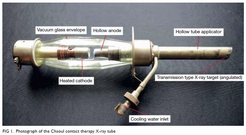
Figure 1. Photograph of the Chaoul contact therapy X-ray tube
Figure 2. (a) Black-and-white photograph of a patient being setup for treatment by the GE Maxima 400 kV deep X-ray machine (courtesy of Mr GF Mauldon). (b) Photograph showing the large GE Maxima-400 X-ray tube (courtesy of Dr CK Law)
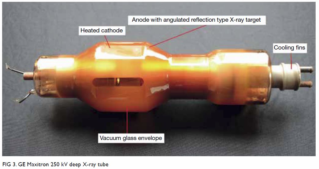
Figure 3. GE Maxitron 250 kV deep X-ray tube
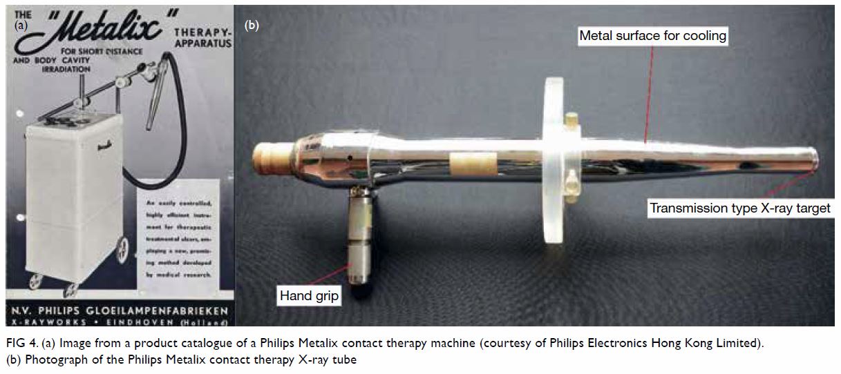
Figure 4. (a) Image from a product catalogue of a Philips Metalix contact therapy machine (courtesy of Philips Electronics Hong Kong Limited). (b) Photograph of the Philips Metalix contact therapy X-ray tube