© Hong Kong Academy of Medicine. CC BY-NC-ND 4.0
REMINISCENCE: ARTEFACTS FROM THE HONG KONG MUSEUM OF MEDICAL SCIENCES
Erisophake: an outdated instrument for cataract surgery
Patrick PC Tong, FCOphth HK, FHKAM (Ophthalmology)
Member, Hong Kong Museum of Medical Sciences Society
 Full
paper in PDF
Full
paper in PDF
An erisophake is a surgical instrument designed
to hold the human lens by suction during cataract
extraction. It was widely employed for cataract
extraction after the 1950s as an alternative to forceps.
Cataract extraction using an erisophake begins
with the opening of a corneal or corneo-scleral
wound. This can be done using either a von Graefe
knife or corneal scissors. The section should open
at least 50% of the circumference of the cornea.1
Peripheral iridotomy or a sector iridectomy is then
performed. The enzyme alpha-chymotrypsin can
be used to loosen the lens zonules. The erisophake
cup is then placed against the anterior surface of
the lens, and negative suction pressure then applied
to hold the lens. The suction can be created by a
spring-operated syringe,2 or a rubber bulb3 (Fig) or
by connection to the operating room suction line.4
The lens can then be brought out by simply sliding
or tumbling it out. The wound is subsequently closed
with silk sutures. Complications of erisophake
cataract extraction include loss of suction, anterior
capsule tear, posterior capsule tear, vitreous loss,
and corneal endothelial damage.5 In addition, the
erisophake is more complicated and difficult to
manipulate than forceps.2
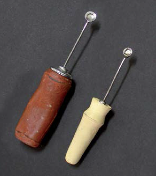
Figure. Two erisophakes (measuring 6.2 and 7.0 cm in length) fitted with rubber bulbs, donated to the Hong Kong Museum of Medical Sciences in 2002 by Dr Kai-hung Lor
Both forceps and erisophake cataract extraction
gradually dropped out of use in the late 1960s
and early 1970s, when cataract extraction could
be more effectively performed using a cryoprobe
(cryoextraction). The cryoprobe can be cooled to
-196°C with liquid nitrogen. The probe is brought
into direct contact with the cataract to freeze it and
then extract it. Cryoextraction of cataract became
even easier with the increasing use of operating
microscopes. When using forceps, an erisophake,
or cryoprobe, the cataractous lens is extracted
together with the lens capsule and the procedure
is called intracapsular cataract extraction. In newer
methods only the anterior capsule is removed with
the cataractous lens, leaving the posterior capsule
intact. These newer methods are called extracapsular
cataract extraction (ECCE).
In the original ECCE, the anterior capsule
was manually opened and the nucleus of the
cataract expressed through a corneal or corneoscleral
wound. Subsequent development of phacoemulsification
involves emulsifying the lens nucleus
using ultrasonic energy. The emulsified nucleus
can then be aspirated through a much smaller
wound. Today, femtosecond laser is used to assist in
phacoemulsification by creating the corneal wound,
making corneal cuts to treat astigmatism, making
the anterior capsule opening, and cutting the lens
nucleus into small pieces before using ultrasonic
energy.
The introduction of phacoemulsification
and femtosecond laser–assisted cataract surgery
has made cataract extraction a minimally invasive
procedure with a corneal incision of 2.8 mm or
less. In addition, the advent of different types of
intraocular lenses has resolved various refractive
problems following cataract surgery. None of these
was conceivable when erisophake cataract extraction
was the norm.
References
1. Esposito AC. The Esposito erisophake and cataract extraction. Br J Ophthalmol 1962;46:697-700. Crossref
2. Lesperance FA Jr. A modification of the erisophake with discussion of technique. Arch Ophthalmol 1963;70:323-7. Crossref
3. Bell AE. A modified erisophake. Am J Ophthalmol 1948;31:610. Crossref
4. Boomer RB. A new erisophake pump. Am J Ophthalmol 1966;61:349-51. Crossref
5. Atkinson WS. Observations on anesthesia for ocular surgery. Trans Am Acad Ophthalmol Otolaryngol 1956;60:376-80.