© Hong Kong Academy of Medicine. CC BY-NC-ND 4.0
REMINISCENCE: ARTEFACTS FROM THE HONG KONG MUSEUM OF MEDICAL SCIENCES
Destructive obstetric instruments: what do they destroy?
KH Lee, MD, FRCOG
Director, Hong Kong Museum of Medical Sciences Society
 Full
paper in PDF
Full
paper in PDF
They destroy the fetus in the uterus.
The instruments shown in the figures were
used more than a hundred years ago to perforate
and crush the fetal head in cases of hydrocephalus
or disproportion, and in prolonged labour when the
fetus had died. The perforator was used to puncture
the fetal skull, release the contents, and reduce its
size (Fig 1). The centre piece of the cranioclast was
used to make the perforation (Fig 2a) and then the
two-side blades approximated by tightening the
screw on the handle to crush the fetal skull (Fig 2b).
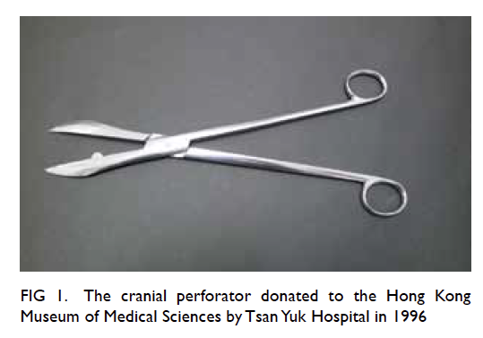
Figure 1. The cranial perforator donated to the Hong Kong Museum of Medical Sciences by Tsan Yuk Hospital in 1996
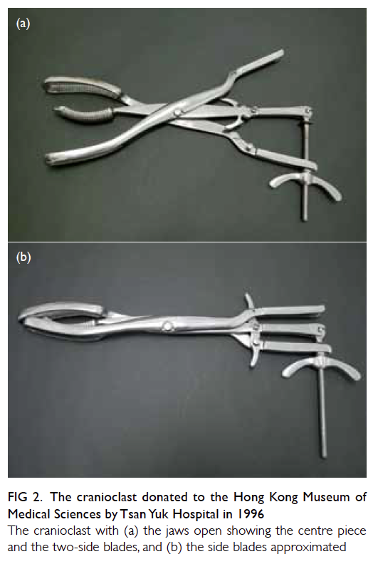
Figure 2. The cranioclast donated to the Hong Kong Museum of Medical Sciences by Tsan Yuk Hospital in 1996
The cranioclast with (a) the jaws open showing the centre piece and the two-side blades, and (b) the side blades approximated
The art of midwifery must be one of the oldest
of acquired skills. In its early days, difficult deliveries
and obstructed labours must have taxed the skills
of the obstetrician. When the fetus was stuck at the
pelvis it would die from the prolonged labour and
asphyxia. Assisted delivery by means of forceps and
caesarean section were not introduced until the 17th
century. Various destructive instruments seem to
have been known for a very long time, but there are
scanty descriptions of them in texts.
Francois Mauriceau (1637-1709), the famous
French obstetrician, was the first to describe
craniotomy and extraction of a dead child. He
invented a perforator and another instrument to
extract a dead baby after making a hole in the head
so that the brain could come out and the bones
collapse. In the 18th century, William Osborn (1736-1808) was a great believer in craniotomy. He claimed
to have once successfully extracted a fetus through
a pelvis with an anteroposterior space as narrow as
three quarters of an inch.1 2
When I was a student in obstetrics at the
new Tsan Yuk Hospital in 1957, I first saw the
instruments shown here. They were placed in the
library of the hospital, moved there from the old
Tsan Yuk Hospital. I never saw them used. They
were donated to the Hong Kong Museum of Medical
Sciences by the Hospital in 1996 at the foundation
of the Museum. The reason why these destructive
instruments became obsolete is obvious: they
were dangerous instruments. Often they not only
destroyed the fetus, but also caused serious injury to
the mother. Since the 20th century, delivery has been
made safe with the use of forceps, vacuum extraction,
and caesarean section. Destructive operations have
been abandoned forever.
Nevertheless we have great sympathy for our
ancestors. In such difficult situations, no intervention
might also be disastrous. The following sad story gives
an account of the plight of those who belonged to the
non-intervention school. Sir Richard Croft (1762-1818) was the obstetrician attending the only daughter
of King George IV, Princess Charlotte, whose son
would have been in line to the throne. The princess
had a very difficult delivery, but Croft allowed her
to go for 2 whole days of labour without assistance,
even denying her any sustenance, although forceps
were at hand. When the child was delivered, stillborn,
it was found to be a large, 9-pound male. The poor
exhausted mother had a retained placenta that had
to be delivered manually, by which time she was very
weak indeed. Croft’s brother-in-law, Baillie, advised
restoratives, and she was given large quantities of port
wine. She died the following morning. Sir Richard
was so criticised for his handling of the case that he
committed suicide by shooting himself.2
References
1. O’Dowd MJ, Philipp EE. The history of obstetrics and gynaecology. New York and London: Parthenon Publishing Group; 1994: 141.
2. Radcliffe W. Milestones in midwifery. Bristol: John Wright & Sons Ltd; 1967: 50.